Author
Gonobobleva Elena Anatolyevna
Leading doctor
Dentist
until January 31
Free* consultation with dentists (generalist, surgeon, orthodontist) More details All promotions
Dental computed tomography
(CT) is a modern method of radiation diagnostics that allows you to obtain a three-dimensional image (3D) of the entire maxillofacial apparatus. Dental computed tomography is currently the most informative study of the condition of the oral cavity and teeth. It helps to identify diseases in the initial stages that are difficult to detect on conventional (2D) radiographs. It is especially important to do a dental CT in preparation for implantation and prosthetics. The resulting 3D image greatly simplifies the production of a three-dimensional model of the jaw.
3D CT – what is this procedure and what is it for?
3D imaging of the jaw is one of the main dental procedures used in diagnosis. The data obtained allows us to assess the condition of a specific maxillofacial area and plan further treatment.
This diagnostic tool is universal; it is used by both therapists and orthopedists, periodontists and implantologists. Using a 3D image, the therapist assesses the condition of the roots and canals of the tooth, and clarifies the localization of the inflammatory process.
An orthopedist can examine the anatomical structure of the temporomandibular joints, an implantologist can examine the volume and density of bone tissue in the area of the upcoming operation, as well as the parameters of the maxillary sinuses (maxillary sinuses).
Sometimes a large part of the tooth remains, as they say, “behind the scenes”, and the dentist simply cannot see it. Then computed tomography comes to the rescue, which allows you to examine the hidden area and prevent unpleasant consequences for the patient.
Until recently, the “gold standard” for instrumental diagnostics was a panoramic image. Today there is a more advanced method in which the maxillofacial area is scanned using computer technology.
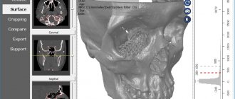
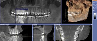
Unique visualization method
A computed tomograph produces a high-quality three-dimensional image of an individual tooth, maxillary sinuses, one or both jaws. Unlike standard panoramic images, a 3D tomogram allows the doctor to see the desired anatomical structures in a virtual section, from any angle.
During the procedure, the doctor can enlarge, rotate and study the maxillofacial area of interest at the required angle, which is unrealistic with conventional radiography.
Computed tomography is an integral and primary stage of examination before implantation. It allows you not only to assess the condition of bone tissue, but also to measure its height, width and density. Moreover, three-dimensional beams help to choose the optimal method for installing implants through preliminary virtual surgery.
3D CT is a multi-purpose and indispensable diagnostic tool that makes it possible to avoid many medical errors and complications. Thanks to this examination, the quality of treatment increases significantly and eliminates unnecessary traumatic operations.
Indications
Computed tomography is prescribed to identify:
- hidden carious lesions;
- defects in the structure of the jaw and dentition;
- fully or partially unerupted teeth;
- dystopic dental units with incorrect location or direction of growth;
- supernumerary teeth;
- damage to the dentition due to jaw fractures and other injuries;
- pathologies of the temporomandibular joint TMJ;
- tumors, cysts and other neoplasms in the jaws;
- condition of periodontal and periodontal tissues in case of gum disease, inflammation in the root area;
- number of roots, canals of teeth;
- cracks in the roots of teeth;
- features of the structure of bone tissue before jaw surgery (installation of implants, bone augmentation).
A 3D photo must be taken before dental implantation. The fact is that the jaw bone is clearly visible on a regular x-ray, but it does not allow assessing the soft tissues. On a three-dimensional tomogram, you can see in detail not only the bone, but also the nerve of the lower jaw, as well as blood vessels.
A 3D tomogram is much more informative than a panoramic image or targeted photographs of all teeth.
Price
The cost of 3D computed tomography of teeth in Moscow averages 2500-5000 rubles, depending on the scope of the diagnosis.
The price for a dental photograph at the Moscow Center for Dental Implantology RUTT starts from 1,300 rubles - depending on the scope of the examination.
Interpretation, assessment or marking for implantation, regardless of the size of the area under study, during treatment in our clinics is carried out free of charge.
CT scans can be taken on any day, including weekends and holidays. You can make an appointment for diagnostics by phone or by leaving a request online.
Free online consultation with a dentist
| Service | Price |
| Description of the radiologist (assessment and/or marking for implantation) regardless of the size of the area being examined during treatment in our clinics | from 0 rub. |
| CT scan | from 1,300 rub. |
| 3D tomography of 1 to 3 teeth (6*6 cm) with recording on CD | from 4,000 rub. |
| CBCT area 7.5*14.5 cm (both jaws) + recording on CD | from 4,000 rub. |
| CBCT of both TMJs + recording on CD and/or sending by email | from 4,000 rub. |
| 3D tomography of 1 jaw with recording on CD | from 5,000 rub. |
| 3D tomography of the temporomandibular joint with recording on CD | from 5,000 rub. |
| Evaluation and marking for implantation of 3 teeth with printing on film | from 5,000 rub. |
| Assessment and marking for implantation of 1 jaw or 2 jaws (on 1 side) with printing on film | from 7,500 rub. |
| 3D tomography of the paranasal sinuses with recording on CD | from 10,000 rub. |
| Linear tomography of the temporomandibular joint with recording on CD | from 12,000 rub. |
| Assessment and marking for implantation of 2 jaws with printing on film | from 15,000 rub. |
| 3D tomography of the 2nd jaw, TMJ, including sinuses (13.00*14.5 cm) with recording on CD | from 20,000 rub. |
Consultation and diagnostics are free!
All prices Promotions
Sign up for a consultation
three ROOTT specialists + diagnostics as a gift
CT scan before implantation
Diagnostics using a computed tomograph before installing implants allows, first of all, to determine whether implantation is necessary at all. The image will give a complete picture, and the doctor will see where the teeth are missing, whether there are problem units, and whether they can be cured.
The 3D tomogram will show:
- hidden carious cavities;
- unerupted and “extra” teeth, which may interfere with the installation of artificial pins;
- properties of roots, canals - curved, narrow and long canals require a special approach, which should be taken into account before implantation;
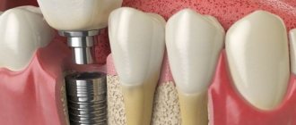
- bone dimensions in height and width, on the basis of which the type and size of the implant is selected;
- condition of bone beams, partitions, voids in the jaw bone;
- the presence of inflammatory processes in the root area - cysts, granulomas, abscesses where implants are planned to be installed. All this needs to be treated or removed before surgery;
- inflammation in the paranasal sinuses and lacrimal ducts, which can become a temporary obstacle to implantation;
- density, size, inclination of the alveolar process, thickness of the cortical bone layer, taking into account which the optimal type of artificial pin is selected;
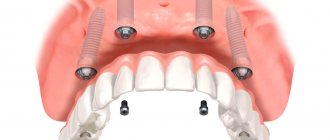
- the physiological structure of the maxillary sinuses, the mandibular canal to determine the angle of inclination of the implant rod;
- defects and anomalies in the structure of the dentofacial apparatus;
- quality of installation, strength of fixation of implants after surgery for their implantation;
- severity and nature of traumatic injuries in fractures.
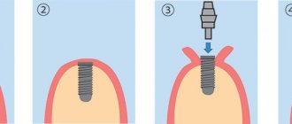
Based on the results obtained, a virtual operation to install the rods is performed. The appropriate size of the titanium pin is selected, its inclination and the point of implantation are determined, bypassing the anatomical structures. Thus, the final outcome of implantation is modeled.
Next, the tomographic data is loaded into the computer, and the program creates a three-dimensional model. The patient’s personal surgical template is printed on a 3D printer - an overlay with guide holes for inserting rods.
During implantation, the template is placed tightly on the gums, and the placement of the pins is carried out with extreme precision.
Multispiral or cone beam?
A multispiral tomograph performs layer-by-layer scanning of an object along a spiral trajectory caused by the continuous movement of the table and the X-ray tube relative to each other.
Most often, MSCT - multislice computed tomography - is used in maxillofacial surgery for facial injuries and pathologies of the temporomandibular joint.
In dental practice, especially when planning implantation, this method is not widely used due to insufficient data accuracy. Since the patient lies down during the examination, the jaw connection is distorted.
In addition, the radiation level of MSCT can reach 1000 μSv, which is unacceptable, since implantological treatment involves more than one procedure over several months.
Cone beam CBCT is a more modern, accurate and safe method compared to MSCT. Its radiation exposure is less, about 25-50 μSv, which makes it possible to carry out the procedure several times a year.
3D image of teeth in dentistry
Diagnosis in dentistry is often complicated by the fact that the doctor is not able to see the oral cavity or an individual tooth in the most detailed angles. Therefore, to obtain an accurate picture of the condition of the dentition, hardware examination methods are used, which allow you to see what is inaccessible with a normal visual examination.
Technologies are constantly improving, and a 3D image of the teeth has been added to the traditional X-ray and orthopantomogram. As a rule, it is computed tomography that is prescribed by most plastic and maxillofacial surgeons, orthodontists and implantologists.
This is a modern diagnostic method that allows a specialist to see a detailed image of the jaw in different projections and from any angle. The examination in 3D format visualizes the oral cavity for a thorough examination of the dentition for the presence of pathologies and assessment of the general condition.
Content
- What does a 3D CT scan of teeth show?
- Indications for the procedure
- Main contraindications
- Can this be done for children?
- Main types of examination
- How do they take 3D photographs of teeth in specialized clinics?
- What are the benefits?
- Where can you take a 3D photo in St. Petersburg?
What does a 3D CT scan of teeth show?
This type of research is considered one of the most informative diagnostic methods in dentistry. With its help, it is possible to obtain the most reliable information about the condition of bone tissue, the circulatory system of the face and soft tissues.
Speaking about what a 3D photograph of teeth shows, we can highlight the following:
- Identification of foci of pathologies in the studied areas using a large-scale image.
- Visualization of the condition of the jaw in three-dimensional format.
- Diagnosis of cysts, tumors and other neoplasms.
- Evaluation of the effectiveness of previous treatment.
- Position of teeth, including unerupted ones.
- Separate areas of the oral cavity at the required angle.
3D dental x-ray allows you to visualize the condition of not only bone, but also soft tissue. This greatly simplifies the process of diagnosing many pathologies and inflammatory processes.
In addition, the resulting images will show the beginnings of teeth, the location of nerves and blood vessels. The popularity of this diagnostic method is due to the fact that without its use, correct installation of orthodontic structures, implantation and planned surgical interventions in the maxillofacial area are impossible.
Indications for the procedure
The use of dental computed tomography is necessary for most orthodontic and surgical procedures. This procedure is also indicated in the following cases:
- Pathologies of the temporomandibular joint.
- Anomalies in the structure of the jaw.
- Fractures of the maxillofacial region (to determine the severity of the condition).
- Preparation for upcoming dental prosthetics.
- Periodontitis and periodontal disease.
A 3D photograph of teeth is taken if there are suspicions of inflammatory processes that cannot be diagnosed by other methods.
Main contraindications
3D computed tomography of teeth is based on x-rays, so this procedure cannot be called completely safe. The radiation exposure during this examination ranges from 0.045 to 0.06 mSv. This is not a very high figure, given that the annual exposure limit is 5 mSv (according to the Russian Ministry of Health).
As for contraindications, they are standard for all types of x-ray examinations. The main limitation is the period of pregnancy (especially in the first trimester). In this case, the situation is considered individually, based on the ratio of harm to the child and benefit to the mother.
If a contrast agent is used (it is not used so often in CT scans of the teeth and jaw), then the following can be added to the contraindications:
- Patients suffering from thyroid diseases.
- Kidney failure.
- Allergy to drugs containing iodine.
The lactation period is not a contraindication, but after the procedure at least 48 hours must pass before the next breastfeeding.
Can this be done for children?
Many parents think that the use of 3D computed tomography is impossible in childhood due to the negative impact on the child’s body. This is not entirely true, because special attention to this study is given specifically in pediatrics.
If you follow all safety rules and do not exceed the frequency of procedures (for children – once a year), then the examination will not lead to any pathological changes in the child’s body.
3D CT in pediatric dentistry allows you to assess the condition of the sinuses, gums and see the rudiments of baby teeth. This method also allows you to identify malocclusion pathologies in a child.
Main types of examination
Three-dimensional images are taken using various types of tomographs. It is extremely important to know which device the examination will be used on, because the dose of radioactive radiation received and the degree of information content depend on this.
There are several main methods:
- Step-by-step computed tomography. It was carried out on old tomographs that produced images of low resolution and detail. Now it is no longer used, because the minimum radiation dose was 1500 μSv (with a standard of 1000 μSv).
- Multislice CT (MSCT). Body scanning is carried out using sensors located on a rotating tube. The table moves slowly through the ring, allowing you to take pictures in several planes at once. This method is characterized by the fact that the detectors are arranged in several rows. Such equipment is better suited for examining lymph nodes, muscles and other soft tissues. The radiation dose ranges from 300 to 400 μSv.
- Cone beam CT (CBCT). This technique is considered the most modern and informative in the field of dentistry. The main advantage is the maximum accurate image of teeth and bone tissue, which cannot be achieved using MSCT. CBCT is also considered the safest type of computed tomography (on average, the radiation dose ranges from 40 to 120 μSv).
How do they take 3D photographs of teeth in specialized clinics?
The average time of the procedure is no more than 1 minute. During this short period of time, the device takes about 200 pictures in various projections.
Before the procedure itself, you need to remove all metal objects in close proximity to the area being examined (chains, piercings, etc.). The examination itself looks like this:
- The patient's head is securely fixed using immobilization devices. This is very important, otherwise the pictures will turn out blurry.
- A protective apron is put on the chest.
- The rotating tube of the tomograph rotates around the head, taking a series of images.
- All information is immediately displayed on the computer monitor connected to the device.
- Further interpretation of the images is carried out by a specialist.
What are the benefits?
Computed tomography is a completely painless procedure that does not cause any discomfort to the patient. The only thing is that he must remain motionless for a short amount of time.
The use of 3D images is due to the following reasons:
- Minimum radiation dose.
- The examination procedure lasts no more than a few minutes.
- Get results quickly.
- Three-dimensional images have a high diagnostic value.
- 3D images eliminate errors in prosthetics and dental surgery.
Where can you take a 3D photo in St. Petersburg?
Diagnostic X-ray - invites you to undergo a 3D CT scan of the jaw if your physician has prescribed this procedure for you. We have no queues, so you can quickly receive images for further delivery to a medical institution.
For the convenience of our clients, we have organized courier delivery within St. Petersburg. You also have access to recording results on digital media, extended consultation and other additional services. You can find out more detailed information on the website.
To make an appointment, you need to call the specified phone number, or leave an online application.
Sign up for a study by phone
+7 (812) 332-52-54
3D CT or panoramic image?
An orthopantomogram allows the doctor to assess the condition of the teeth, root canals, and soft tissues. With its help, hidden inflammations, abscesses and abnormally located teeth are revealed.
However, a panoramic photo does not give a 100% accurate picture; the error is about 20%. Even a slight shift causes the focal spot to shift, and the image is compressed or stretched.
Due to the difference in the refraction of X-rays by tissues of different densities, it is impossible to assess the properties of the cancellous bone layer, since it is simply not visible behind the denser periodontium.
A two-dimensional orthopantomogram is, in fact, an auxiliary technique that gives a general idea of the condition of the oral cavity and identifies mainly obvious pathologies. It does not show the configuration and structure of the alveolar process at the desired level.
The advantage of a three-dimensional 3D tomogram is that it produces not one flat photo, but several consecutive images from different angles.
The doctor sees and evaluates all necessary objects located at any depth, from all sides and at different angles.
Contraindications for 3D dental imaging
The radiation exposure of CT scans ranges from 0.045 to 0.06 mSv. This is relatively little, given the recommendations of SanPiN, which indicate the upper possible threshold of radiation exposure for research purposes equal to 1 mSv per year. However, if you compare a CT scan with a dental x-ray, where radiation occurs in the range of 0.002 - 0.003 mSv, a 3D photograph of teeth no longer seems so harmless. In this regard, there are a number of contraindications to CT scanning. If we are talking about simple tomography, then, as a rule, it is not done during pregnancy, especially in the first trimester. The doctor will always consider the need for radiation exposure for a pregnant patient based on the balance of benefit to the mother and risk to the fetus. If tomography with contrast is necessary (for better visualization of soft tissues and blood vessels, it is rarely used for CT of the jaw), then the number of contraindications expands. It should not be given to pregnant women, nursing mothers, or to persons suffering from thyroid disease, severe diabetes mellitus, renal failure, or an allergy to iodine. If you have any allergies or have ever experienced drug intolerance, be sure to tell your doctor.
How to make a 3D tomogram
Usually the procedure is performed standing, the patient bites a small flat plate with his teeth and stands without moving for 15 to 30 seconds. The device makes several rotations around the head, managing to take about two hundred pictures in various projections.
In 10-15 minutes, the information is processed and transferred to electronic media.
We invite you to make a three-dimensional tomogram in our clinic using the latest generation dental tomograph. Sign up for the procedure online or by phone at a time convenient for you.
How is the research going?
The examination is carried out in 1-3 minutes:
- The doctor takes the patient to the CT scanner and helps fix the head in the desired position. Depending on the design of the tomograph, the examination takes place while sitting or standing.
- A small plate covered with a disposable cover is placed in the mouth, which fixes the position of the jaws.
- The scanning arc of the tomograph makes one revolution around the patient's head. The exposure time to X-ray radiation does not exceed 20 seconds.
The specialist writes the obtained data onto a CD and, if necessary, prints it onto film.