Computed tomography (CT) allows you to most accurately diagnose the condition of the patient’s oral cavity. That is why this procedure must be done before implantation. Only based on the results of such diagnostics can the dentist make a conclusion whether implantation is indicated or, conversely, contraindicated for a particular patient.
CT allows you to obtain a three-dimensional image of the jaw, and in great detail. This is a kind of successful alternative to an x-ray. In dentistry, spiral or multispiral dental tomographs specially created for this purpose are used. They scan tissues and transmit data to a monitor.
Advantages of computed tomography
The results of computed tomography make it possible to detect any pathologies in the oral cavity at the earliest stage of their development. Thanks to this, treatment can be prescribed on time and, accordingly, proceed faster than with an advanced problem. For successful dental implantation, three-dimensional photographs are simply necessary for the dentist; this is the only way to choose an error-free option for installing implants. Statistics show that in 65% of cases of implant failure, dentists did not have three-dimensional photographs of the patients’ jaw bones. On the other hand, if such images are available, successful implantation is observed in 98% of cases.
Benefits of CT
- Minimum X-ray exposure. CT scan is indicated even for young children.
- Diagnostics takes from 8 seconds to 5 minutes.
- There is no need to decipher the diagnostic result.
- The highest quality and resolution of images.
- Adjustable image contrast allows you to reveal the smallest details.
- The ability to study the problem area in three planes.
- Ability to change the scale of the image or part of it.
- The ability to see the location of nerves and thereby optimally select places for bone drilling.
- The ability to quickly make copies of a photo.
- The ability to detect complications invisible to the eye.
Computed tomography is a completely painless procedure. It does not even cause the slightest discomfort in the patient. Depending on the type of tomograph, diagnostics can be performed while the patient is sitting, lying, or standing. No preliminary preparation is required to carry it out. A computed tomography scan can be done even when the patient comes only for a consultation. Pictures can be received instantly. Computed tomography is important for both the patient and the dentist; there is no more accurate diagnosis.
Pro Surgical 3D
Free program
Pro Surgical 3D is another free program that specializes in viewing medical images from CT and MRI scanners.
Pro Surgical 3D allows you to quickly view patient images, simplifying the workflows associated with 2D and 3D CT and MRI examinations. The program provides treating physicians with a variety of information to develop optimal treatment plans for patients. And helps patients better understand their health status and treatment options.
Among the functionality and features of the program, we note:
- The program is designed primarily for viewing DICOM images from CT and MRI scanners, but it also supports common non-DICOM file formats: NifTi (.nii), Visualization Toolkit (.vtk) and ANALYZE (.hdr).
- Ability to import DICOM from PACS (local and online database), CD/DVD, USB and local computer.
- Ability to load DICOM images into PACS directly from CD or local computer.
- Easy to use search of patient metadata.
- Performing 3D reconstruction and volumetric visualization (3D rendering).
- Multiplane and oblique cutting.
- Instant interactive surface extraction and export to STL and PLY formats.
- Simple anonymization and de-identification of patient images.
- Adjust the brightness and contrast of images.
- Creating and saving screenshots.
- Function for parallel comparative assessment of pre- and postoperative scans.
- Availability of tools to perform various measurements.
Disadvantages of Computed Tomography
Alas, this seemingly completely harmless procedure has its drawbacks. They consist in the fact that CT has a number of contraindications. These include the following reasons:
- an allergic reaction to a contrast agent that is used to pre-treat the patient’s oral cavity;
- pregnancy at any stage;
- breastfeeding a baby;
- the impossibility of diagnosing excessively active young children;
- panic fear of closed spaces;
- diabetes;
- kidney diseases;
- thyroid diseases.
The introduction of a contrast agent into the oral cavity allows you to obtain the highest quality image of the oral cavity. However, as a last resort, you can do without this, which experienced dentists sometimes do.
A CT scan of the oral cavity can be done no more than twice a year. Moreover, over the next year it is necessary to refrain from this procedure.
What determines the radiation dose during CT?
The amount of radiation a person receives during a tomography depends on three main factors:
- Scan area. It's not about whether it will be a targeted shot or a panoramic shot, but about the part of the body that is to be examined. It has been scientifically proven that individual tissues of the body react differently to exposure to x-rays. One of the smallest doses is obtained when examining the dental system.
- Scan time. The time required to obtain CT data is taken into account. The use of cutting-edge equipment makes it possible to reduce the exposure time to X-rays to a few milliseconds.
- Type of equipment. Since tomographs are used in different fields of medicine and for different purposes, they are set to a specific time and intensity of radiation. High-field tomographs, better known as MRI, are considered the most powerful. And small-sized dental (intraoral) portable scanners are gentle.
Manufacturers of medical dental equipment know that CT scanning of teeth and jaws is required more often than tomography of other organs. That is why the devices used in modern clinics provide the minimum possible radiation exposure to the body, equal to approximately 0.039-0.06 mSv. This allows not only to repeat tomographic studies several times a month, but also to carry out diagnostics that are as harmless to health as possible, even for children.
How is a tomographic image taken?
A three-dimensional dental tomograph consists of a scanner and a computer. Scanning of the oral cavity is performed using very weak x-rays.
Currently, the industry produces two types of tomographs for dentistry. Tomographs of the first type have a scanning device in the form of a cylinder, through which a table with the patient moves. Tomographs of the second type are equipped with a head stand. The stand is installed on the rotating part of the device. If the first type of tomograph is universal, then the second is designed specifically for the needs of dentistry.
Both types of tomographs work on the same principle. The scanner takes from two hundred to six hundred images of the patient’s oral cavity within one hundred seconds. All of them are sent to the computer in electronic format. The program installed on it analyzes the images and produces an accurate image of the area under study. A three-dimensional image is formed by superimposing layers of different thicknesses on top of each other. Each such layer is separately saved as a DICOM file and can be examined separately.
The tomographic diagnostic procedure itself is not at all burdensome and is done in the following sequence.
- The patient is freed from all metal objects that he has with him.
- The patient places his chin on a support.
- The patient remains completely motionless for several seconds.
That's the whole procedure. If the diagnosis requires the administration of contrast, the patient must not eat or drink anything for 4 hours before the diagnosis.
Syngo fastView: instructions for patients
Syngo fastView does not require installation. It is enough to launch the disk on your computer or open the flash drive that you received at the medical center.
Find out how to run a program from a DVD.
There are several ways to run a program from disk. Make sure your drive supports DVDs. Insert the study disc into the drive.
1. Wait for the pop-up window to appear, click on it once with the left mouse button:
Another pop-up window will appear, select “Run autorun.exe” in it:
2. If the pop-up window from step 1. does not appear or appears and disappears, open any folder on your computer and select “disk drive” in the left side menu. Please note that your drive may be called differently, for example, “DVD R drive (D:)”, but its name will definitely contain the word “disk drive” (or “drive” if you use Windows in English):
Click on this menu item once with the left mouse button, in the right window select the line “AUTORUN.EXE”, click on it twice with the left mouse button:
3. If there is no item in the left menu with the word “drive” (or “drive”), select “This computer” (“Computer”), click on it once with the left mouse button:
In the window on the right, select your drive, double-click on it with the left mouse button:
Next, select the line “AUTORUN.EXE”, double-click on it with the left mouse button:
Completing any of the three steps will lead you to a pop-up program window. Click "OK" and wait for the program to load:
Find out how to run a program from a USB flash drive
If you received your study on a USB flash drive, you do not need special equipment to view the images. Insert the flash drive into an available USB port on your computer and follow the instructions below.
1. Most often, the flash drive is detected and opened automatically. You will see a folder named "1". Enter it. There you will find program and research files. Select the file “autorun.exe”, double-click on it with the left mouse button:
2. If the flash drive does not open automatically, open any folder on your computer, in the left menu find the “My Computer” item (or “Computer” if you are using Windows OS in English). Click on it once with the left mouse button. In the right window, among the devices and disks, find the flash drive with the research results, click on its designation twice with the left mouse button:
You will see a folder named "1". Enter it. There you will find program and research files. Select the file “autorun.exe”, double-click on it with the left mouse button.
3. If in one of the previous paragraphs, when you click on the “autorun.exe” file, the program does not start, go to the “syngo_fV” folder, scroll through the files in it to the end, find the “syngo_fV.exe” file, double-click on it with the left mouse button :
Following these instructions should lead you to a program pop-up window. Click "OK" and wait for the program to load.
Open CT images on your computer
When the program loads, two windows will automatically open: 1 - Syngo fastView - the main program window (opens in full screen by default); 2 — fastView Browser — window of the built-in browser (loader) of files with research.
If the fastView Browser
with study files did not open, in the upper left corner of the
Syngo fastView
, click: “Patient” → “Open Patient Browser” (Patient → Open patient browser).
In the window that opens, in the “Source” section, click on the rectangle with three dots:
Step 1: In the pop-up window that opens, find the DICOM folder under the line with the word “disk drive” or “drive” (and if you received the research results on a USB flash drive, then find a similar folder on the flash drive) and select it with one click of the left mouse button. There is no need to open the folder. Step 2: Click the "Add" button.
What exactly does CT help with successful dental implantation?
Computed tomography is not a mandatory procedure when preparing a patient for dental implantation. You can do without it by trusting the experience of the dentist. However, in this case, there is a high probability of unsuccessful implantation. Simply put, there is no complete guarantee that the implant will grow into the bone safely.
Computed tomography allows the dentist to obtain:
- comprehensive information on jaw bone density;
- a complete picture of existing foci of inflammation and defects in the oral cavity;
- an accurate picture of the location of the maxillary sinuses, nerves and blood vessels;
- the ability to accurately calculate the size of implants and the optimal depth of their immersion into the bone;
- the ability to accurately determine the optimal angle of inclination of the implant in relation to the dentition;
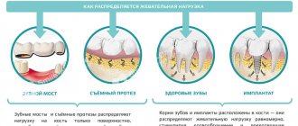
- the ability to calculate the maximum permissible mechanical load on the implant.
Agree, each of the listed points is extremely important for successful implantation. Under no circumstances should any of these points be neglected.
Indications
Determining whether a patient has indications for this tomography is usually determined by a dentist, oral surgeon or orthopedist-traumatologist. Computed tomography of the jaw is primarily intended to identify bone diseases and pathologies of the maxillofacial area, including:
- injuries of the upper and lower jaw
- dislocations and subluxations;
- osteomyelitis
- osteonecrosis
- osteoarthritis and osteoarthritis.
Computed tomography data will additionally be able to show some dental defects and dentition anomalies, bone cysts. Tomograms can be used to first approximate the condition of the temporomandibular joint, but its pathologies are better visible on MRI of the temporomandibular joint. In the field of maxillofacial surgery, MSCT is used to create implants and maxillofacial structures.
Individual application
Dental implantation is not some cookie-cutter procedure. On the contrary, it requires an exclusively individual approach, since no two identical jaw bones exist in nature.
The key to successful implantation is the complete surrounding of the implant surface with bone tissue. The implantation should be completed without the slightest gap between the bone and the artificial root. This process occurs differently for each person.
The dentist’s task, among other things, is to choose the right place to drill a hole for the implant. In this place, the density of bone tissue should be maximum, but it is far from being the same throughout the entire volume of the jaw.
No less important is the choice of the angle of inclination of the implant. If there is an error, the crown will not fit into the overall shape of the remaining dentition.
The drilling depth must also be calculated as accurately as possible to avoid damaging the nerve.
Without computed tomography data, it is extremely difficult to do all this.
What is dental tomography?
When performing any type of CT scan, the principle of different conductivity of X-ray rays by different tissues of the body is used: cavities, bones, muscles, ligaments, etc. In this case, the rays penetrate through any tissue of the body and are captured using a special detector. After a series of layer-by-layer images, a 3D computed tomography model is built using a computer.
Most often, dental tomography of teeth is prescribed before prosthetics, dental implantation, or surgical intervention on the jaw.
The X-ray method does not provide all the advantages that modern computed tomography can provide. Dental CT allows you to study various features of dentofacial anatomy: the structure of teeth and canals, the condition of the jaw bones.
CT results are indispensable for orthopedists, pediatric dentists, orthodontists, and surgeons. For example, for the most accurate production of any prosthesis, the doctor often needs data on the exact dimensions of the jaw anatomy and the most detailed location of the teeth. Only in this case can the prosthesis be manufactured with maximum comfort for the patient.
If, when using implants, the artificial root is implanted incorrectly (inaccurate dimensions or in the wrong location), then the prosthesis will be uncomfortable for the patient or will quickly fail.
Therefore, CT in many clinics is a mandatory preparatory procedure for any type of dental prosthetics (removable or dentures).
Dental tomography is also indispensable for orthodontists, as it ideally shows the exact location of the teeth. In case of surgical pathologies, this method allows you to fully examine the existing dentofacial injuries in all projections.
Risks and dangers of dental implantation
The most risky is dental implantation in the lateral parts of the upper jaw. This is due to the presence of the maxillary sinuses in this area. The trouble is that after the loss of teeth, these sinuses tend to increase in size. It is almost impossible to find out their size without a computed tomography scan. Problems in the maxillary sinuses can significantly complicate dental implantation in the upper jaw, and therefore diagnosis of the sinuses is done as carefully as possible.
If there is chronic inflammation, tumors, polyps or cysts, dental implantation is not performed until all these problems are eliminated. If this is not done, then the implants in the bone tissue simply will not take root and will be rejected by the body. An opinion on the readiness of the upper jaw for implantation is given by two specialists - a dentist and an otolaryngologist. This approach minimizes the likelihood of implant failure.
Work with the “Image” tab: change the size and position of the image as you see fit
Also in the “Image” tab you will find the following tools for working with images: 1 - Pan - Moving (Panning) 2 - Zoom - Scaling 3 - Magnify by 1.25 - Increase up to 125% 4 - Magnify by 0.8 - Reduce up to 80% 5 - Home View - Default View
When you click on the “Magnify by 1.25” icon (3), the image will be enlarged by 125% of the previous value. You can only enlarge the picture up to a certain limit.
If the enlarged image extends beyond the visible area, move it around to see all parts. To do this, use the move tool - click on the “Pan” icon. Please note that the cursor will change from an arrow to a hand:
Hold down the left mouse button and drag the image across the screen in any direction. You can move not only an enlarged image, but also an image of any other size.
To reduce the image, click on the “Magnify by 0.8” icon (4) as many times as necessary - each click will reduce the image to 80% of the previous value.
You can also quickly zoom in and out of images using the zoom tool. To do this, click on the “Zoom” icon (2), hold down the left mouse button and move the cursor up and down in the Image Display Window. When the cursor moves up, the image will increase, and when you move down, it will decrease.
To restore the image to its original form after applying any of the resizing functions, click the house icon (5):
Hold down the left mouse button and drag the image across the screen in any direction. You can move not only an enlarged image, but also any other size.
To reduce the image, click on the “Magnify by 0.8” icon (4) as many times as necessary - each click will reduce the image to 80% of the previous value.
You can also quickly zoom in and out of images using the zoom tool. To do this, click on the “Zoom” icon (2), hold down the left mouse button and move the cursor up and down in the Image Display Window. When the cursor moves up, the image will increase; when moving down, the image will decrease.
To restore the image to its original form after applying any of the resizing functions, click the house icon (5):
What are the consequences of not having a three-dimensional image?
Refusal to conduct a computed tomography scan can lead to very sad consequences. The fact is that without a three-dimensional image, the dentist makes a decision almost intuitively, and this, of course, is a risk. The consequences may be as follows:
- nerve damage;
- inflammation of the gums due to uneven load on the implants;
- premature wear of the structure;
- the likelihood of resorting to treatment for problems with the oral cavity;
- risk of re-installation of the implant.
Save DICOM images in a convenient format
With Syngo fastView, you can save images from DICOM files in other, more familiar formats: JPEG (plain image) or BMP (bitmap). To do this, select the “Patient” tab in the main menu, and in it the “Save as...” item:
In the pop-up window, select the file type from the drop-down menu (JPEG or Bitmap (BMP)), select the folder on your computer where the image will be saved, click “Save”:
The image will be saved with service information. To save only the image, without data on it, before saving, click on the “Show/Hide Image Text” icon, see the item “Hide service information in the image”
CT scan after implant placement
Many patients have questions about whether a CT scan can be performed after implants, crowns or pins have been installed. This question is asked for a reason. Many people know that when performing an MRI, artificial materials present in the oral cavity create a “phonic” sound and thereby confuse dentists. Nothing like this happens with a CT scan. Moreover, it is advisable to do a computed tomography scan before and after dental implantation. This will allow, firstly, to verify the success of the operation, and secondly, to notice in time possible problems with implantation.
However, if there are metal crowns in the oral cavity, it is better to refuse computed tomography. The fact is that metal interferes with the normal operation of the scanner. As a result, the picture turns out to be of poor quality. The same applies to metal pins.
Features of irradiation during CT and not only
The operation of the tomograph is based on the effect of x-rays. Without knowing all the features of the procedure and equipment, some still believe that dental CT is harmful to health, since the body is exposed to radiation. At the same time, few people remember from a school physics course that a person is exposed to radiation everywhere, even outside the X-ray room, for example:
- Outdoors on clear days. The sun is the largest natural source of radiation.
- At home, being near switched on household appliances: TV;
- refrigerator;
- various kinds of gadgets, etc.
To understand what kind of radiation exposure a person receives during a CT scan of the jaw and whether it is harmful, it is worth learning a little more about the radiation itself. It is measured in sieverts (Sv), millisieverts (mSv) and microsieverts (µSv). According to SanPiN standards, the following are safe for a person undergoing preventive medical examinations:
- 1000 μSv per year for an adult;
- 300-400 µSv per year for children under 15 years of age.
Taking into account the fact that one x-ray examination is performed with a radiation dose from 2-3 μSv (sighting shot) to 16-18 μSv (panoramic shot of the jaw), it is permissible to take 300-500 targeted shots or 60-70 shots of the jaw per year. These figures reflect average data without reference to any specific type of equipment. But they also show that it is not harmful to the body to receive only a few microsieverts for a detailed diagnostic picture.
You might be interested in:
Dental diagnostics
Computed tomography of teeth and jaws
Orthopantomogram of teeth