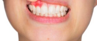
A fistula on the gum (also called a fistula) is an opening in the gum from which pus is released, often with blood. A fistula appears against the background of caries, periodontitis, and also due to poor-quality dental services. Over a long period, the fistula develops asymptomatically, but subsequently can lead to severe complications. Therefore, if a purulent formation appears on the gum, you should immediately contact your dentist.
Fistula on the gum: causes
A fistula on the gum is a hole that forms as a complication after inflammatory pathologies, including untreated ones. It can also occur after dental intervention (poorly provided services). Very often, a fistula is formed as a complication of long-term chronic periodontitis.
Also, the reasons for its appearance may be:
- damage to the tooth root;
- advanced caries;
- pulpitis;
- inflammatory processes of the cyst;
- problems with the growth of wisdom teeth;
- improper teething in children;
- granuloma - inflammatory processes in the tissues of the periosteum and the apical region of the tooth root (accompanied by an increase in temperature, enlarged lymph nodes and other signs).
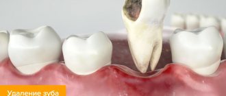
Fistula in the soft periodontal tissues after tooth extraction
The formation of a hole after tooth extraction is normal. As a rule, the hole heals within one to two weeks. If the operation was performed on wisdom teeth, the tightening process may take several weeks. Impaired socket tightening can occur due to infection or in case of injury to the area where the tooth was removed.
In a situation where the hole has tightened and a gap has formed in the periodontium, it may indicate that the tooth was not completely removed. Small fragments of the tooth gradually begin to decompose, forming a fistula.
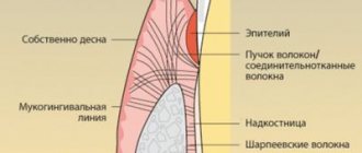
How a fistula is formed
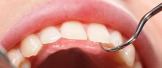
The process of fistula formation looks like this. A small hole appears in the gum near the base of the tooth. Its color stands out a little against the background of healthy tissues - the color is rich pink or red. At the same time, the remaining teeth remain healthy and do not hurt.
At first, the fistula looks like a small swelling, then it grows and resembles a pimple or an abscess. The last stage - the seal opens, after which mucus or pus comes out of it. Then the wound becomes covered with a scar, but does not disappear. In the future, the development of the fistula can become chronic - it will open several times a year and exude pus. During exacerbations, mild pain will be felt.
The appearance of a hole in the periodontium in a child
The formation of a hole in the gum near a tooth in a child can be due to reasons such as: periodontitis caused by complicated caries, or injury to the oral mucosa. With periodontitis, the lesion reaches the jaw bone. A cavity appears inside the periosteum, which is filled with pus. When there is an excess of pus, it breaks out through the fistulous tract, thereby bringing severe pain.
Frequent mechanical damage to the mucous membrane and gums causes infection to enter the oral cavity. Bacteria entering the periodontium cause inflammation and fistula formation.
Symptoms of a fistula: how dangerous is it?
Fistula manifests itself with a number of symptoms:
- unpleasant odor from the mouth;
- tooth mobility;
- feeling of foreign taste;
- mucous membrane is bluish or, conversely, pale in color;
- discharge of purulent mucus, often with blood;
- painful sensations during mechanical contact (chewing food, drinking hot drinks, brushing teeth).
The disease may also be accompanied by symptoms not directly related to the oral cavity. It is manifested by such signs as apathy, general weakness, and fever.
Despite the small area affected, the fistula poses a certain health hazard. Thus, purulent discharge can penetrate the lymph or blood, which will lead to diseases of the internal organs, pathological processes in facial tissues and even loss of teeth. Therefore, even at the first relatively mild symptoms, it is important to consult a doctor.
It is worth keeping in mind that the disease is asymptomatic for a very long time. Moreover, if treatment is delayed, this can lead to partial death of the periosteum. And then the patient will have to remove not one, but several teeth at once.
Prognosis and possible complications
If the fistula is detected and treated in a timely manner, the prognosis is favorable. If you follow all the specialist’s recommendations, within a week the resulting wound will completely heal and heal.
If proper treatment is not carried out, the following consequences are possible:
- abscess formation;
- phlegmon;
- osteomyelitis;
- tooth loss.
Preventive measures
- regular teeth cleaning;
- carrying out professional hygiene in the dental office;
- preventive examinations by a dentist every six months;
- daily consumption of fruits and vegetables.
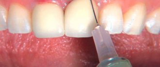
Diagnosis and treatment of fistula
To carry out a diagnosis, you must contact your dentist. The doctor orders an x-ray and conducts a visual examination. Treatment is carried out in a clinical setting. Therapy depends on the cause of the fistula. For example, fillings are used to treat caries, and when an infection is detected, appropriate medications are used. The dentist can also perform treatment with ultrasonic waves or laser.
In some cases, a fistula appears due to poorly done filling of the canal. Then the patient is given an anesthetic, the filling is removed, the dental canal is cleaned and then a new high-quality filling is performed.
If the cause is a cyst, it is removed, which also requires the intervention of a dentist. For recovery, the use of antimicrobial agents, antibiotics, and antihistamines is indicated. If swelling occurs, the doctor may prescribe rinsing with a salt solution.
What reasons can lead to its appearance?
The main cause of epulis on the gums is considered to be constant mechanical impact (rubbing) of soft tissues.
Injury to one area can cause gum swelling:
- poorly installed seal;
- uneven edges of the opposite tooth, which is destroyed or chipped;
- tartar;
- poor quality or incorrectly installed prosthesis;
- agonist tooth for malocclusion;
- adjacent teeth when teeth “pile up.”
Supragingival tissue occurs and, as a consequence, injury to epithelial tissue as a result of a burn, bruise or constant ingestion of irritating foods. In pregnant women, tumors occur due to hormonal imbalances. Epulis on a child’s gums can form due to general causes or disturbances in the eruption of permanent teeth.
Is treatment possible at home?
A fistula can only be treated in a clinic. But if it is impossible to urgently visit the dentist, for example, the pain occurred at night, it is recommended to rinse the mouth with the following means:
- infusion of chamomile;
- infusion of oak bark (dry raw materials can be purchased at a pharmacy);
- if there is purulent discharge, the mouth should be rinsed with a solution of salt and soda or antibacterial agents.
The described measures help relieve pain, but do not eliminate the cause of the disease. Therefore, in any case, you need to see a dentist.
What is epulis
A tumor on the gum is characterized by a benign nature. It is classified as a group of periodontal diseases. Epulis is also called “giant cell granuloma” or “supragingival”. Epulis looks like a lump that is attached to the gum in the area of the interdental space on a stalk. A formation is formed from the epithelium of the alveolar process.
In diameter, an epithelial tumor of the gums can reach a size of more than 3 cm. The shade of a giant cell granuloma can be different: bluish, brown, red-brown, or the color of the gums. Ulcers may appear in the affected area.
If the epulis on the gum has a diameter of up to 2 cm, does not cause pronounced symptoms and progresses slowly, then the pathology is classified as a benign form. With rapid growth of the supragingival tissue, severe pain, inflammation and swelling of the gums, there is a risk of developing a malignant tumor.
According to statistics, the pathology is most often diagnosed in pregnant women, but supragingival formation also occurs in women who are not pregnant. Less commonly, the disease occurs in men and young children when replacing milk teeth with permanent ones.
Prevention of fistulas
The main measure for preventing fistulas is maintaining oral hygiene. It is necessary to undergo an annual examination by a dentist and, if necessary, begin treatment for caries and periodontitis in order to prevent the development of chronic diseases.
It is also advisable to be treated by experienced, qualified dentists, since poor-quality services can also lead to the formation of a fistula. After the intervention, you need to monitor your well-being to prevent the development of a fistula.
Symptoms
Signs of fistula formation include:
- aching pain that increases in the evening;
- burning and itching of gums;
- swelling in the affected area;
- gum hyperemia;
- increased body temperature;
- general weakness, chills;
- increased irritability;
- apathy.
The appearance of symptoms of pathology is individual for everyone. Some may experience very severe pain and discomfort in the mouth, while for others the symptoms may be almost invisible.
Diagnostic methods
Typically, parents or a dentist detect fistulas at a very early stage. They are localized on the outer or inner side of the gums and reveal themselves by noticeable swelling and the appearance of a whitish lump.
It is very important not to self-treat when a problem is discovered. If your baby has baby teeth, improper treatment can pose a risk to the formation of the dentition. When the teeth are molars, there will also be a lot of problems, and there is a risk of needing extraction.
At the dentist's appointment, the doctor will carefully examine the child and take an x-ray. The location of the inflammation, its nature, and estimated depth are taken into account. Already at the first visit, the doctor begins to develop treatment methods to try to solve the problem without surgical intervention.
Main treatment methods
The answer to the question of how to treat a fistula on the gum in a child is given by the dentist after a careful examination of the patient and collection of anamnesis. It is important that the doctor considers the process comprehensively, using not only medication, but also therapeutic methods. Sometimes proper local surgical intervention is also necessary. Let's talk about each of the listed measures in more detail.
First aid methods
When parents and a child visit the dentist, they need to answer questions about how long ago the first symptoms appeared and whether the child has other concomitant diseases. At the first stage, the symptoms of the disease are relieved, and then a deeper plan is drawn up to eliminate the root cause of poor health.
There are several basic first aid methods:
- Relieving pain syndrome. If the baby has a high fever and all signs of inflammation, you can give him antihistamines - Nurofen, Cifecon, paracetamol.
- Reducing swelling. It is best to use safe gels, such as Kalgel.
- Removal of pathogenic bacteria. Most often, this reduces inflammation. It is recommended to rinse or carefully treat the affected area with Miramistin or Chlorhexidine.
First aid helps to slow down the spread of the inflammatory process and reduce it. But you should take medications only with a clear understanding that the baby is not intolerant to them. It is best to consult a doctor.
Subsequent drug treatment will be based on taking properly selected antimicrobial drugs, antiseptics, anti-inflammatory and antihistamines.
Surgical intervention
Surgery is usually the last option to correct the situation. Doctors resort to it when other means are completely ineffective.
The main reason for prescribing such treatment is the widespread spread of soft tissue inflammation. The area becomes more and more extensive, and pus is actively flowing from the fistula.
In this case, the doctor removes the fistula and scrapes out the affected tissue. Today, laser processing is used for this purpose, which automatically seals small vessels. The likelihood of infection is minimal and the procedure itself is much faster and painless than using traditional surgical instruments.
Next, depending on the situation, the question of the need to remove the tooth is considered. Often it can be preserved and done with simple filling of the canals. A more detailed answer to the question about treatment methods can only be given after a careful examination of the patient.
Factors contributing to the appearance of a fistula tract
Among the provoking factors that are important to exclude:
- poor oral hygiene;
- presence of untreated caries;
- pulpitis;
- malocclusion;
- frequent stress;
- unbalanced diet;
- physical fatigue;
- deficiency of vitamins and minerals;
- habit of eating a lot of sweets;
- smoking.