This article provides a detailed step-by-step description of anesthesia techniques using the STA (Single Tooth Anesthesia) system from Milestone Scientific (USA).
This article provides a detailed step-by-step description of the methods of anesthesia using the STA system (Single Tooth Anesthesia – anesthesia in the field of a single tooth), the company Milestone Scientific (USA).
The history of local anesthesia in dentistry goes back more than 150 years. However, it took decades for dentists to move from the Dr. Luer syringe and Novocaine to a disposable syringe with needles of different sizes and a solution of lidocaine as an anesthetic. It took several more decades to switch to the use of carpule syringes and carpule forms of anesthetics. However, despite all the innovations, the doctor was still limited to two traditional methods of pain relief: infiltration and conduction. The method of intraosseous anesthesia developed in recent years actually turned out to be quite traumatic and risky, since the anesthetic solution could enter the bloodstream, causing various complications, especially in patients at risk. Even intraligamentary anesthesia could not become a full-fledged replacement for mandibular anesthesia due to the short duration of pain relief, the complexity of implementation and the high incidence of side effects, such as injection pain, postoperative discomfort, soft tissue damage and bone resorption. All these side effects are mostly associated with the inability to control the speed of supply of the anesthetic solution, the accuracy of its administration and the pressure created in the tissues.
In recent years, computer technologies for supplying anesthetic solution have been increasingly developed. The ability to control the feed rate, which does not exceed the patient's pain threshold, the pressure created in the tissues and the position of the needle in the tissues, makes computer-controlled anesthesia the technique of choice in everyday dental practice. Modified intraligamentary anesthesia has made it possible to abandon conduction techniques in almost all clinical situations, thanks to the ability to accurately, computer-controlled injection of more anesthetic (which increases the duration of anesthesia) with minimal pressure (which reduces the number of postoperative complications) and low delivery speed (which makes the procedure virtually painless).
This article provides a detailed step-by-step description of anesthesia techniques using the STA (Single Tooth Anesthesia) system from Milestone Scientific (USA).
Milestone Scientific has developed a pre-injection technique that is highly recommended when using the STA System. The technique consists of three stages:
- Puncture
- preliminary anesthesia according to the type of topical anesthesia by pressing the cut of the needle with the tip of a cotton swab to the surface of the tissue with the supply of an anesthetic solution - Penetration
- slow two-way rotation of the needle as it moves through the tissue until it reaches the injection site - Injection of a standard amount of anesthetic solution
at a constant rate not exceeding the patient's pain threshold
This method provides a virtually painless injection for every patient.
All stages are easy to use and differ slightly from traditional techniques, however, the doctor is freed from the need to independently control the rate of anesthetic supply and pressure in the tissues - this function is performed by a computer, which greatly facilitates the procedure and makes it simpler and more predictable.
Infiltration anesthesia
- Press the bevel of a 30G 1″ or 30G 1/2″ needle against the mucous membrane at the injection site and apply pressure using a cotton swab.
- Deliver anesthetic at a ControlFloTM rate of 8–9 sec.
- Slowly rotate the needle to pierce the first layer of soft tissue.
- Slowly advance the needle until the anesthetic injection site is reached.
- Once the needle reaches the site of injection of the anesthetic, perform an aspiration test. If blood is observed in the tip, reposition the needle and repeat the aspiration test.
- If the aspiration test is negative, continue injecting anesthetic at the ControlFloTM rate for 5 to 6 seconds, then change to the RapidFloTM rate and inject the usual amount of local anesthetic as if using a traditional carpule syringe.
- After injecting the recommended amount of anesthetic solution, wait 5-6 seconds and slowly remove the needle, trying to avoid any drops of anesthetic getting into the patient’s mouth.
- The effect occurs much faster than when using a traditional carpule syringe.
Areas of application and contraindications
Clinical situations in which intraperiodontal anesthesia is recommended include:
- Removal of pulp and extraction of dental elements;
- Therapy aimed at treating caries in superficial and medium forms of development;
- Planning the installation of artificial crowns;
- Preparation of tooth enamel in the preparation of supporting units for dentures;
- Treatment of inflammatory processes localized in the tissues of the oral cavity.
In addition, the technique is recommended for the treatment of pregnant and young patients, which is due to the absence of pain and side effects.
Among the contraindications that limit the possibility of anesthesia in accordance with the protocol under consideration, it is worth highlighting:
- The presence of purulent formations in the periodontal structure;
- Formation of gumboil, cysts and periodontal pockets;
- The need for treatment or other procedures with the fangs.
The last limitation is due to the lower probability of a successful outcome of anesthesia, resulting in the achievement of an analgesic effect only in four cases out of ten. Such low rates are associated with the peculiarities of the anatomical structure of this category of teeth, which have an elongated root part.
Anesthesia of the anterior and middle superior alveolar nerve
- Place a 30G 1/2″ needle at the injection site, centered between the midpalatal suture and the free edge of the gum on a line drawn between the premolars.
- Press the bevel of the needle against the mucous membrane and press on it with a cotton swab.
- Deliver anesthetic at a ControlFloTM rate of 8–9 sec.
- Slowly rotate the needle left and right to penetrate the first layer of soft tissue.
- Slowly advance the needle while continuing to rotate it.
- Once bone contact is achieved, position the needle at a 90° angle to the premolars and perform an aspiration test.
- If blood is observed in the tip, reposition the needle and repeat the aspiration test.
- If the aspiration test is negative, continue injecting anesthetic at the ControlFloTM rate. Inject the usual amount of local anesthetic as if using a traditional carpule syringe: when using 4% articaine with 1:200,000 epinephrine, the recommended dosage for adults is approximately 1/3 to 1/2 a carpule.
- After injecting the required volume of anesthetic, wait 6 s and slowly withdraw the needle using a bilateral rotation technique, taking care to avoid dripping anesthetic into the patient's mouth (Fig. 1).
The area of anesthesia extends from the medial buccal root of the first molar to the central incisor, including the palatal tissue in this quadrant.
Rice. 1
AMSA (Anterior Middle Superior Alveolar) technique
Materials and methods
78 children of different socio-economic classes aged 4.1–12.8 years (average age – 8.8±2.45 years) were selected for the study.
Before the eruption of permanent teeth, intraligamentary anesthesia was performed using the KSMA-ILA method on 76 primary first molars and 90 primary second molars (166 teeth in total). 72 of the selected children had also participated in previous studies examining the effectiveness of CCMA-ILA in primary molars [2–4].
The dynamics of long-term treatment results were observed taking into account the age of the children after 0.5–8 years. Erupted permanent premolars were analyzed for the presence of developmental disorders of hard tissues after previously performed CSMA-ILA. Similar premolars that were not subjected to CCMA-ILA served as controls. The teeth of the control group were either not treated during the formation of rudiments, or were anesthetized using standard infiltration or mandibular anesthesia.
Diagnosis of developmental disorders of hard tissues in erupted teeth
A registration form was developed that included the following information: date of birth, gender, age at the time of KSMA-ILA, type of tooth treated (deciduous first or second molar of the upper or lower jaw), type of local anesthesia performed (traditional infiltration or mandibular, KSMA-ILA ), dental treatment (restorations, metal crowns, pulpotomy, pulp extirpation).
The type of developmental disorders of the hard tissues of permanent premolars was recorded according to the Clarkson and O' Mullane classification [7], which includes the following parameters: a) type of defect: chalky spots (white-cream or yellow-brown), diffuse chalky spots, hypoplasia, etc.; b) length of the defect: less than 1/3, about 1/3 or c) localization of the defect: cervical, incisal, occlusal, tubercular; d) area of disturbances in the development of hard tissues:
The children were seen by two pediatric dentists (MA and SB - M.A. and B.S.) who initially treated them.
To identify the findings, the examiners used 54 slides of teeth with clinical enamel development disorders, kindly provided by Professor Haim Sarnat, former head of the Department of Pediatric Dentistry at Tel Aviv University. Each examiner independently studied and scored all slides. In case of disagreement, the controversial issue was discussed before a common decision was made.
The study protocol was approved by the ethics committee of Tel Aviv University.
Statistical analysis
Differences in groups were analyzed using the McNemar test. Basic calculations were performed using Compare 2 version (Sagebrush Press, Salt Lake City, UT, USA, 2001).
Anesthesia in the area of the incisive foramen
- If desired, apply topical anesthesia to the incisive papilla.
- Press the bevel of a 30G 1/2″ needle against the gingival papilla perpendicular to its direction using a Q-tip.
- Deliver anesthetic at a ControlFloTM rate of 8–9 sec.
- Slowly rotate the needle to penetrate the gingival papilla.
- Hold the needle in place for 5 to 6 seconds while continuing to inject anesthetic at the ControlFloTM rate. Wait until the gingival papilla turns white.
- Place the needle vertically to improve its penetration into the nasopalatine canal.
- Continuing to slowly rotate the needle left and right 180°, advance it into the nasopalatine canal. Make stops as you advance the needle so that the anesthetic solution always advances in front of the needle tip.
- Upon reaching the inner bone wall of the nasopalatine canal, withdraw the tip back 1 mm and perform an aspiration test. If the aspiration test is positive, reposition the needle slightly.
- If the aspiration test is negative, maintain needle position and inject 1/3 to 1/2 carpule of 4% articaine with 1:200,000 epinephrine at ControlFloTM rate.
- Stop the anesthetic injection, wait 6 s and slowly remove the needle, being careful to avoid dripping anesthetic into the patient's mouth (Fig. 2).
The area of anesthesia extends from canine to canine of the upper jaw, including the tissues of the palate.
Rice. 2
PASA (Palatal Anterior Superior Alveolar) technique
Advantages of the technique
Among the positive aspects that characterize intraligamentary anesthesia are:
- Convenient protocol implementation that does not require special skills;
- Effective action of the composition, guaranteeing the required result in 99% of cases;
- Safety and painlessness of the procedure for patients;
- Promptly achieving the desired effect.
The technology provides correct pain relief, allowing for almost any type of dental intervention.
Mandibular anesthesia
Press the bevel of a 27G 11/4″ needle against the mucosa at the injection site and apply pressure using a Q-tip.
- Deliver anesthetic at a ControlFloTM rate of 8–9 sec.
- Slowly rotate the needle to pierce the first layer of soft tissue.
- Using a bilateral rotation technique (rotating the needle left and right up to 180° to prevent deflection during penetration), slowly advance the needle until it reaches the anesthetic injection site while delivering the solution at ControlFloTM speed.
- Once the needle reaches the injection site, perform an aspiration test (release the pedal or, if the cruise control function is activated, press the pedal) to ensure that the tip of the needle is not in the lumen of a large blood vessel. If blood is observed in the tip, reposition the needle and repeat the aspiration test.
- If the aspiration test is negative, continue injecting anesthetic at the ControlFloTM rate for 5 to 6 seconds, then change to the RapidFloTM rate and inject the usual amount of local anesthetic as if using a traditional carpule syringe.
- After injecting the recommended amount of anesthetic solution, wait 5-6 seconds and slowly withdraw the needle using a bilateral rotation technique, trying to avoid dripping the anesthetic into the patient's mouth (Fig. 3).
If repeat injection is necessary, TurboFloTM speed can be used.
Rice. 3
Mandibular anesthesia
Intraligamentous or intraligamentous anesthesia
The method of intraligamentary anesthesia is about a century old. Its development in 1907 is associated with the name of the French dentist Noque. The method owes its second birth to the improvement of local anesthesia, or more precisely to the invention of Lafargue (1965) of an injector that creates a sufficiently high pressure (35-70 kg/cm2) during injection to promote the solution into the periodontal tissue.
Intraligamentous anesthesia consists of anesthetizing one tooth by injecting an anesthetic solution into the periodontal gap. The designation “intraligamentous anesthesia” is not entirely correct, since the injector is not inserted directly into the ligament. But still, most authors use this term.
A special feature of ILA is the fact that the anesthetic is injected under higher pressure than with conventional anesthesia. If it is sufficient, then only a small part of the solution is distributed along the slit-like periodontal space, while the main part of the liquid penetrates into the intraosseous space of the alveolar bone through the openings of the Lamina cribriformis. From here it spreads to the periapical region, which proves the intraosseous nature of this anesthesia.
In many properties, intraligamentary anesthesia stands out among other types of infiltration anesthesia, namely:
- the latent period is minimal: anesthesia occurs in the 1st minute from the moment of injection;
- the maximum effect develops immediately and lasts until the 20th minute;
- The anesthesia technique is quite simple and easy to master;
- performing ILA is almost painless;
- absence of numbness of soft tissues during and after injection.
This property is very important not only for adult patients whose professional activities involve speech stress. We consider this method especially useful in pediatric practice, since: a) the formation of a hematoma and postoperative chewing of a numb lip, tongue or cheek are prevented; b) bite correction after therapeutic interventions is relatively easier to carry out; c) potential toxicity of the drugs is unlikely due to the minimal amount of solution used.
Types of injectors
Intraligamentary anesthesia is safer and easier to perform with special injectors. The requirements for them must be the following: - create and maintain a sufficiently high pressure during injection; — have a system for dosed removal of the solution; - have an angular attachment or a rotating head to change the angle of the needle in relation to the tooth; — made of material that can withstand various sterilization methods; - be easy and convenient to work with.
The anesthesia injectors used use the muscular strength of the doctor's hand, which allows the development of strong pressure using a reducer. We will provide brief characteristics of the new generation of syringes that are currently used for intraligamentary anesthesia.
Syringes STERINJECT and PERI-PRESS (company 1KB PRODUCTS) have an anatomical cross shape, equipped with a latch held by 4 fingers. The syringe allows you to use the maximum muscle strength of the doctor's hand, forward movement is carried out using a comb system that allows for the greatest increase in force. Each press of the lever releases 0.2 ml of anesthetic solution.
CITOJECT syringe (Bauer, Germany) - in the form of a fountain pen; The comb connected to the piston operates with a latch that releases 0.2 ml of anesthetic solution in the Ligmaject syringe and 0.06 ml in the Citoject syringe.
The PAROJECT syringe (Ronvig, Denmark) is a hollow metal cylinder, no larger than a fountain pen. Each piston step removes approximately 0.06 ml of anesthetic.
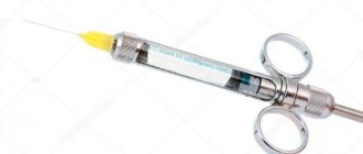
The uniqueness of the domestic IS-01-1-MID injector is that its tip has a rotating head, which allows you to create any angle of the needle during anesthesia. The needle recommended for injection (Fig. 3) has a diameter of 0.3 mm (its length can be 10, 12 or 16 mm). Its peculiarity is the ability to bend without breaking. The inner diameter of the cannula (0.03 mm) convincingly proves that the needle does not enter the periodontal fissure (the width of which is 0.05-0.36 mm in the middle part of the root), and the solution flow under pressure is pushed into the fissure.
The carpuled local anesthetic solution used for ILA (volume 1.7 and 1.8 ml) must contain an amide anesthetic and necessarily a vasoconstrictor.
Method of intraligamentary anesthesia
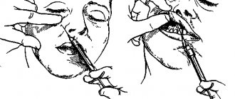
After plaque removal and antiseptic treatment (for example, 0.06% chlorhexidine bigluconate solution) of the entire surface of the tooth and the gingival groove around it, the anesthetic solution is injected under pressure into the periodontal space. The needle slides along the surface of the tooth and, at an angle of 30 degrees to the central axis of the tooth, pierces the gingival groove and penetrates to a depth of 1-3 mm until tissue resistance is felt. Then maximum pressure is developed by pressing the syringe handle for 7 seconds, the solution is injected.
Proper needle placement is indicated by strong tissue resistance. In rare cases, when the needle is inserted correctly, there may be no fluid flow from the needle. This is possible in the following cases: when the needle is pressed very tightly against the root surface or alveolar wall; when the needle is blocked. In the first case, you should change the position of the needle, in the other, check whether the solution flows through the needle.
It is very important to monitor the flow of anesthetic from the needle: if a drop of anesthetic appears in the area where the needle is located, this indicates that the needle is incorrectly positioned and the solution is leaking out. In this case, it is necessary to change its position. A clinical sign of properly administered anesthesia is ischemia (blanching) of the gums around the anesthetized tooth.
The number of injections depends on the number of tooth roots. To anesthetize a single-rooted tooth, 0.12-0.18 ml of solution is required. The main requirement is to slowly introduce the solution. When using an injector with a 0.06 ml dispenser, this amount of solution is injected within 7 seconds. For a single-rooted tooth, this injection is repeated 2-3 times with an interval of 7 seconds. At the end of the injection, it is not recommended to remove the needle immediately, but wait another 10-15 seconds. so that the solution does not come back out.
Anesthesia is carried out from the approximal surfaces of the tooth (medial and distal), i.e. at every root. Thus, to anesthetize a single-rooted tooth, 0.12-0.18 ml of anesthetic is sufficient, and for double-rooted teeth - 0.24-0.36 ml, for three-rooted teeth (for upper molars, an anesthetic is additionally administered at the palatal root) - 0.36-0 .54 ml.
Clinical guidelines for the use of anesthesia
During conservative interventions (dental treatment for caries and pulpitis), as well as preparing teeth for a crown during anesthesia, it is necessary to carefully insert the needle into the periodontium, to a depth of no more than 2-3 mm and release the solution very slowly, strictly observing pauses between the administration of each dose solution.
To remove a tooth, intraligamentary anesthesia does not require gentle measures; in this case, both deeper insertion of the needle and faster administration of the solution are permissible. In case of insufficient effectiveness of ILA in the treatment of acute and chronic forms of pulpitis, an anesthetic solution can be injected intrapulpically, using the same injector with a needle.
The previously opened area of the pulp is anesthetized by application. The effectiveness of intraligamentary anesthesia is high - from 89% for therapeutic to 94% for orthopedic and 99% for surgical interventions. It should be noted that ILA is not effective for all groups of teeth, namely: in 46% of cases, anesthesia of the canines on the upper and lower jaws is ineffective; the effectiveness of anesthesia of the upper central incisors is slightly higher. The success of anesthesia is likely influenced by the root length of these groups of teeth.
Advantages of the intraligamentary method of anesthesia
- High percentage of successful pain relief from 89% in therapeutic practice to 99% in surgical practice. The exception is anesthesia of canines and sometimes central incisors of the upper jaw (46%).
- Virtually painless anesthesia.
- The anesthetic effect appears almost immediately (after 15-45 seconds), which saves time for the doctor and the patient.
- The duration of intraligamentary anesthesia is sufficient for basic outpatient dental interventions (from 20 to 30 minutes).
- Minimal use of an anesthetic (0.12-0.54 ml for anesthesia of one tooth) and a vasoconstrictor, which is especially important in persons with concomitant pathologies.
- Anesthesia is devoid of the disadvantages inherent in conduction anesthesia, such as long-term disruption of nerve conduction, long patent period, contracture, etc.
- Possibility of replacing conduction anesthesia when performing interventions on the front teeth of the lower jaw, without resorting to bilateral conduction anesthesia.
- Treatment in one visit to teeth in 4 quadrants of the jaws, using a minimum volume of anesthetic solution, without causing discomfort to the patient during the injection.
Indications for intraligamentary anesthesia
- Dental treatment for caries, acute and chronic pulpitis.
- Depulpation of intact teeth for prosthetic purposes.
- Tooth extraction for chronic periodontitis.
- Preparation of hard tooth tissues under the crown.
Contraindications to intraligamentary anesthesia
- Presence of a periodontal pocket, unless tooth extraction is required.
- The presence of acute inflammatory diseases of periodontal tissues.
- Treatment and removal of teeth for acute and exacerbation of chronic periodontitis.
- History of endocarditis.
Intraligamentary anesthesia is a promising, highly effective, safe and easy-to-use method of anesthesia, providing adequate anesthesia for almost all outpatient dental procedures. Anesthesia is acceptable for the patient, since at the end of the intervention not only the functions of the dental system are not impaired, but the injection itself does not cause negative emotions.
ILA can serve as both a primary and an additional method, the development and application of which will improve the effectiveness of pain relief during dental interventions.
General principles of pain-relieving procedures
In order for local anesthesia in dentistry to bring the expected effect and not become a source of complications, it is important for the doctor to follow certain principles of its implementation:
- You should first assess the patient’s condition and find out if there are any allergic reactions to painkillers.
- The correct choice of anesthetic drug and place for its administration.
- The use of exclusively sterile preparations that are compatible with oral tissues.
- The temperature of the solution for administration should be close to normal human body temperature.
- The rate of drug administration should be minimal, and the patient should not experience any unpleasant sensations (burning, itching, pain) during the process.
- Only sharp needles should be used to avoid tissue injury.
- The area of the upcoming injection must be pre-treated with an antiseptic.
- The injection should not be unexpected: the patient must be prepared for local anesthesia.
Possible complications
Despite the simplicity of the procedure, it must be performed by an experienced, highly qualified doctor. If a mistake is made, there is a possibility of adverse consequences and complications.
Such conditions include:
- Bacteremia . During the injection, in 70% of patients, microorganisms from the gingival groove are pushed into the bed of the vessel. This risk can be reduced by high-quality antiseptic treatment of the furrows.
- Injury to the ligamentous apparatus . It manifests itself as temporary pain when percussion or pressure is applied to the treated unit during the first 24-36 hours after treatment. This happens if the anesthetic was administered very quickly.
- Local necrosis of the mucosa at the site where it was pierced with a needle . It develops as a result of incorrect anesthesia, i.e., when the solution is quickly administered, the needle is incorrectly positioned, or the volume of the drug is incorrectly calculated (its excess).
The dentist’s knowledge of the anatomical structure and features of the maxillofacial apparatus, high-quality antiseptic treatment, and modern equipment will help eliminate these complications.
Reviews
Opinions about intraligamentous anesthesia for the most part are only positive, both from dentists and from their patients.
Having a minimum number of contraindications, this technique is effective, safe and most convenient, and also facilitates easier treatment.
If you have experience in dental treatment under intraligamentary anesthesia, and you would like to express your opinion about its effectiveness and feasibility, share it in the comments to this article.
If you find an error, please select a piece of text and press Ctrl+Enter.
Tags intraligamentary anesthesia
Did you like the article? stay tuned
No comments yet
Conductor technology
Conduction local anesthesia in dentistry is used when infiltration anesthesia is ineffective (for example, if volumetric manipulations on the lower jaw are to be performed), when several teeth need to be treated simultaneously, or a long-term intervention is expected. In this case, the anesthetic solution is supplied to the branches of the nerve innervating the area of the upcoming intervention. The drug can be administered endoneurally (directly into the nerve, used rarely and for special indications) or perineurally (next to the nerve, so that the solution gradually permeates the nerve fibers, the most commonly used method of administration). There are also extraoral and intraoral methods of performing the injection - the doctor decides which method to choose in a particular case.
Depending on the site of administration of the anesthetic drug and the branches of the nerves that are to be blocked, the following types of conduction local anesthesia in dentistry are distinguished:
- Infraorbital – anesthesia of the branches of the infraorbital nerve.
- Palatine – blocking impulses from the greater palatine nerve.
- Tuberal - blocking impulses from the superior posterior alveolar nerves.
- Mental, or mental – anesthesia of the mental nerve.
- Mandibular - blocking impulses of the lower alveolar and lingual nerves in the lower jaw. One of the varieties is considered to be torsal anesthesia (blocking the branches of the lingual, inferior alveolar and buccal nerves).
- Incisive – blocking impulses from the nasopalatine nerve passing in the incisive canal.
Typically, the conduction technique requires preliminary preparation of the patient for local anesthesia - applying the anesthetic by application method to numb the injection site, as well as explaining to the patient how best to position his head for this or that type of administration of the anesthetic drug.
Features and mechanism of action
Anesthesia has several properties that allow it to remain the most popular and in demand among patients and dentists.
These properties are:
- short latency period - usually the effect of the drug begins from the first minute after injection;
- the maximum effect of the anesthetic appears immediately and lasts up to 20 minutes;
- sufficient duration;
- ease of technique;
- absence of severe pain;
- after therapeutic manipulation, bite correction can be performed;
- hematomas and tissue numbness do not occur.
The above features are important when treating young patients who are very afraid of the doctor and pain.
The very name of the technique reveals the mechanism of its action. An anesthetic drug is injected into the bone tissue under pressure through the dental ligament (soft tissue of the alveoli).
It is the pressure that allows the drug to quickly reach the desired area and spread to the root tip.
At the same time, not the mucous membrane and most of the gums are anesthetized, as with traditional methods, but only the unit needed by the doctor.