The most common dental diseases that a child can develop are caries and pulpitis. However, there are more serious pathologies, and parents need to be aware of them.
One of these diseases is edentia, which is characterized by the absence of teeth. This disease affects both primary and permanent teeth and can develop at any age. There are four main types of edentia:
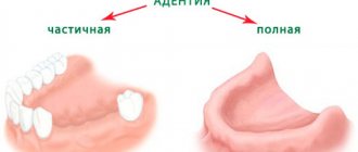
- depending on the number of missing teeth - complete or partial. The first type is uncommon, diagnosed in only 3-5% of people;
- in accordance with the causes of the disease - primary (congenital) or secondary (arising during life).
Edentia of twos can also occur - the absence of paired front teeth located in the central part of the jaw.
Reasons for the development of edentia
It is difficult to name the exact causes of this pathology, because to date the disease has not been sufficiently studied. According to existing hypotheses, the first manifestations of complete or partial adentia in a child occur during the period of intrauterine development. They are associated with disturbances in the development of the ectodermal layer and the formation of organ roots. Edentia of two can also be caused by endocrine pathologies of the fetus, as well as genetic reasons.
As for secondary adentia, experts identify several variants of the clinical picture of this disease, which is diagnosed relatively often. According to statistics, tooth loss is most often caused by the following factors:
- caries;
- other advanced oral diseases, in particular periodontitis and periodontal disease. Lack of treatment leads to destruction of the masticatory organ;
- other pathologies that cause a general deterioration in the body’s condition. As a rule, they are associated with the functioning of the endocrine and immune systems;
- age-related changes that lead to deterioration in health, including tooth loss;
- hereditary factors
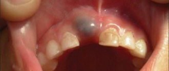
- mechanical injury, for example, a strong blow. In this case, the tooth may fall out either immediately if the main impact was on it, or later if the gums were damaged or the tooth remained in the jaw but was severely damaged. Mechanical trauma is one of the most common causes of adentia, including in children. In a small child, it can lead to developmental defects in baby and permanent teeth.
Causes of loss of chewing teeth
In childhood, the main reason for the loss of chewing teeth is the absence or death of their rudiments. There may also be hereditary or external factors that have a direct impact on the fetus even at the stage of formation of its entire skeletal system. Parents should very carefully monitor the health of their children’s baby teeth, because if timely treatment is not carried out, the consequences can be very dire – including the premature loss of permanent ones.
In adult patients, the main cause of early tooth loss is various dental problems: pulpitis or periodontitis, as well as diseases of periodontal tissues, which begin with inflammation of the gums - periodontitis, less often - periodontal disease. In addition, these diseases can develop against the background of hormonal or age-related changes, diabetes mellitus, and metabolic disorders in the body. Loss of chewing teeth can also occur due to trauma to the jaw system or after prolonged wear, for example, of bridges.
Symptoms of adentia in children and adults
Diagnosis of this disease takes a long time. But there are a number of symptoms indicating its presence. It is especially important to take them into account when determining dental edentia in children. These factors include:
- reduction of jaws (both or just one);
- signs of retraction of the cheeks and lips;
- deformation of the alveolar processes;
- the appearance of wrinkles and folds approximately in the places where the twos are located;
- manifestations of muscular atrophy of the oral cavity;
- change in jaw angles.
All these symptoms can lead to the formation of a malocclusion. Malocclusions, in turn, cause certain shifts of the teeth and the formation of gaps in the jaw. In some cases, growths may form that affect the appearance and make chewing difficult.
Complete secondary adentia can be caused by caries, periodontitis, or surgical intervention. The doctor may also notice scars from injuries that led to tooth loss. Another symptom that explains the loss of masticatory organs is the presence of cancer.
It is worth noting that there is a steady downward trend in the age of edentulous dental patients. Poor ecology leads to the fact that young people are increasingly faced with this problem.
Diagnosis and detection of the disease
Edentia is a serious problem, and to solve it it is very important to promptly and accurately identify the presence of pathology and its type.
The first stage of diagnosis is to conduct a visual examination. Based on the results obtained, the doctor determines whether adentia is primary or secondary.
Then it is necessary to take an X-ray of the jaw, especially if the initial examination showed that the disease is congenital. This can only be confirmed with an x-ray, in which a specialist will check for the presence of dental follicles or identify signs of abnormal development.
When identifying the causes of adentia in children, at the initial stage of diagnosis it is necessary to find out the structure of the root system and find out how things are under the gums. Therefore, in the treatment of childhood adentia, the method of panoramic radiography is used. It allows the doctor to obtain a detailed picture, including information about the structure of the bone tissue of the alveolar processes.
Why is timely diagnosis so important?
Timely detection of adentia is of such high importance for one serious reason. The fact is that before starting any therapeutic actions, it is necessary to diagnose and eliminate all existing pathologies.
First of all, these include inflammatory infections of the oral cavity. During treatment, it is recommended to preserve the roots of the teeth as much as possible, since the absence of roots can lead to the loss of the entire masticatory organ. In addition, it is necessary to carry out complete disinfection of the oral cavity.
If the doctor suspects the development of acquired adentia, it is necessary to find out the cause of the disease as soon as possible. This is due to the fact that some pathologies can lead to complications during prosthetics. To eliminate these risks, the dentist checks for the following symptoms during the examination:
- exostoses (benign growths on bones consisting of bone or cartilage tissue);
- unremoved roots covered with mucous membrane;
- tumors and inflammatory processes;
- infections of the oral mucosa.
Types of adentia and their diagnosis
General symptoms indicating the presence of the disease were discussed above. But there are also a number of signs characteristic of different types of adentia. Their knowledge can also help to identify pathology in a timely manner.
Primary complete edentia
The rarest type of disease and at the same time the most serious. With primary complete adentia, the patient does not even have the rudiments of teeth and their roots. This pathology leads to serious consequences, namely:
- facial symmetry is disrupted;
- the correct formation of the alveolar processes of the upper and lower jaws is difficult;
- the secretion of mucus and saliva in the mouth slows down, which can cause additional inconvenience to the patient.
One of the symptoms of primary complete adentia may be paleness of the mucous membranes. The absence of baby teeth in children can also be easily determined by palpation. If such signs are present, it is necessary to urgently take appropriate measures.
Primary partial edentia
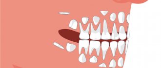
It is expressed in the congenital absence of some teeth in the patient. If partial adentia is detected in a child, the x-ray will show gaps in the root system. It should be noted that the absence of wisdom teeth is not considered edentulous.
As the disease develops, diastemas and tremata are formed - spaces between the chewing organs. In simple terms, the teeth begin to “creep”: if you look at a photo of edentia in children, you will notice large gaps between the teeth.
Primary partial adentia can be symmetrical or asymmetrical. In the first case, there are no paired teeth - doubles or fangs. Asymmetrical adentia means the absence of teeth in different places of the jaw.
Secondary complete edentia
This type of disease is diagnosed when it is acquired. Its symptoms can be observed on all jaws or only on one. Both primary and permanent teeth may be missing. Secondary complete adentia is diagnosed when all the chewing organs have fallen out or needed to be removed due to another disease.
One of the main unpleasant consequences of this pathology is jaw deformation. It becomes closer to the nose, soft tissues begin to retract. Also, this type of disease is characterized by muscle atrophy, leading to disruption of the shape of the entire jaw or one or both alveolar processes.
In addition, the loss of all teeth immediately leads to everyday problems - the patient will not be able to chew independently, and will also begin to swallow some sounds.
Secondary partial edentia
This is the most common type of disease, in which several masticatory organs are missing from the dentition. The loss of canines, twos or other teeth often leads to increased sensitivity of bone tissue. This is due to the gradual wear of the side walls of adjacent teeth.
With this complication, the patient faces discomfort, due to which he gradually stops eating solid food, which is difficult to chew. Both children and adults face this problem.
Possible consequences of edentia
Tooth loss is a serious disease that can lead to various unpleasant consequences, including the development of other serious diseases. Among them are the following:
- impaired jaw development, which leads to difficulty pronouncing various sounds;
- the patient is forced to exclude solid foods from his diet, which causes problems with intestinal motility and disruption of the gastrointestinal tract;
- development of persistent psychological discomfort. The patient’s self-assessment is distorted and depression may develop;
- in the complete absence of teeth, the jaw develops abnormally and can put pressure on any area of the head, thereby provoking the appearance of temporal tumors.
Shift order: norm and pathology
The pathological process can be caused by various reasons. In order to deal with them, consider the normal replacement scheme. The roots of baby teeth are small. They gradually resorb (dissolve), mobility appears. The cutting order is as follows:
- Central lower incisors, first molars on both jaws.
- Central upper and lateral lower incisors.
- Upper lateral incisors.
- Lower canines.
- Premolars on both jaws.
- Upper canines.
- Second lower molars.
- Second upper molars.
- Third molars (wisdom teeth).
When the first molars erupt, no replacement occurs, as the jaw grows and the molars simply grow in place. Therefore, sometimes the patient has a question about how to distinguish a baby tooth from a molar that has already grown. “Baby teeth” are usually visually different. They have a smaller crown. If in doubt, you should consult a doctor. Only on the basis of an examination will a dentist be able to determine whether an adult’s teeth have completely changed.
Root resorption is stimulated by their contact with the crown of the growing permanent root. If there are no rudiments, resorption may occur upon contact with adjacent teeth, but sometimes this does not happen and the root is not destroyed. There is no change. As a result, an adult remains in a row of milk teeth that have not fallen out, which are called persistent.
For normal replacement, it is necessary that the rudiments of permanent teeth be formed, and they be at a depth sufficient for their crown to come into contact with the root of the milk teeth. If this does not happen, the loss is delayed or does not occur at all. Violation of the formation of primordia can occur both in the prenatal period and as a result of previous diseases or inflammatory processes in the oral cavity.


