Diseases of the temporomandibular joint are difficult to diagnose and treat due to the fact that they manifest themselves with a variety of symptoms. The clinical picture is very complex and similar to diseases that fall under the competence of neurologists, otolaryngologists and psychiatrists. Doctors at the Yusupov Hospital use modern diagnostic methods to accurately determine the cause of painful dysfunction of the temporomandibular joint.
The Yusupov Hospital employs professors and doctors of the highest category. They examine patients using the latest equipment from leading European, American and Japanese manufacturers. For laboratory research, laboratory technicians use high-quality reagents to obtain accurate test results. Patients undergo the most complex examinations at partner clinics
What is arthrosis of the TMJ
Arthrosis of the TMJ is a disease that destroys the components that form the joint (Greek arthron joint, suffix oz - destruction). First, the articular cartilage is destroyed, then the following occurs in the articular elements:
- proliferation (tissue growth);
- calcification (calcium redistribution) and ossification of cartilage;
- hyperplastic (proliferation) and destructive (destruction) processes in the epiphyseal parts of bones (these are the rounded ends of the bones - the head and fossa);
- reactive-inflammatory (from the word “response”) changes in the synovial membrane;
- fibrosis (overgrowth of connective tissue) with hardening of the joint capsule, which affects nearby muscles, tendons and ligaments.
With the destruction of cartilage, its shock-absorbing functions are reduced, and impacts are transmitted directly to the bone. Patients involuntarily increase the destruction by reacting emotionally to events - they clench their teeth, not daring to say too much, with a “stony” face and tense muscles, compressed blood vessels and stress hormones, they face the blows of fate. The amount of nutrients decreases, the TMJ would be happy to recover - but there is no building material. Instead, the epiphyseal sections of the bone are flattened under pressure, and bone growths appear on them.
Then the joint enlarges, compressing the nerve endings located nearby. The pain radiates to the ear, back of the head, and teeth. When the jaw moves, a specific clicking sound appears (occlusion-articulation syndrome).
ICD codes M.19. 0 (1, 2, 8 – last digit changes)
Differentiation of diagnosis
The symptoms of pathological processes in the joint, which provides movement of the lower jaw, are similar to the manifestations of other diseases. Doctors, using special examinations, must accurately differentiate the diagnosis so as not to miss the development of the following serious pathologies:
- myocardial infarction, which is also characterized by pain radiating to the neck, lower jaw and shoulders;
- otitis media of unknown etiology can be suspected when there is severe pain and hearing loss;
- cerebrovascular accident may occur with dizziness, flashing of flies and nausea;
- cervical and thoracic osteochondrosis are similar in localization of pain;
- pinching of the facial nerve is also caused by unilateral tension of the facial muscles and swelling;
- complicated diseases of the gums and teeth, which are accompanied by inflammation and immobility of the lower jaw.
To avoid making an incorrect diagnosis, the doctor carefully studies the patient’s life history and illness, conducts a visual and palpation examination, and also prescribes the necessary tests.
[slide-anything id=”3470"]
Causes of arthrosis of the temporomandibular joint
Arthrosis can be triggered by a one-time injury (compression, blow, bruise), as a result of which cracks and erosions appear on the articular surfaces. The disease is caused by a fracture of the condyle and condylar process if the fusion is incorrect.
Other reasons:
- prolonged stress;
- consequence of acute traumatic arthritis;
- birth trauma (arthrosis develops due to improper application of forceps);
- underdevelopment of the jaw (microgenia);
- sudden removal of molars (accident, fight);
- errors during dental prosthetics;
- impaired coordination of muscle contractions during dislocation and subsequent sharp (jerky, zigzag, circular) movements of the jaw;
- complete absence of teeth;
- deep bite;
- introduction of drugs into the joint cavity (for example, hydrocortisone, glucose solutions, novocaine).
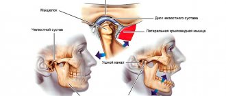
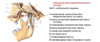
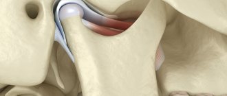
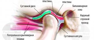
Structure of the TMJ
Etiological factors of arthrosis (without which the disease does not develop):
- infections;
- metabolic disease;
- injuries;
- atherosclerosis of the terminal branches of blood vessels;
- prolonged spastic contraction of the lateral pterygoid muscle (responsible for moving the jaw forward and to the side).
Even children are diagnosed with TMJ arthrosis. In newborns, the disease develops as a result of birth trauma. Dysfunction in the joint due to various malocclusions is observed in 40% of children from 4 to 14 years old, but in only 1% x-rays reveal coracoid (myogenic) arthrosis.
During menopause, the likelihood of developing arthrosis due to endocrine disorders increases. With age, it is possible to develop senile, i.e. invaluable arthrosis, when cartilage tissue cannot recover, dries out and collapses.
At risk are people whose professional activities involve inadequate load on the joint (violinists), or those suffering from spasms of the masticatory muscles (bruxism).
Why do patients experience TMJ dysfunction?
Pathology, first of all, arises against the background of problematic functioning of the complex (hinge-shaped) joint connecting the upper and lower jaws.
There can be several causes of TMJ pathology:
- malocclusion;
- constant stiffness of the facial muscles as a result of neuropsychic or physical stress;
- absence of a chewing tooth or several units of chewing groups in the mouth;
- tendency to increased tooth wear;
- mechanical injury;
- poor-quality installation of prostheses;
- problems with the spine (for example, scoliosis);
- the habit of chewing on one side, thereby loading the TMJ;
- birth injuries;
- mistakes when installing fillings and treatment in the past;
- incorrectly selected orthodontic treatment in the past;
- anatomically incorrect structure of the joint (for example, when the shape and size of the articular head and the articular fossa of the TMJ do not correspond to each other).
Symptoms of TMJ arthrosis
Information about arthrosis of the temporomandibular joint on the Internet is 50% far-fetched descriptions of arthrosis of large joints, 30% is outdated data and obvious nonsense. And only 20% is true. Alas, texts are written by people without medical education, copying not from special educational literature or monographs, but from each other. Therefore, trust only trusted sources, and treat your health where there are no such ignorant things on the clinic websites.
First signs
A person may assume that he has arthrosis of the jaw when, after visiting doctors and following their recommendations, pain in the back of the head, ear, when chewing, hearing loss on one side, clicking, etc. does not go away.
Due to the structural features of the joint, the body manages to turn on the compensatory mechanism, so there is no long-term aching pain; due to the medications taken, it successfully disappears for a while.
Obvious symptoms
There are only 2 obvious symptoms (but it is also impossible to say 100% that this is arthrosis):
- displacement of the jaw to the side;
- pain when chewing.
You need to see a doctor immediately.
Prevention of TMJ dysfunction
What to do to prevent the primary or secondary development of joint dysfunction?
- Have regular dental checkups (at least twice a year);
- take care of your teeth and oral cavity;
- reduce excess loads;
- do not expose the body to stress and anxiety;
- treat caries, pulpitis and gum diseases in a timely manner;
- wear orthodontic systems when indicated for bite correction;
- perform implantation and installation of dentures by experienced doctors;
- correct orthopedic problems in a timely manner;
- treat factors that increase the risk of joint dysfunction (bruxism, neurological diseases, etc.).
How dangerous is the disease?
TMJ arthrosis is silent and unnoticeable; people live with the disease for years without even knowing about the problem. But in vain.
Degrees of TMJ arthrosis
In the Russian Federation, the Kosinskaya classification of arthrosis has been adopted, which takes into account both symptoms and radiographic data. However, the TMJ is an exception to the rule: the joint “hangs”, held by muscles and ligaments, and does not experience weight loads comparable to other joints.
When at stage 1 according to Kosinskaya the joint space narrows, the pressure on the jaw simultaneously increases, which leads to problems with the teeth, but maintains the distance. The process is gradual, so this moment can be recorded on an MRI, but since there are no symptoms characteristic of the disease in the initial stage, it cannot be said unequivocally that this is stage 1 arthrosis. Only at stage 2, when symptoms appear (pain, facial asymmetry, etc.), and the patient finally consults a doctor, is a diagnosis made.
Stage 3 according to Kosinskaya: absence of joint space, sclerosis, necrosis, inability to open the mouth, chew and speak.
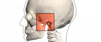
Damage to the TMJ by arthrosis
Possible complications
Arthrosis is not only a problem of the joint. Compensatory, in an effort to maintain chewing function, the body redistributes the load, which leads to tooth loss and rapid wear.
The previous diseases will be reflected in TMJ synovitis, and then the inflammatory process will affect the ear and nose (with decreased hearing, nasal congestion on one side), a headache will appear, which can radiate to the neck, back of the head and not stop.
The face will lose symmetry and become pasty (the skin appears loose, finely swollen, and grayish in color). Feeding is possible only through a tube; already at the second stage the ability to fully open the mouth is lost
Any localization and form of arthrosis has serious complications, so you should not delay treatment.
See how easily the disease can be cured in 10-12 sessions.
Exacerbations
Osteoarthritis is not arthritis; a chronic disease does not have periods of exacerbation. But this does not mean that the pain will be equally aching. The inflammatory process (cold, infection, virus) spreads to the joint with the development of synovitis. Swelling and pain appear, which can appear at any radial point (from the teeth to the back of the head). The source of inflammation expands, the oral cavity, ears, and breathing through the nose are at risk.
You need to understand that the brain is located nearby. And you shouldn’t wait for necrotic tissue to give rise to oncology.
Advantages of the TMJ Treatment Center Partner-Med
Experience at least 5 years
Only doctors with 8-30 years of experience practice at the Partner-Med clinic
15 year labor guarantee
Only a qualified dentist gives an extended guarantee of up to 15 years on his work.
Study abroad
Our doctors annually improve their qualifications in specialized clinics in Europe.
Chief Physician, Dmitry Nikolaevich Salatsky
Sign up for a gnathological consultation
+7
Types of arthrosis of the temporomandibular joint
For treatment to be effective, it is important to understand that there are several types of arthrosis of the lower jaw.
Deforming arthrosis
Osteoarthritis of the TMJ usually develops after injury. The clinical course depends on the nature of growth and the location of osteophyte proliferation (towards soft tissues or the articular cavity). If bone growth is directed to soft tissues, the disease is asymptomatic for a long time. If the osteophyte grows into the cavity of the glenoid cavity, local acute pain appears, which occurs with limited jaw movement. Clicking and crunching are dull, and sometimes popping sounds appear.
The joint becomes deformed with the growth of the condyle, changes occur in the synovial membrane and are accompanied by hemorrhagic synovitis. The reason for this is irritation of the TMJ, caused by the multiple presence of dead and rejected cartilage cells (intra-articular detritus). The synovial villi on the inner lining of the joint enlarge and fat is deposited in them. Occasionally, they degenerate, forming islands of bone and cartilage tissue (metaplasia), which are separated from the articular surface and form intra-articular free bodies.
Please note: this is not salt, it is osteochondral tissue. Therefore, folk remedies for arthrosis, which can still help with gout, do not work.
Viral and infectious diseases during this period inflame the joint membrane, accelerating the destruction of cartilage and bone.
Facial asymmetry does not appear in all patients diagnosed with arthrosis deformans. This depends on the compensatory capabilities of the neuromuscular complex and on the functional grinding of the articular surfaces.
Sclerosing arthrosis
Not only vessels can be sclerotic. With arthrosis, the 2 upper layers of bone become sclerotic (bone tissue is replaced by dense connective tissue). In this case, some compaction of the head occurs, followed by expansion. Since replacement is a slow process, the body manages to compensate for the changes. Therefore, the disease goes unnoticed in the initial stages.
Neoarthrosis (post-infectious arthrosis of the TMJ)
The disease is a consequence of an acute inflammatory process in the TMJ, with repeated acute respiratory viral infections and with the presence of dysfunctional jaw syndrome (luxation, neuromuscular, occlusal-articulatory). It is asymptomatic. With exacerbation of chronic inflammation, the following is noted:
- dull, aching pain that intensifies when moving the jaw;
- crunch;
- clicking in the HFNS.
X-rays show usuria (disappearance of osteochondral tissue), defects in the articulating surfaces of bones, and sometimes the complete absence of condyles.
Myogenic arthrosis of the TMJ
In orthopedics, there is a separate type of deforming arthrosis of the TMJ, myogenic. Its difference: a beak-shaped bone growth on the anterior surface of the condyle.
X-ray shows myogenic arthrosis, the contours of the articular surface due to osteophyte resemble a bird
Myogenic arthrosis occurs due to prolonged spastic tension of the lateral (lateral) pterygoid muscle. Its middle bundles are attached to the anterior-inner surface of the condyle and its process. Prolonged muscle spasm leads to a lack of coordination of muscle contractions, the bone beams change direction, stretch, positioned along the direction of the tendon traction. If the spastic contraction of the muscle continues, the bones that form the joint will begin to break down.
Differences from other forms:
- the condyle always has a beak-like shape;
- bone growth (osteophyte) is always localized in a specific place;
- no restrictions on jaw movement;
- the disease occurs without facial asymmetry.
The initial stages of the disease are asymptomatic. The osteophyte grows gradually on the anterior surface of the condyle, does not rub against hard tissues, and forms a bed in soft tissues. In the joint area, nutrition is disrupted, there may be a slight swelling on the face, spider veins - but very often this is explained by fatigue, overload, without paying attention to the TMJ. Painful symptoms occur at the moment of dislocation, subluxation of the lower jaw. Since the movement of the jaw in such cases is atypical, the osteophyte injures the soft tissues, irritating the nerve endings - severe pain appears (it hurts to chew hard food), severe swelling, clicking, mild swelling and paleness of the skin flap (pastyness). At the moment the mouth opens, the jaw begins to shift to the side.
Metabolic arthrosis
This is a rare type of disease that occurs when salt metabolism in the body is disrupted. The reason is needle-shaped crystals of uric acid settling in the TMJ. In patients, large joints are first affected; they suffer for a long time from metabolic polyarthritis, the visual manifestation of which is “gouty bumps” on the joints.
Symptoms:
- significant deformation of the head of the lower jaw, detected by palpation;
- asynchronous movement of the condyles when opening and closing the mouth;
- hinge movements on the side of the diseased temporomandibular joint;
- crunch;
- local dull pain;
- when opening the mouth, the jaw moves to the side;
- Lateral position of the head leads to facial asymmetry.
On radiographs with metabolic arthrosis, the condyle is covered with whitish needle-shaped curls of various shapes that are not permeable to x-rays.
Crunching in joints - when to worry
Intra-articular injections of hyaluronic acid
Senile arthrosis of the TMJ
Senile, or invaliable, arthrosis occurs with age. “Aging” of cartilage tissue occurs in 3 stages:
- cartilage tissue becomes soft and loose;
- loses some of the water, dries out, becomes denser;
- The smooth surface disappears, the cartilage becomes fragile and becomes covered with cracks.
After 60 years, bone exposure begins. Patients feel uncomfortable chewing and clicks are noted in the TMJ. The x-ray shows subtle changes.
Diagnosis of TMD
This disease is difficult to diagnose. Moreover, difficulties in making a diagnosis occur not only among dentists, but also among doctors of other specialties. Very often, for this reason, the disease is detected late, and treatment becomes lengthy and quite difficult.
Based on the fact that the symptoms of TMD are closely related to occlusion and prosthetics, diagnosis and treatment should be carried out by a dentist specializing in this field: a gnathologist or a neuromuscular dentist.
To make a correct diagnosis, consultations with several specialists in different areas of dentistry will be required.
Diagnostics
In the initial stages, arthrosis of the jaw is asymptomatic (more precisely, if there is pain, discomfort - they are attributed to a cold, problems with teeth, inflammation of the facial nerve, etc.). When constant pain appears, the face loses symmetry, it is impossible to chew - the patient begins to visit doctors.
Remember: at the slightest suspicion of TMJ arthrosis, you should consult a doctor; it is impossible to make a diagnosis yourself (if you are not an orthopedist or a healer).
In the clinic to confirm the diagnosis you will need:
- take blood tests (clinical - to identify an infectious-inflammatory process, biochemistry - for arthrosis, biochemical parameters should be normal);
- take an x-ray in 2 projections (the image clearly shows the deformation of the osteophyte, the narrowing of the joint space, but the articular cartilage is not displayed in the image, and it is impossible to assess the degree of destruction of the TMJ in the early stages);
- undergo an MRI or computed tomography (MRI uses magnetic waves, and computer tomography uses X-rays, so in the early stages, MRI is an advantage).
Occasionally, an ultrasound of the joint is prescribed. In addition, a personal examination is required, because often it is necessary to treat not only arthrosis, but also to remove defects in the dentition, and to treat the accompanying inflammation of nearby tissues.
Splint therapy with an occlusal splint for the treatment of TMJ dysfunction
This type of dental treatment is a preparation for further correction of the bite with a brace system.
At this stage, a splint splint or a so-called occlusal splint is placed in the patient’s oral cavity, which is fitted by a specialist. It is made individually for each person. The plastic splint is designed to reprogram the muscles, after which the resulting position of the jaws is stabilized by a brace system.
Features of splint treatment:
- along with the gradual correction of the position of the jaws, the splint splint gradually wears off;
- recommended for constant wear (both day and night);
- It’s comfortable to talk to her, you just need to get used to her;
- as the splint corrects the position of the joint itself and the work of the masticatory muscles, the person does not experience pain or discomfort;
- caring for the splint during treatment is extremely simple - it is removed only when brushing your teeth, washed with warm water and toothpaste or soap;
- Putting on the splint is very simple and quick.
In some cases, wearing a so-called TMJ mouth guard is prescribed.
Quite often, TMJ dysfunction is treated through systematic orthodontic expansion of the upper jaw. As a result of this treatment, the width of the upper row of teeth is normalized, not only articular pathology goes away, but also the face becomes symmetrical and more harmonious.
The result of treatment with a splint or other orthodontic systems should be an ideal position (occlusion) of the jaws with correct fissure contacts. After wearing splints, prosthetics or artistic restoration are usually recommended as the final aesthetic stage of treatment.
Treatment of TMJ arthrosis
Treatment of TMJ arthrosis is complex, regardless of the stage of development of the disease. The disease cannot be overcome with one method or remedy.
Medicines for the treatment of TMJ arthrosis
In the early stages, arthrosis of the TMJ is asymptomatic, but dysfunctional syndromes may appear. Therefore, treatment should be aimed at normalizing the functioning of the lower jaw. To do this, use myogymnastic exercises (only after consultation with a doctor) and physiotherapy.
Then the position of the articular heads is normalized, the integrity of the dentition and bite are restored. For pain, clicking, crunching, and asynchronous contraction of the masticatory muscles, permanent splints, braces, and bandages are used.
At the same time, the doctor prescribes medications to restore cartilage tissue, relieve inflammation and improve metabolism around the joint.
For arthrosis of the jaw joint, consultation with a psychotherapist is indicated, because Chronic muscle spasms are always associated with problems in relationship with the world.
Medication
To restore cartilage tissue, chondroprotectors are prescribed:
- glucosamine, stimulates the production of key elements in cartilage tissue, restores the articular surface, protects from destruction;
- chondroitin sulfate, increases the ability of cartilage molecules to retain water (especially important for senile arthrosis of the temporomandibular joint), neutralizes the influence of enzymes that destroy cartilage, and stimulates the formation of collagen.
But if the cartilage is completely destroyed, chondroprotectors are not effective.
To relieve muscle spasms, the doctor prescribes mydocalm and sirdalud.
Remember: you cannot use medications on your own without a doctor. Muscle spasm is a protective reaction; without it, the TMJ will begin to deteriorate at an accelerated pace.
Drugs of this group, muscle relaxants, are used only with the simultaneous use of chondroprotectors and orthopedic treatment (splints).
Corticosteroids quickly relieve pain during synovitis, intra-articular injection only relieves inflammation, BUT the next dose is less effective (3-4 injections is the maximum), and the hormone destroys and does not heal articular cartilage. Therefore, for arthrosis of the temporomandibular joint without inflammation, drugs are not used with proper treatment.
The hyaluronic acid preparation “Ostenil mini”, a 1% solution of sodium hyaluronate (10 mg of active substance in 1 ml syringe), is also called “liquid prosthesis”. It restores the joint more effectively than chondroprotectors. 1-2 injections per year (3-4 years) are enough. There are only 2 drawbacks:
- there should be no inflammation in the joint, drugs with HA are instantly destroyed in such an environment, and the treatment will not be effective;
- this is an expensive drug (however, it is better to use it than to go under a scalpel).
For post-infectious arthrosis of the TMJ, Movalis (selective anti-inflammatory) is prescribed to suppress inflammation, as well as:
- Brufen;
- indomethacin;
- methindol;
- butadione;
- rheopirin;
- sodium salicylic acid;
- antibiotics (in the presence of low-grade fever).
Please note: long-term use of non-steroidal anti-inflammatory drugs has a negative effect on articular cartilage.
Electrophoresis with medical bile, bischofite, dimexide (compresses are also made from them), as well as with salicylic sodium (10%), lidase is indicated. Mud therapy helps a lot.
For post-infectious arthrosis, treatment is physiotherapeutic (electrotherapy with potassium iodide solution (5%) and novocaine solution (2%)). Recommended ointments are apisatron, vipratox, and an analgesic mixture.
For myogenic arthrosis, they practice novocaine blockade with vitamins B1, B12, massage using anesthetic ointments, UHF.
Metabolic arthrosis of the jaw joint is myogymnastics and the use of a splint. At the same time, salt-removing therapy (delogil, collection of salt-removing herbs) is prescribed.
Chondroprotectors: what are they, how to choose, how effective are they?
Joint pain at rest
If the condyle in the temporomandibular joint is excessively enlarged, surgical and orthopedic treatment is performed.
In addition, at any stage and for almost any type of arthrosis, vasodilators xanthinol nicotinate and pentoxifylline are prescribed, which relieve spasm of small vessels and improve blood circulation in the joint. At the same time, slight redness of the face and a feeling of heat are the norm.
Therapeutic ointments and creams do not cure advanced arthrosis of the jaw joint, but their use relieves pain, relieves swelling, and improves tissue nutrition. Finalgon and Nicoflex increase blood circulation, relieve pain and partially relax the spasmed muscle. Creams based on bee venom additionally improve the elasticity of ligaments, but due to the large number of allergic reactions, they must be used with caution. Ointments containing non-steroidal anti-inflammatory drugs (Voltaren-gel, Fastum, ibuprofen, indomethacin, etc.) are less effective than medications, but do not have as many contraindications.
Orthopedic treatment
Orthopedic devices help redistribute the load in the joint and straighten the jaw. Using splints and a sling bandage:
- functional rest is created in the joint;
- traumatic factors are eliminated;
- the activity of the chewing muscles and joints is restored.
When treating arthrosis, the dentition must be restored. Wearing mouth guards, braces, and teeth grinding are practiced.
How to treat TMJ arthrosis with exercise therapy
Physical therapy for arthrosis of the jaw joint is useful only after permission and under the supervision of a doctor.
The joint is destroyed from the inside, and the destruction of it and nearby tissues, as well as compensatory muscle spasm, intensifies with movement. Stupid exercises can cause harm, because... unknown until images are received:
- how arthrosis develops;
- what type is it;
- where are the osteophytes directed?
The joint is already receiving load - we talk, eat, opening and closing our mouths. And moving the jaw from side to side will add subluxation and swelling.
At home you can do:
- soft massage with a sponge using rotational movements around the joint to stimulate lymph outflow and blood circulation;
- gently tap a bag of raw peas or beans around the joint;
- stroke the cheek from the nose to the bridge of the nose, applying slight pressure with the palm of your hand.
Nutrition, diet
The development of arthrosis of the temporomandibular joint is not associated with dietary habits (you just don’t need to crack hard nuts so as not to break your teeth). However, it is important to pay attention to the amount of water entering the body. The individual need for clean water is calculated using the formula: 1500 ml + 20 ml per kg (over 20 kg). For example, with a weight of 60 kg, the amount of liquid is 1500 ml + 40 * 20 ml = 2300 ml
When edema occurs, diuretic herbs and herbs (birch, linden, clover inflorescences, mistletoe branches, etc.) are used.
Traditionally, for problems with joints, it is recommended to eat more vegetables and fruits (vitamins and minerals), as well as jellied meats, jelly (some patients have a special craving for soft cartilage, pig ears, etc.).
Eat a varied dietC
When pain occurs (stage 2), it is painful to eat. Food should be soft and pureed. These are juices, pureed soups, ready-made baby food in jars. Sometimes you have to feed through a tube - do not bring yourself to this state, at the first unpleasant sensations, consult a doctor.
If an operation has been performed on the joint, the food for the first time should be dietary. Food should be pureed, spicy, spicy and salty foods should be excluded.
Folk remedies
Among the folk remedies for arthrosis of the jaw, a compress with bischofite or medical bile helps a lot. But due to the fact that it is necessary to align the joint, this is a temporary measure to alleviate the condition. You still have to go to the doctor.
A compress is made only when there is no inflammation, swelling on the face or viral infectious diseases. First, place a warm (not hot!) heating pad on the sore side of the face for 3-5 minutes to warm the joint and slightly relax the spasming muscles. Then put gauze on it (attention: no colorful synthetic rags), soaked in a bischofite solution, cover first with parchment paper (cling film), then with a flannel cloth (terry towel). The compress should be kept for 1-1.5 hours, for people with sensitive skin no more than 20 minutes. If there are no negative reactions, the procedure time is increased. Course of home treatment: 10-15 compresses every other day.
A compress with medical bile cannot be used if you have pustular rashes, acne, rosacea, or rosacea. 6 layers of gauze are soaked in bile and a “sandwich” is made in the same way as with bischofite. However, they keep it for 30 minutes maximum. Course of treatment: daily for 2-3 weeks.
In case of cardiovascular insufficiency or hypertension, such procedures without medical supervision are prohibited.
When diagnosed with “metabolic arthrosis of the jaw joint,” herbal preparations that remove salts are taken. For example, collection (all herbs 100 g, grind in a meat grinder into powder):
- mint;
- buckthorn;
- dandelion;
- immortelle;
- juniper fruits;
- celandine;
- buckthorn;
- chicory (herb);
- yarrow;
- sage (leaves);
- burdock.
1.5 tbsp. collection, brew 1.5 tbsp of boiling water and infuse. Drink 0.5 tbsp. 3 times a day before meals.
This is the only type of arthritis where herbal treatment is effective. However, you can drink herbs to strengthen the immune system and for prevention during epidemics of viral infections.
Surgical operations
Surgical intervention is indicated:
- during ossification;
- with further destruction if conservative treatment does not produce results.
The joint or part of it is removed, replaced with an artificial implant or your own graft (usually part of the fibula).
Surgical treatment of TMJ dysfunction
Surgical treatment of TMJ dysfunction involves endoprosthetics or arthroscopy.
Arthroscopic surgery involves the use of a thin tube with a video camera at the end. With its help, it is possible to visualize the condition of the patient’s joint and monitor the progress of the operation. During arthroscopy, the surgeon removes adhesions, can correct the position of the jaw disc, and apply targeted pressure to areas of inflammation. The dentist also thoroughly rinses the jaw joint with antiseptic compounds to disinfect the area of inflammation. Anti-inflammatory drugs are administered. Thus, the blocking of movements is removed, the characteristic clicks go away, and the patient’s condition gradually normalizes.
As one of the options for solving the problem, a dental surgeon may recommend endoprosthetics. We are talking about replacing parts of the joint with implants. For example, a carbon fiber endoprosthesis is often installed.
During arthrocentesis, the surgeon cleans the joint by piercing it with a special needle, passing a sterile fluid through the joint cavity.
Arthroplasty involves realigning the joint.
Approach to treating the disease in our clinic
Our clinic is an example of integrative medicine: a synthesis of Eastern and Western approaches to treatment. In addition to neutralizing the causes of the disease and restoring the functionality of the HFNS, we restore the disturbed energy balance of the body and the integrity of its structure. Therefore, patients have the strength to cope with the disease and recover much faster than using only the usual medical protocol. All patients are different, so the appointment after the examination is individual.
We combine proven techniques of the East and innovative methods of Western medicine.
Read more about our unique method of treating arthrosis
First aid
If by all indications you have TMD, then to improve chewing function and reduce pain you need to apply a compress to the sore spot: for example, a plastic bottle with hot water, which should be wrapped in a warm, damp towel to avoid burns.
You can reduce inflammation and pain with ice. A bag or plastic bottle with ice should be wrapped in a cloth and applied to the sore joint. You can hold it for no more than 10-15 minutes, then take an hour break.
Analgesics will also help in the fight against pain.
To give your jaw a rest, you should eat soft foods. Don't open your mouth wide to take a big bite.
General clinical recommendations and prevention
With arthrosis of the temporomandibular joint, it is necessary to reduce the load on the joint. To do this, you need to restore the integrity of the dentition and periodically wear braces. If you are involved in (and cannot quit) contact sports (boxing, martial arts), be sure to wear sports mouthguards.
To restore blood circulation in the joint, it is recommended to slowly (!) open and close your mouth (without sudden or lateral movements).
You will also have to get rid of habits that create additional stress on the joint:
- chew gum vigorously;
- support your cheek with your palm;
- chew seeds, nuts, hard cartilage.
Osteoarthritis of the jaw joint is called a disease of suppressed emotions. The illness can be a consequence of divorce, dismissal, or critical life situations. The most severe forms develop in nice and non-conflict people who keep their own emotions to themselves. You need to learn to enjoy life and stop seeing the world in gray colors.
Classification of TMJ diseases
1. Musculo-articular dysfunctions associated with dysfunction of the masticatory muscles:
- muscle contracture;
- hypertrophy of individual masticatory muscles;
- myositis.
2. Associated with functional and morphological disorders in the joint:
- incorrect position of the head and disc of the joint;
- subluxation or dislocation of the articular head;
- subluxation or dislocation of the articular disc with reduction;
- thinning and perforation of the disc;
- hypermobility of the articular head;
- prolapse (prolapse) of the articular disc (disc dislocation without reduction);
- diseases caused by inflammation of the tissues of the joint capsule, synovial membrane, and retrodiscal zone (arthritis);
- chronic arthritis, arthrosis;
- ankylosis.
3. Anomalies and acquired diseases of the TMJ.
Frequently asked questions about the disease
Who treats arthrosis of the temporomandibular joint?
The treatment is complex. If there is no gnathologist in the medical institution, treatment is carried out by a surgeon or orthopedic traumatologist. In this case, a dentist, a neurologist, an otolaryngologist and, if necessary, a rheumatologist and an infectious disease specialist must be involved.
Is it possible to cure TMJ arthrosis?
If bone growths have begun, the process can be stopped, but it will not be possible to defeat the disease when the joint is young and healthy. But if you start treatment at least at stage 2 of the disease, you will be able to get rid of the symptoms, stop the destruction and even restore cartilage tissue.
Why is arthrosis of the TMJ dangerous?
Deformation in the joint leads to facial asymmetry, secondary inflammation spreads to the nasopharynx and ear. Due to spasmed muscles, teeth wear out and fall out. The skin on the face becomes pasty and ages quickly.
What is the difference between arthrosis and TMJ arthritis?
Arthritis is an inflammatory process in the temporomandibular joint of infectious-allergic, traumatic, autoimmune, etc. origin, which in advanced cases can lead to arthrosis. For example, a purulent infection (purulent otitis, boil in the ear canal, flu, sore throat, mumps, etc.) infects the joint fluid. The inflammatory process spreads to the joint capsule (the local temperature rises, the blood vessels of the heads of the bones grow and dilate). The purulent process then dissolves the cartilaginous surface and meniscus, and then destroys the bone tissue, leading to arthrosis. Arthrosis destroys the joint asymptomatically at the first stage and without an acute inflammatory process. The cartilage loses moisture, dries out, and cracks. The bone then grows, changing the structure of the joint.
Literature
- Evdokimenko P.V. Arthrosis
- Petrosov Yu. A., Kalpakyants O. Yu., Seferyan N. Yu. Diseases of the temporomandibular joint
Themes
Arthrosis, Joints, Pain, Treatment without surgery Date of publication: 10/08/2021 Date of update: 11/01/2021
Reader rating
Rating: 4.67 / 5 (3)
Prognosis for the treatment of TMJ dysfunction
Treatment of TMJ dysfunction cannot be delayed. The disease will not go away on its own! It can smoothly flow into further destructive processes, “resulting” in arthrosis of the jaw joint and other serious consequences. If nothing is done, ankylosis of the joint may occur, that is, its complete immobilization, and the person ceases to “own” his jaw.
At the same time, comprehensive treatment of the problem with strict medical supervision provides positive prospects for development and complete recovery.
Pain syndrome and restrictions in movements of the lower jaw
Before
Frontal photograph of the bite
Lateral photograph of the bite (right). Absence of tooth 4.5, absence of tooth 1.4 are noted with replacement of the defect with a removable partial apparatus
Lateral bite photograph (left)
MRI slice of the left TMJ. There is a structural change in the shape of the joint head. Anterior disc dislocation without reduction (without restoration)
After
Frontal photograph of occlusion after joint surgery
Lateral bite photograph (right)
Lateral photograph of the bite (left). There is a rise in the height of the bite on the operated side
MRI section of the left TMJ after surgical reduction of the articular disc. The correct position of the disc is noted
Frontal photograph of the bite in the mouthguard (Splint)
Lateral photograph of the bite in the mouth guard (Splint) (right view)
Lateral photograph of the bite in the mouthguard (Splint) (left view)
Specialists:
Nazaryan David Nazaretovich
Description:
A typical complaint for patients with TMJ problems is pain and restrictions in the movements of the lower jaw. Such complaints were also present in this clinical case. The entry MRI of the TMJ shows a pronounced structural change in the shape of the head of the left joint and an articular disc displaced forward without the ability to restore its position.
We carried out preliminary Splint therapy in preparation for joint surgery (we made a transparent rigid mouthguard for the lower dentition) and assessed the result over time with a series of control MRIs of the TMJ. Using the example of a section from one of these studies, one can clearly compare the picture before and after surgery. The patient also noted positive dynamics regarding pain symptoms already at the stage of wearing a mouth guard. In the postoperative period, pain was no longer observed. The range of movements of the lower jaw was restored. Of course, the closure of the teeth changed after placing the disc in the correct position. We will require orthodontic and orthopedic treatment to support the stability of the TMJ.
Indications
Characteristic symptoms that serve as the basis for diagnosing TMJ dysfunction are:
- The appearance of extraneous sounds - clicks and crunches - during jaw movements.
- Vague or localized pain during exercise.
- Involuntary jaw spasms leading to clenching of teeth.
- A feeling of constant stress on the jaw caused by muscle hypertonicity.
Late stages of pathology development are characterized by problems with full mouth opening, articulation disturbances, noticeable thickening of tissues and slow jaw movement during movements. Dislocation or blocking of the joint is possible, as well as headaches, hearing loss and malocclusion.
Diagnosis of TMJ dysfunctions and cervical dysfunctions
1. Introduction
Pain in the face, head or neck is a universal complaint of patients seeking help from a dentist. In some cases, the source of this pain remains undetected even after all the usual clinical and instrumental diagnostic procedures have been carried out. Diagnosis of common craniofascial-cervical dysfunction and therapeutic interventions are usually aimed only at eliminating the symptoms, namely pain (Asch and Ramfjord, 1995; De Vocht, 2003).
Some of these craniofascial-cervical dysfunctions fall under the category of temporomandibular dysfunctions (TMJDs). TMJ dysfunction is a complex disease whose origins are not yet fully understood (Asch and Ramfjord, 1995; Vigere, 1995; De Vigere, 1996; Gross, 1996; Liu, 1999 Alcantara, 2002; Landulfo, 2004). Most patients with TMD complain of pain in the masticatory muscles, describing symptoms of muscle damage (Wieser, 1995; De Vigere, 1996; Sato, 1998; Liu, 1999; Riho, 2000; John, 2003; Suvinen, 2003; Landulfo, 2004).
Patients who receive custom occlusal splints also do not receive complete healing, and muscle pain becomes a big problem for them. The main complaints of these patients are pain in the neck. In fact, some association between neck pain and TMJ dysfunction has been demonstrated previously (Ciancaglini, 1999 and Alcantra and Vischer, 2002), with some overlap of stomatognathic and cervical symptoms in both TMJ and cervical dysfunction. dysfunction (De Vigere, 1996). The stomatognathic apparatus includes the head and upper cervical structures related to the digestive system, including the oral cavity, teeth, jaws and gums, as well as the tongue, salivary glands, pharynx, muscles of mastication and TMJ. TMJ dysfunction also involves the neck muscles, and in these patients the head position is altered compared to healthy people of the same age and gender (Lee, 1995), although the difference may be quite small (Visher, 2002). In healthy people, the cervical and mandibular muscles act in a coordinated manner as a complex structure, and disruption of one part of this structure causes disruption of the other (Erickson, 2004).
Analysis of EMG indicators in the study of the muscles of the neck and head allowed us to take a deeper look at the work of this complex structure, both normally and in pathology, in the works of Pino, 2000, Ferrario, 2002 and Suvinen, 2003. In the analysis of masticatory function In patients with intra-articular deformities of the TMJ, in previous studies, EMG research has proven to be a useful diagnostic tool (Sato, 1998; Pino, 2000; Landufo, 2004), allowing one to quickly obtain valuable and reliable information about the condition of patients with chronic cervical pain (Falla, 2004) . But in no case was the stomatognathic function in patients with TMJ disorders compared with the performance of those patients in whom neck pain was the main symptom.
In the present study, EMG indicators of patients in two groups were analyzed: patients with TMD and patients with neck pain. The working hypothesis was that patients of the two analyzed groups do not differ in the analysis of EMG characteristics of the masseter and temporal muscles during a standardized test of clenching of teeth. To obtain an external indicator of the norm, the data obtained were also compared with the indicators of healthy control group patients with good occlusion.
2. Materials and methods
2.1. Patients 133 patients aged from 18 to 67 years were examined, 38 of whom were referred to the dental clinic for treatment of myofascial pain syndrome due to complaints of pain in the maxillofacial and cervical region. As a result of a therapeutic examination and radiographic analysis of the condition of the cervical spine and stomatognathic apparatus (De Wither, 1996; Bogduk, 1999) and discussion of subjective information collected when describing the medical history, patients were divided into 2 groups: (a) patients with TMD (17 women and 7 men, aged 21-66 years, average 35, DM 14) and (c) patients with neck pain (11 women and 3 men, age 30-67 years, average 48, DM 17). The therapeutic examination included palpation of the TMJ area, masticatory and neck muscles, assessment of joint and cervical spine movements, as well as examination of the oral cavity, teeth and supporting structures. In patients with TMJ dysfunction, internal disorders with or without visible changes, capsulitis, synovitis, myalgia of the masticatory muscles with articular dysfunction, arthrosis, arthritis, with or without arthralgia were identified. Pain in the TMJ area and masticatory muscles, both spontaneous and occurring in patients during palpation, ranged from moderate to severe, with limited mouth opening and laterodeviation of the lower jaw when opening the mouth. In all observed cases, the duration of pain was no more than 6 months. The subjects also had clicks in the TMJ area. On the other hand, patients with cervical pain had only craniocervical pain without symptoms of TMD (Asch and Ramfjord, 1995; De Wither, 1996; Bogduk, 1999; Vischer, 2002). Patients with cervical pain reported pain concentrated in the neck (moderate to intense, aggravated by palpation), which indicated myalgia, and was combined with a limitation in the range of motion of the head and neck. There was no history of traumatic injury; An X-ray examination of the spine did not reveal any anatomical abnormalities.
All patients who fell out of one category or another were excluded in this study, and the list was replenished with other patients. None of the subjects had systemic diseases of the musculoskeletal system, vestibular or neurological problems, or anatomical disorders in the cervical spine. All patients had complete dentition, at least 24 elements (all teeth natural or with partial fixed dentures), without crossbite. The patients did not have periodontal problems or acute inflammatory diseases of the oral cavity.
Also, 95 patients in the control group were examined (28 women and 67 men, aged 18-22 years, average 20, diabetes 2), all of them had a full set of their teeth (at least 28) in the absence of crossbite, periodontal problems or acute inflammatory diseases of the oral cavity. Contraindications to the participation of patients in the control group were symptoms of TMJD (muscle or joint pain, clicking in the TMJ, limited mouth opening, mandibular laterodeviation when opening the mouth) in the present or past, as well as cervical problems (including traumatic) and systemic diseases. EMG data obtained from a study of control patients were used as external indicators of normality in a healthy occlusal state.
2.2. Electromyographic study and measurements
- Tool
During the study, EMG indicators of the masseter and anterior temporal muscles were measured using disposable bipolar surface electrodes made of silver or silver chloride with a diameter of 10 mm and an interelectrode distance of 21 mm. One electrode was fixed on the forehead as a reference electrode. Bipolar surface electrodes were glued to the area of the muscle bundle parallel to the location of the muscle fibers (Ferrario, 2002): on the temporal anterior - vertically along the anterior edge of the muscle; on the masticatory - parallel to the muscle fibers. The patient's skin was pre-cleansed with alcohol at the site where the electrodes were applied to reduce the skin's resistance, and recording was carried out 5-6 minutes later, after the skin was well moistened with conductive gel.
EMG indicators were recorded using a FREELY portable electromyograph from De Gotzen (Milan, Italy), supplemented with special software. The analog EMG signal was amplified (gain 150, bandwidth 0-10KHz, input peak-to-peak 0 to 2000mV) using a high-rejection differential amplifier (CMRR -105dB within -60Hz, input impedance 10 CQ) and digitized with a resolution of 12b and a frequency of 2230Hz A/D. The digitization process used a 30 Hz high-pass filter and a 400 Hz low-pass filter, as well as a 50-60 Hz band noise limiter. The signal was averaged to 25 ms, and muscle activity was estimated as the average root of the amplitude squared (RMS, mV). The EMG signals obtained during recording were saved by the program for subsequent analysis.
- Standardized recording (teeth clenching on cotton rolls)
The first measurement in the process of each study was a standardizing recording made when the patient clenched the teeth as hard as possible for 5 seconds (MVC) on cotton rolls of the same density. During the recording process, patients were asked to sit upright with their head level (without support) and maintain a natural position while clenching their teeth as tightly as possible.
Two 10-mm thick cotton wool rolls were placed between the patient's second to first molars. This recording made it possible to obtain reference EMG indicators for subsequent comparison and normalization of data. For each of the 4 analyzed muscles (masseter and temporalis on the right and left), the result was an averaged EMG indicator (RMS), which was taken as 100% in further calculations.
All EMG data obtained during further research (with maximum clenching of the teeth by the same patient, but directly with the occlusal surfaces of the teeth, without ridges) were compared with this initial value and expressed as a percentage (mV/mV*100).
- Direct maximum teeth compression (no rollers). (MVC)
EMG indicators were recorded for 5 seconds. The patient was asked to clench his teeth with maximum effort in his usual occlusion with full contact of the upper and lower teeth and maintain the maximum level of muscle tension throughout the entire 5 seconds of recording.
For each patient, the best result (the one where the EMG signal was most constant) was selected, then 3 seconds of the test were selected from it by an automatic data processing program, and the EMG potential was normalized as described previously (the amplitude of the EMG signal when recording the test without rollers was compared with the average amplitude of the EMG signal of the normalized recording on the rollers). The average values of total muscle activity (right and left masseter and temporalis muscles) were then processed by the program as a region of standardized EMG potentials (with normalized RMS amplitude) over a period of time (mV/mVs%) (Ferrario, 2004). Muscle activity has already been used as a general index of the work of the masticatory muscles, both in dynamics and during static work (Ferrario, 2004, 2006).
EMG signals of paired muscles were compared using the percentage overlap factor POC,(%) (Ferrario, 2000). POC is an index of the symmetry of the distribution of muscle activity due to occlusion, determined in calculations by superimposing the EMG amplitudes of the right and left sides of the masseter and temporal muscles over a period of time: the overlap zone is the percentage of total EMG amplitudes. This coefficient varies from 0% to 100% - with good symmetry of the work of paired muscles, POC is about 100% (EMG signal amplitudes completely overlap each other). The mean POC for the masseter and temporalis muscles was determined for each patient.
Discussing the direction in the lateral plane of muscle fibers relative to the skull (fixed bone) and mandible (mobile bone), the two muscles in the present study act in opposite directions: the superficial part of the masseter muscle pulls cranio-caudally and anteriorly-posteriorly, while the anterior temporalis – cranio-caudal and posterior-anterior. The masseter muscle and the temporalis muscle on the opposite side form a muscle pair (for example, the right temporalis and left masseter) (Ferrario, 2000). If one pair of muscles is activated, unbalanced by the contraction of another muscle pair on the opposite side, a potential lateral displacement of the mandible occurs. Torque coefficient (%) (Ferrario, 2000) is calculated by superimposing the right temporal plus left masseter normalized EMG amplitude with the left temporal plus right masseter normalized EMG amplitude: the overlap area is defined as the percentage of total EMG amplitudes. The torque is distributed from 0% (presence of lateral displacement forces) to 100% (absence of lateral displacement).
The reproducibility of EMG measurements has already been tested in our laboratory by repeating the analysis on seven randomly selected patients (Ferrario, 2006). For all EMG variations, intraclass correlation coefficients were greater than 0.63, indicating good measurement accuracy, without random errors (paired student test, P greater than 0.05).
2.3. Information analysis
The resulting statistical data were processed by computer both in the control group of patients and in the other two. Prevalence by age and sex in the three groups was compared with the same-variance analysis (by age), applied to post-hoc tests (Tukey truly significant difference), and the x-square test (sex prevalence). EMG differences in measures (POS, TC, and activity) were compared among the three groups using a generalized linear model (GLM), which used age, sex, and age * sex interaction as factors in the variance analysis. The model separated the effects of age and gender from important differences in EMG parameters in all three groups. SAS statistical design was used.
Linear discriminant analysis was performed between the two groups of patients using individual muscle activity and POC. Discriminant functional analysis (Leason, 1961; Walker and Kowalski, 1974) allows you to differentiate two populations by calculating the function L=Lx*X+Ly*Y, where X and Y are two independent deviations measured in populations, and Lx and Ly are significant discriminant coefficients. The analysis also introduces a threshold value of Lo for distinguishing between the two populations, together with a probable error for classifying a new single individual match with Lo. The threshold value Lo was chosen to minimize the number of misclassified patients. The value of the discriminant analysis was calculated at its rejection (Leason, 1961). The significance was 5% (P less than and equal to 0.05) for all statistical tests.
3. Result
Patients in the control group were significantly younger than those in the other two (analysis deviation, P less than 0.001). Post-hoc test revealed a significant difference in all three groups. Also, sex prevalence in all groups was significant (x-square test, P less than 0.001). To discuss these differences, EMG measures were then compared across the three groups using GLM variance analysis. Patients with TMD had standardized muscle activity (combined by four muscles) at a maximum tooth clenching of 75 mV/mVs%, that is, their muscle electrical potentials were approximately one-fourth less when clenching without rollers (directly on the occlusal surfaces) than with rollers (Figure 1). In the group of patients with cervical pain, compression without cushions was carried out at greater muscle potentials than with maximum compression on cushions, and the value of standardized muscle activity was more than 100% (precisely 124mV/mVs%) (see Table 1).
In the control group of patients, teeth clenching with or without bolsters represented the same muscle activity (standardized muscle work 95mV/mVs%). The activity score in all three groups was significantly different (P=0.002) as determined by GLM analysis variance. The model found that the effect of sex (P=0.656), age (P=0.237), and the interaction between age and sex (P=0.092) were all insignificant, while the effect of group assignment was significant (P less than 0.001). ).
Significant differences between groups were also revealed in muscle symmetry (POC index, the value of the anterior temporal and masticatory muscles, P less than 0.001 analysis fluctuations) and torque (TS index, P less than 0.001): control patients were more symmetrical and there was no torque, while while patients with TMD were less symmetrical and had a large torque value. A significant effect by group (P less than 0.001) remained even when factors such as gender (P = 0.981 for ROS, P = 0.589 for TS, both not significant), age (P = 0.004 for ROS, P = 0.005 for TS, both significant) , and the sex*age combination (P=0.008 for ROS, significant; P=0.945 for TC, not significant) were outside the GLM analysis deviations.
Linear discriminant analysis examined between the two groups of patients was high (F=28.2;35 free degrees, P less than 0.001). Using the function L=0.1002POC + 0.0478 muscle activity and the threshold value L=13.173 for all discriminants, a single, new patient can be placed in both the TMD and neck pain groups with an error of 18.2%. In the present 38 patients, the sensitivity of the division was 0.86, with a specificity of 0.92.
Table 1
Maximum possible tooth clenching in TMD and neck pain and in the control group of patients (value and standard deviation) P, possible error: age, typical analysis of variability (2; 125 degrees of freedom), values with different exponents (a, b, c) post-hoc test; EMG variability, GLM analysis decomposed for age, sex, and sex-age ratio, actual group difference
Fig. 1 Standardized activity, symmetry (ROS) and torque (TS) in patients with TMD, cervical pain and the control group (value +1SD). ***P less than 0.001 (GLM analysis of variability).
4. Discussion
EMG testing of the masticatory muscles is a method that can be easily used to quantify patient performance in dentistry. Among all the muscles involved in raising the mandible, the masseter and temporalis muscles are the muscles that are most often identified during clinical examination, since they are located most superficially and are more detectable for EMG studies. On the other hand, the medial and lateral pterygoid muscles can also be assessed using a needle. Indeed, the determination of stomatognathic dysfunctions and several cephalic dysfunctions, analysis of the masseter and temporalis muscles provides quantitative functional information with minimal discomfort for the patient and without invasive and dangerous procedures (Wieser, 1995; Sato, 1998; Liu, 1999; Burnet, 2000; Pino, 2000 ; Ferrario, 2002, 2004; Suvinen, 2003; Landufo, 2004). Unfortunately, as emphasized by some researchers, the simplicity, low cost and speed of the study, but there are also limitations that are discussed and eliminated (De Luca, 1997). With the exception of technical artifacts (instrumental noise), the thickness of the subcutaneous fat layer, crosstalk from other muscles. However, a correct EMG determination should only be presented with standardized (normal) potentials to eliminate underlying biological and technical noise (De Luca, 1997). In the present study, to reduce the variability of each patient, an EMG protocol was used that included a normalized recording (maximum tooth compression on cotton rolls, which preceded the recording of the test during natural closure, the same electrodes, conductors, EMG machine, the same skin area) that should limit the influence of biological and technical interference (De Luca, 1997; Burnet, 2000). Indeed, the height of the cotton roll can slightly change the vertical dimension (and, accordingly, the length of the muscle fiber and the interelectrode distance), but when the teeth are compressed, the rolls become thin, which makes this effect insignificant. The resulting standardized EMG potentials are limited only to muscle contraction, which is related to the occlusal surface (Ferrario, 2000, 2002).
Standardized EMG potentials allow us to measure the actual influence of morphology on stomatognathic function (Wieser, 1995; Sato, 1998; Liu, 199; Wurnet, 2000; Pino, 2000; Ferrario, 2002, 2006; Landulfo, 2004). From standardized electrical potentials produced by single muscles of mastication, muscle activity (integrated over time) can be calculated to determine the actual voltage produced by the muscles (Sato, 1998; Burnet, 2000; Ferrario, 2004-2006).
Several previous scientific studies have analyzed the EMG characteristics of patients with TMJ disorders. The masticatory muscles of symptomatic patients with TMD were more hypertonic at rest, less efficient, and fatigued more quickly when compared with healthy controls of the same age and sex (Liu, 1999; Pino, 2000). In general, contraction of the masticatory muscles caused a decrease in electrical potentials (Vieser, 1995; Sato, 1998; Pino, 2000), chewing efficiency decreased, and the maximum compression of the teeth in the bite was also significantly less (Sato, 1999). In the present study, bite force was not measured, but EMG activity at maximal clenching can be considered a useful approximation (Vogle and Glaros, 1995; van Kempen, 2002).
Chronic musculoskeletal dysfunctions of the head and neck, often without specific anatomical changes, can be used for objective differential diagnosis (Bogduk, 1999; Visher, 2002), and functional determinations can reveal useful information (Falla, 2004). A recent scientific study found that patients with craniomandibular disorders and patients with problems in the cervical spine did not differ in head position in space (Visher, 2002). On the other hand, no information on EMG in dental patients with neck pain as a principle symptom has been discussed so long ago.
We analyzed the indicators for patients from the two groups of this study; the indicator of standardized muscle activity obtained during the test with the maximum possible clenching of the teeth differed significantly from those obtained in patients in the control group. In patients with TMD, maximal compression on cotton rolls (standardized recording) was recorded with significantly greater EMG potentials than maximal compression directly on the occlusal surface. Also, the standardized activity of the masticatory and temporal muscles in these patients was significantly unbalanced, both from different sides (asymmetry) and between muscle pairs (torque). Asymmetrical normalized muscle activity and unbalanced muscle pairs could potentially displace the mandible to one side, and generate greater force on the half arch and TMJ than on structures on the opposite side (Ferrario and Sforza, 1994). Asymmetry (side) and instability (muscle pairs) during normal muscle activity often result from functionally unstable occlusion when the maxillary and mandibular teeth contact during clenching and swallowing (Ferrario, 1999). In agreement with the literature (Ferrario, 2000), and compared with control patients, patients with TMD have a functionally unstable occlusion (Landulfo, 2004). Clenching teeth on cotton rolls reduces the proprioceptive signal from unstable occlusion and causes patients to clench their teeth with greater activity of the masticatory muscles. Even if the important role of occlusion in the development of signs and symptoms of TMD in patients is still questionable, in some patients the position of altered occlusion may serve as a trigger for abnormal muscle activity (Ferrario, 2002).
On the other hand, in patients with cervical pain, cotton rolls inhibit muscle contraction, and greater activity was found in intertubercular closure. A possible explanation for this inhibition was that the bolsters produced further damage that increased with true non-occlusal and non-articular problems. Indeed, their POC and TC coefficients, even the lowest ones compared to those calculated in the control group, were within normal limits (more than 85% for the POC index, and more than 90% for the TC index, Ferrario, 2006).
To easily distinguish between patients in the two groups, linear discriminant analysis was used. This analysis not only allows the difference between two groups to be detected using a linear combination of variables (two in the present study) (Walker and Kowalski, 1974), but also provides a possible change in the classification of each patient when the same indicators are measured (Leason, 1961). EMG takes readings from the masticatory and temporal muscles with a standardized clenching of the teeth and calculates muscle activity, and the POC index makes it possible to distribute each patient either into the group of patients with TMD or with cervical pain, with an error of 18.2%, i.e. the number of misallocated patients may be less than 2 in 10. Also, the sensitivity and specificity of the test, as presented in the present patient group, was good.
It was mentioned that the patients analyzed were a good case study and extrapolation of the presented results to the general population should be done with caution. Moreover, patients in the control group were younger than patients in the other groups, and men: women were related differently. A linear model analysis was used to compare the EMG values of the three groups using these differences for discussion, and a significant effect of age was found on the ROS and TC indices, and a significant interaction of age and gender for the ROS. On the other hand, the effect of gender was small. Discussing these effects, differences in EMG measures remained significant among the three groups. Indeed, the use of standardized potentials has reduced inter-individual variability (De Luca, 1997; Burnett, 2000; Ferrario, 2000).
Literature data on the influence of gender and age on EMG parameters are very scarce. In comparison with the present findings, Dr. Ferrario reported minor differences (by sex) in normalized EMG potentials recorded during maximal clenching (2000,2006) for a young healthy population. In patients with good occlusion, age has minimal effect on normalized EMG indices: in the control group, the mean age was 53 years, the latest scientific report reported a standardized activity rate of 104.9 mV/mVs% (SD 28.9) POR rate of 87% (SD 0.9) and TC rate 90.8% (SD 0.4) (Ferrario, 2004). On the other hand, Ueda (2002) found that the masticatory muscles in women were more fatigued than in men, and a small effect of age was reflected in the muscle gain during incisal biting at different closure forces according to Fogle and Glaros (1995). Indeed, given the differences in experimental design and change analysis, it is difficult to come to a definitive statement on this issue.
5. Conclusion
The EMG interface of the right and left masseter and anterior temporalis muscles is represented by a specific protocol that provides standardized information that is used for quantitative expression across the two groups. The test is simple, inexpensive, quick to perform and non-invasive, does not cause discomfort to patients and has no side effects.
Discriminant analysis of standardized information obtained during the test makes it possible to differentiate patients with TMD and patients with cervical pain. These two groups of patients require different treatment, and a quick diagnosis will allow the right decision to be made to correct the problem. Patients with low standardized muscle activity and reduced right-left muscle symmetry during maximal clenching are more prone to TMD, and the first step in their treatment is the construction of a stabilizing splint (Ferrario, 2002). Indeed, when treating patients with TMD, occlusal splints have a therapeutic effect in most clinical cases: it is a conservative and reversible treatment that reduces pain in most cases (Nemkowski, 1992; Asch and Rumford, 1995; Ferrario, 2002). On the other hand, patients with high standardized muscle activity and almost normal right-left muscle symmetry with maximum tooth clenching are more prone to cervical problems, and treatment with occlusal splints will most likely not be successful. In this case, working with a physical therapist and chiropractor will be helpful, and manual therapy combined with a tailored exercise program can be successful. An occlusal splint should only be used to avoid the biomechanical effects of occlusal stress on the neck.
Article provided