What is retention
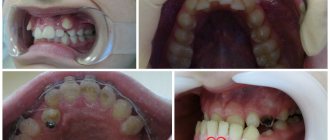
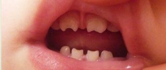
Retention is a delay in the growth of baby or molar teeth. With this pathology, the tooth may erupt, but not completely, but can be barely visible above the gum, and it can also grow only under the gum, not showing out at all. First of all, this disease affects the second premolars and third molars located on the lower jaw, as well as the canines of the upper jaw. Maxillary canine impaction is much more common in women. This pathology can occur on one side of the jaw or on both sides at once. Canine impaction in the lower jaw is a very rare occurrence. Impaction of primary teeth is also extremely rare. Such disorders in children can be caused by an acute lack of vitamins in the body or serious pathologies during teething. Impaction of primary teeth can be caused by severe rickets.
Why is it dangerous
Taking into account the number, location, size of the jaw and the age of the patient, the negative consequences are:
- delayed eruption of primary teeth;
- the formation of a diastema - a gap between the front incisors;
- damage to the mucous membrane leading to inflammation;
- inability to comply with hygiene measures;
- violation of the chewing process and digestive problems;
- the impossibility of proper oral hygiene and, as a consequence, the spread of carious bacteria.
Types of pathology
Teeth eruption disorders can be of two types: complete and partial, and the tooth, respectively, impacted and semi-impacted. The last type means that the tooth has erupted a little, i.e. visually observed above the gum. The impacted tooth is completely hidden by the gum and is not accessible to palpation. According to the depth of their occurrence, such teeth can be tissue embedded (the tooth is located in the gum tissue) or bone embedded (lies in the jaw bone). Such teeth can be located:
- Angularly, i.e. at an angle.
- Vertical.
- Horizontally.
Sometimes there are so-called reverse impacted teeth, most often these are the lower eighth teeth. In such teeth, the upper part is turned towards the jaw, and the roots are turned towards the alveolar edge. There are also symmetrical, unilateral or bilateral tooth retention.
Symptoms of the disease in children
The first supernumerary teeth in children appear before birth or in the first six months of life. The main inconvenience they cause is difficulty in feeding.
Polyodontia of primary teeth in older children occurs with symptoms similar to the eruption of regular teeth. In this case it is observed:
- temperature increase;
- swelling of the gums in the place where the tooth should erupt;
- pain;
- excessive salivation;
- swelling of the nasal mucosa;
- loose stool.
Symptoms are especially severe when extra teeth appear in the upper palate.
If hyperdontia makes itself felt in a two-year-old child, this can interfere with the formation of normal speech. In turn, due to injury to the tongue and mucous membranes, some kind of inflammation constantly appears in the oral cavity.
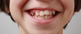
When supernumerary teeth appear in very noticeable places in school-age children, ridicule towards the patient may occur, which is fraught with the development of psychological problems and complexes in the future.
Why does a canine or molar not appear?
The concept of retention refers to the anatomical specificity of the jaw or an anomaly in the formation of the tooth germ. Experts believe that this pathology arose in modern society as a result of eating too soft food, i.e. people have practically stopped chewing solid food. Other reasons why retention may occur:
- improper feeding of the child;
- reduced immunity associated with exposure to infections;
- delay in replacing milk teeth with molars;
- the presence of supernumerary teeth that prevent the eruption of a permanent tooth;
- incorrect location of the permanent tooth germ in the jaw bone. With this pathology, the crown of the tooth is directed towards the root of the adjacent tooth, thereby preventing its appearance and the eruption of neighboring teeth;
- bad heredity.
Causes of hyperdontia
There is no consensus among experts about the reasons for the appearance of extra teeth. There are several versions:
- hyperdontia is a manifestation of atavism;
- the embryonic plate has split into too many tooth germs;
- genetic and hereditary factor.
It is not possible to prevent hyperdontia, so we recommend that parents pay more attention to the dentist’s findings. Timely measures are a guarantee of successful treatment.
The main symptoms of the pathology
Retention in dentistry is quite common, and there are some signs by which this pathology can be detected:
- pain in the gums, radiating to the ear and temporal part;
- regular injury to the same place in the oral mucosa;
- numbness and swelling;
- painful sensations when opening the mouth and while chewing food;
- mobility or displacement of teeth;
- deterioration of general health due to the inflammatory process (fever, weakness, chills, etc.);
- the appearance of a cyst or abscess.
What can happen if you refuse the help of a specialist?
If the tooth root remains under the layer of gum tissue, decay can spread to neighboring units. In this case, treatment will be longer and more expensive. The main symptoms of this problem are a strong odor from the mouth and periodic pain that radiates to the head. Harmful microorganisms will begin to accumulate under the gums, which can lead to the development of caries.
In 99% of cases, the patient has a small sac with purulent filling at the apex of the root. After some time, it inevitably turns into a painful swelling called gumboil. The only correct solution when gums grow on a tooth is to quickly contact an experienced dentist. It will not only eliminate inflammation, but will also be able to save the destroyed unit.
previous post
What happens if you swallow toothpaste?
next entry
Elimination of anomaly
Treatment of retention is a rather complex process that requires qualified assistance from dentists of several specializations. Treatment is selected individually for each patient and depends on the clinical picture of the oral cavity. The first question that a dentist needs to decide is whether to remove or save a diseased tooth. There are several treatment methods, most often this is a surgical intervention in which a specialist cuts the gingival hood so that nothing prevents the tooth from erupting outward. Surgical procedures of this kind are used in situations where the tooth grows correctly and does not interfere with neighboring teeth. In other cases, most often, the tooth is removed. Removing an impacted tooth is a rather difficult operation. The procedure is carried out under local anesthesia, after which the gum is cut, and the specialist uses a bur to create access to the tooth, after which it is completely removed. Then a special medicine is placed into the tooth socket. If necessary, sutures are applied and removed ten days after the procedure. Swelling may occur after removal of an impacted tooth. Most often, after removing such a tooth, the doctor prescribes antibiotics and painkillers. Also, the patient, for the first few days, needs to stop eating solid foods, hot and cold foods and drinks.
Treatment of supernumerary teeth in children
Babies sometimes develop their first extra teeth immediately after birth. In this case, breastfeeding is a difficult test - the mother's nipple is constantly injured, and the baby's mucous membrane is damaged. In this situation, the teeth need to be removed urgently. If the supernumerary tooth erupted during the period when the child’s milk teeth were not replaced by molars, it will be removed. And to correct the bite, the intervention of an orthodontist will be required.
Difficulties arise if the extra tooth turns out to be impacted or semi-impacted - that is, it did not erupt and remained under the gum. Then dentists will prescribe special procedures:
- vibration stimulation;
- stimulating massage;
- electrical stimulation.
This will help speed up teething. Then the dentists decide whether to remove such a tooth or leave it. Sometimes doctors decide to save extra teeth if they do not interfere with the remaining teeth erupting in their place and do not spoil the bite. Modern techniques quickly solve the problem of hyperdontia if you consult a doctor at an early stage. Therefore, Family Dentistry specialists recommend visiting the dentist twice a year and teaching children to do this from an early age.
Prevention after surgery
The rehabilitation period after removal of an impacted wisdom tooth lasts longer than after a normal one. Healing of the gum socket can take up to 2-3 weeks. During this period, it is important to follow the doctor's recommendations to prevent complications. It is necessary to carefully but delicately carry out hygienic care of the oral cavity. For the first days after the intervention, take painkillers and follow a gentle diet. Too hard, burning foods can damage the gums and cause tissue inflammation.
Dentists also recommend rinsing the mouth after each meal using antiseptic solutions, herbal decoctions, or just water. During the week, you must avoid physical activity, visiting the gym, baths, saunas. If any complications occur, you should not self-medicate; you must immediately seek help from a doctor.
If it hurts, what should you do?
In case of acute pain in the area of the impacted tooth or after extraction, you can take an anesthetic. For this purpose, non-steroidal anti-inflammatory drugs are best suited - Nimessil, Ibuprofen, Paracetamol, Ketanov. At home, you can also carry out hygienic cleaning and rinsing the mouth with antiseptics. For a professional examination and treatment, you must visit a dentist.
Incorrect growth of wisdom teeth
70% of people who turn to the clinic for help with the eruption of a wisdom tooth are diagnosed with its incorrect location. Often the concept implies a horizontal tilt, excessive grinding into the seven, or the growth of the eighth tooth into the root part of the neighboring one.
- The growth of a tooth under the gum harms the rest. The sooner a person shows interest and finds out exactly how even an unerupted wisdom tooth is located in the jaw, the easier it will be to eliminate the problem when it is discovered. It happens that the figure eight harms the healthy seventh tooth, while still hidden under the gum. While growing, the third molar seems to be trying to push out the neighboring one or rests against its walls, causing damage. It is better to remove such a wisdom tooth in advance, otherwise the risk of damage to a healthy molar increases. Later, you will have to get rid of both the third and second molars, which have lost functionality.
- A similar situation develops when a wisdom tooth erupts at an angle. As it continues to grow, it will begin to prop up the seven. A particularly alarming picture emerges if there is a lack of space on the jaw. Then the dentition loses its alignment. More often this problem occurs in women.
- The recumbent position of a wisdom tooth is also considered unfavorable , especially when the tooth is turned towards the adjacent seventh tooth. A person sometimes argues that preserving his own, albeit pathologically growing, tooth may be useful in the future, when the time comes to install a crown on the tooth. And the recumbent tooth takes up a lot of space in the row, shifting the others and causing their uneven arrangement.
- The fit of the figure eight to the second molar , even with an anatomically correct location, is also not considered a good sign. Excessive crowding of teeth in the depths of the oral cavity almost always causes the development of caries.
- At first glance, the direction of the wisdom tooth towards the cheek However, this is an imaginary impression. A tooth growing sideways regularly causes microdamage to the vulnerable mucous membrane of the cheek. Often leads to gum inflammation and pericoronitis. The seriousness of the situation is enhanced by the fact that the mucous membrane of the cheeks is not rich in pain receptors, so a person sometimes does not even feel the harm being caused.
- As a result, the cheek is damaged from the inside, and the wounded area becomes a gateway for infection and becomes severely inflamed. Gradually, the cells of the mucosa, under the pressure of what is happening, are transformed and replaced by foreign, denser tissue. This indicates the emergence of neoplasms at the site of permanent injury - benign or carcinogenic.
Dentists recommend removing wisdom teeth that are positioned incorrectly in advance. This information is available to the patient after undergoing an X-ray examination. The picture immediately shows how the figure eight is located inside the gum; it becomes possible to predict the result when it erupts.
Ultrasonic removal of wisdom teeth using a piezotome
You don’t even need to wait for the mentioned process to begin; the modern level of development of dentistry allows the doctor to intervene while the tooth is “sleeping” and perform an operation to extract it in adolescence. In youth, such manipulation is tolerated much easier, and tissue regeneration occurs faster and more completely. And by the time you turn twenty-five, the undesirable scenario of the eruption of a crooked wisdom tooth will be a thing of the past.
Make an appointment with surgeons at Dr. Lopaeva’s clinic
Make an appointment
Or call +7(985)532-21-01
Why is retention dangerous?
A partially erupted tooth is covered with a “hood” of adjacent tissues, under which food debris collects and colonies of pathogenic microflora develop, which can provoke purulent inflammation. With complete retention, another problem arises: a tooth embedded in the tissues constantly affects its “neighbor,” causing it to move from its correct position. The situation may sooner or later become more complicated:
- caries of adjacent teeth and resorption of their roots;
- malocclusion, crowded teeth and other dental anomalies;
- pulpitis and periodontitis;
- periodontal cyst;
- pericoronitis (inflammation of the gingival “hood”) and its complication, periostitis;
- purulent lymphadenitis;
- inflammation of the trigeminal nerve;
- abscess and phlegmon.
Therefore, when diagnosing retention, the patient is offered the optimal way to solve the problem.
What can be done about an impacted tooth?
- According to indications, based on the general clinical situation, removal of an impacted wisdom tooth may be recommended (especially for patients undergoing treatment with braces for an abnormal bite).
- In some cases, not accompanied by obvious problems, when removal is not necessary, the doctor will suggest observing the tooth.
- Healthy impacted incisors and canines located in the smile zone, subject to certain conditions, can be “pulled out” from the dental tissues and returned to the dentition.
A wisdom tooth remains in the gum - is this critical?
Retention or partial eruption is characterized by incomplete emergence of the crown above the gum. Only 1-2 bumps or part of the tooth crown may appear. Retention can be independent or together with dystopia - incorrect positioning of the molar. Symptoms of the disease will depend on the individual clinical situation.
Depending on the location, retention can be vertical, horizontal or angular. With a vertical crown, the tooth crown is located normally in the bone, in accordance with other teeth. Horizontal is characterized by the location of the tooth perpendicular to the others, horizontally to the arch of the jaw. With angular retention, the crown is tilted to the side. According to the depth of its location, the tooth can be covered only by gum, but by a bone plate.
Retention is accompanied by the ingress of food debris and plaque under the gum. This causes inflammation, redness, tissue swelling, and pain. Pericoronaritis may occur - acute inflammation of the gums and mucous hood above the crown of the impacted tooth. The disease can be serous and purulent, causes acute pain, hyperemia, tissue swelling, and makes it difficult to open the mouth.
In most cases, a tooth in the gum is a source of various complications: pericoronitis, the formation of caries and its complications, cysts, stomatitis, periostitis, abscess. Therefore, in case of pathology, dentists recommend removal.
To date, exact methods for preventing retention are unknown. General measures include proper hygienic care of the oral cavity and monitoring the correct development of the child’s jaws and bite. As well as timely and correct orthodontic treatment of malocclusions.
Indications for removal
Retention can manifest itself as a stage of eruption. Over time, the crown completely appears above the gum and the tooth can function normally. Therefore, the decision on the need for removal should be made by a doctor only after examining and diagnosing the condition of the dental system. Indications for surgery are the following clinical cases:
- Constant pain;
- Complications of eruption;
- Trauma to an adjacent molar;
- Pressure on the dentition and its deformation;
- Horizontal or angular position of the third molar in the jaw;
- Dental crowding, which can be aggravated by wisdom teeth;
- Orthodontic and orthopedic indications, that is, the need for extraction to correct the bite or prosthetics.
How is retention diagnosed?
Impacted teeth are often detected by dentists in children. They are brought to the appointment by parents who have noticed that after a baby tooth fell out, a permanent tooth did not erupt in its place. Quite often, the problem is revealed during medical examinations of children aged 11–12 years - when the eruption of permanent teeth is delayed. So, if by this time there is still a primary canine in the oral cavity, the child is prescribed an x-ray.
The ideal option for diagnosing tooth impaction is an orthopantomogram. A panoramic photograph of the jaws clearly shows all the upper and lower teeth, located both physiologically and abnormally. Some patients aged 20–30 years have baby teeth, the roots of which have not been resolved due to retention of permanent teeth. Using an overview image, the doctor determines the type of retention, assesses the complexity of the situation and develops a treatment plan.


