Molars are divided into canines, incisors, premolars, and molars. The first three types of teeth erupt in place of similar milk teeth. In turn, molars do not have predecessors and appear behind the temporary ones, therefore their second name is accessory.
Each jaw has 16 teeth - four incisors, two canines, four premolars and six molars. In some cases, third molars, better known as wisdom teeth, do not erupt because they do not have buds in the jaw - then instead of 16, a person has 14 teeth above and below. Experts explain this phenomenon by a reduction, or simplification, of the dental system caused by changes in dietary style.
The structure of the human jaw: teeth and their formula
When citing one number for any tooth, the dentist means its anatomical and functional affiliation. If you conditionally divide the dentition into two halves and start counting from the first incisor, you will get 8 teeth in each direction (top and bottom). The first incisor is a one. The second, accordingly, is a deuce. And so on until the last molar, which is called the figure eight. If the doctor says - bottom seven on the left, then we are talking about the bottom left second molar. As you can see, everything is very simple!
This can be expressed schematically as follows (permanent teeth):
For baby teeth, everything is the same, only the teeth are designated by Roman numerals (there are no premolars in the primary dentition):
As for the two-digit designations, they were invented in order to simplify the written recording of medical history. The above is preserved, but a number appears before the number indicating which jaw and on which side the tooth is located. Conventionally, the oral cavity is divided into segments - upper right (number 1), upper left (number 2), lower left (number 3), lower right (number 4). As you noticed, the report is conducted starting from the top right and clockwise. The segment is written first, and then the tooth number. For example, the third canine from the top right would be designated tooth 13. The lower incisor on the left is 32 teeth. In children, the segments are designated by numbers 5 to 8, so as not to confuse the primary bite with the permanent one. Let's take the same third fang on the right. In a child, it will be designated as tooth 53. And so on by analogy. The main thing is to always start the report from the right side at the top, then you won’t get confused. These are not snails with 25 thousand teeth! Can you imagine their dental formulas?
Classification
In orthodontic practice, various assessment criteria are used to determine which dental bite is correct and how to correct existing anomalies. The ideal situation, in which nothing needs to be corrected, is characterized by the following indications:
- Semi-elliptical shape of the upper and parabolic shape of the lower arc.
- Small - no more than a third - overlap of the vestibular surface of the mandibular elements.
- Contact of antagonists during closure and absence of obvious gaps.
Compliance on all points is quite rare, but in most cases the deviations are minimal and do not require medical intervention. Often the cause of imbalance is not congenital defects, but bad habits, some of which are formed at an early age.
Types of correct bite
In accordance with the generally accepted classification, there are four main types of occlusal relationships, characterized by the absence of problems and not having a negative effect on the jaw region.
Orthognathic
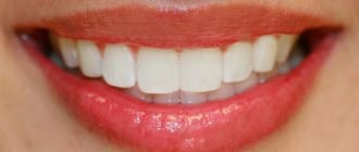
The optimal, from an aesthetic point of view, is a condition in which all elements of the dentition have an even shape, are located in the correct manner, without gaps or deviations from the midline.
Straight
A common phenomenon, the main feature of which is the lack of overlap - the cutting edges of the incisors meet along a line, which causes a specific type of smile, but is not considered as a pathology.
Biprognathic
Another type of even dentition without threes and diastemas, the distinctive feature of which is a slight deviation of the vertical growth vector of the units. With this development, the crowns are slightly tilted forward.
Progenic
In a normal state it is not accompanied by abnormal manifestations. The identifying feature is a slight frontal protrusion of the lower units.
Anatomical structure of the tooth
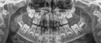
We figured out the order of the teeth. Further - more interesting! After all, a tooth is a unique organ, designed by nature to perform very important functions, primarily chewing. To be firmly held in the jaw bone, you need a root. The longest root is at the canine. Teeth can be single-rooted or multi-rooted. The structure of the upper teeth (molars) is somewhat more complex than that of the lower ones, since they have 3 roots. There are especially many variations in the number and type of roots in “wisdom teeth.” But the visible part of the tooth protruding above the gum is called the crown. It has different shapes in the incisors, canines and molars to perform the functions of biting, crushing or chewing food. The boundary between the crown and the root is the neck of the tooth. It is usually not visible, but it can become exposed due to certain diseases (periodontal disease, etc.). An x-ray is a kind of photo of the structure of the tooth and will give you an idea of what the state of affairs is in the oral cavity.
Eruption of canines in infancy
The order of formation of the primary occlusion provides for the appearance of fangs after the incisors and the first chewing units have erupted. The upper elements emerge at the age of 1.2-1.5 years, which is slightly earlier than the period of formation of the lower molars. Late eruption is determined by the deep position of the units in the structure of the jaw, which also determines the characteristic painful sensations that may occur during growth.
To relieve the discomfort experienced by the child, the following techniques are recommended:
- Massage of gum tissue - before the procedure, you need to thoroughly wash your hands, then use your index finger to massage the area above the canine for a couple of minutes. It is recommended to repeat the massage several times a day;
- The use of teethers that provide a cooling effect - a product filled with distilled water does not pose a danger to the baby’s health even if the baby manages to chew the shell;
- Applying anesthetic gels that relieve inflammation within a few minutes;
- For nasal congestion, use children's nasal medications that promote vasoconstriction;
- At elevated temperatures (more than 38 degrees) - take fever medications, available in the form of syrup or suppository.
It is worth emphasizing that the use of medications must be agreed with a doctor, who will determine the appropriate list, as well as the duration of the course.
Histological structure of the tooth
The special hardness of teeth is due to their structure. The outside of the crown is covered with enamel, the basis of which is hydroxyapatite crystals. 96% of the enamel consists of inorganic substances, which is why it is so hard (which, however, does not mean that experiments can be carried out on teeth, testing their strength). But dentin, which is located under the enamel, has much less inorganic substances - about 70%. It is softer, and therefore is destroyed by caries more than enamel. Inside the tooth there is a cavity filled with a neurovascular bundle, or pulp. The entire rich spectrum of pain during the deepening of the carious process is associated with it. In addition to nerve fibers, the pulp also contains a large number of blood vessels, through which infection from the affected tooth spreads throughout the body. This is why it is so dangerous to leave dental diseases untreated.
Function and role of fangs
The main function of the fangs is to tear and hold food. In addition, canines are the teeth that are least susceptible to caries. These are the most powerful and strong teeth, which bear quite a large load. If a person suddenly lost his fangs, then:
- perhaps he would have problems with diction;
- the entire load would fall on the weaker neighboring teeth, which in the near future would either become deformed or become loose;
- the dentition would lose its aesthetic appeal;
- the process of infection of teeth with caries has accelerated, since it is the fangs that are the barrier to the “creeping” of this problem from the molars that are most susceptible to it to the incisors;
- Tooth enamel is the hardest human tissue the body produces, but even it can wear down and wear down over time. Fangs are an excellent shock absorber for the contact of the upper and lower jaws; in their absence, tooth enamel would wear down several times faster.
The structure of baby teeth
A temporary bite differs from a permanent bite not only in the number of teeth (in a temporary bite there are 20). Baby teeth have a bluish tint to the crown, which is also much wider than the root. Children have less enamel mineralization, so caries affects them very quickly, and the pulp takes up more space compared to permanent teeth. The canals of baby teeth are wider, easier to pass for instrumentation, and the roots themselves have a rounded shape. The structural features of baby teeth determine their rapid destruction in case of infection penetration and very severe pain in the child, so in childhood, visit the dentist in a timely manner.
Knowing the structure of teeth will help you quickly find a common language with the dentist and feel more confident when discussing treatment issues.
You will be able to better navigate the manipulations performed by the doctor and clarify all the points that interest you with an understanding of the essence of what is happening. This article is for informational purposes only, please consult your doctor for details!
International Viola system: a convenient diagram of the arrangement of a person’s teeth by numbers
The described method is very convenient and most common in dentistry. It has received international recognition and has been generally accepted among dentists since 1971, called the two-digit Viola system. The convenience of such a system lies primarily in the fact that there is no need to create a special map of a person’s teeth, the numbering is easily calculated in the mind and information about the condition of certain dental units of the patient can be easily conveyed in an oral conversation, by telephone or by e-mail.
However, many people, having read this far, may say: but our teeth were counted completely differently, and the map contains completely different designations! That’s right, because in addition to the Viola system, there are several other systems that can be used by dentists.
Molars
These are large molars. There are twelve molars in total, including wisdom teeth, six on each jaw. The crown of large molars is cuboidal. On the chewing surface of the crown there are from 3 to five tubercles. The presence of so many tubercles on the chewing surfaces of large molars provides numerous points of contact when closing the jaws and facilitates the grinding of food. They are located in the chewing zone and are used for primary food processing.
Internal anatomy of the tooth
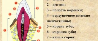
What structure do teeth have in an adult, what parts do they consist of, and what can cause so much pain? If you look at the crown from the outside and mentally “climb” inside, the tooth consists of several layers.
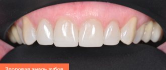
- Enamel. This is the outer layer of the tooth and the hardest part in the human body, consisting of mineral prisms. Its thickness is minimal in the neck area and becomes as strong as possible on the “working” surface of the tooth.
On the outside, the enamel is covered with pellicle - a special organic film, without which acids have a negative effect on the teeth, dissolving enamel minerals.
- Dentine. This is the structural basis of the tooth, a hard, durable tissue that consists of a collagenous base substance that forms the tubules. Odontoblast processes are located inside the dentinal tubules.
Dentin is the primary tissue of the tooth.
In lower vertebrates (fish, amphibians), teeth consist only of dentin. In higher vertebrates, starting with reptiles, enamel and cement appear in the teeth “Anatomy of human teeth” Dr. med. Gaivoronsky I. V.
- Pulp. It consists of loose connective tissue in which nerves and blood vessels pass.
- Root. The part of the tooth, normally not visible to the eye, is located in the alveolar socket of the jaw. On the outside, the root is covered with a protective layer - cement, which is a complex of lime salts, collagen fibers and special cells called cementocytes. The structure of cement is similar to bone tissue, but it does not contain blood vessels or living cells, so it does not grow or renew itself.
- There are many connective tissue fibers around the root of the tooth. They are called periodontium or periodontal ligament. These are the “ropes” that hold the tooth root in the bony alveolus.
Dental formula
Dental patients look with curiosity at the notes on the examination card, on which numbers, corners, and sometimes letters are written. We are talking about a system of numbering teeth without naming them: that is, a diagram written on paper, where each place corresponds to a tooth.
Thus, one of the popular formulas for adults in Russia consists of four blocks [1]
| 87654321 | 12345678 |
| 87654321 | 12345678 |
The teeth are numbered from front to back as they are seen by the dentist examining the oral cavity. Using this diagram of teeth, he describes their general condition using symbols:
- dash—tooth missing;
- the number is circled - treatment is required;
- the figure remains unchanged - a healthy tooth.
Roman numerals are used to designate baby teeth.
| V IV III II I | I II III IV V |
| V IV III II I | I II III IV V |
There are other recording methods, but their general essence is the same - to clearly show the condition of the dentition, so that all specialists involved in the treatment of the patient can immediately see problem teeth and build the correct tactics of help.