53349
Crooked, unhealthy teeth can ruin the appearance of an otherwise attractive person. Oral health is important not only for our attractiveness, because the main function of teeth is to grind food. The condition of the stomach and intestines depends on this, which directly affects health and life expectancy .
Teeth of ancient people
The dentofacial apparatus of prehistoric and modern humans differs significantly. Ancient people had more than 36 teeth, protruding fangs and a massive jaw. This was explained by the need to chew rough food and raw meat. With the addition of thermally processed foods to the diet, the dentition began to change. The canines were the first to transform, becoming aligned with the bite line. Then the jaw arch narrowed, the interdental spaces disappeared, and the teeth themselves decreased in size. Currently, 32 teeth in humans are the norm, but third molars are considered to be an atavism.
Interesting fact!
The teeth of ancient man cannot be called aesthetic, but they were healthy. According to scientists, cavemen never suffered from caries and other oral diseases.
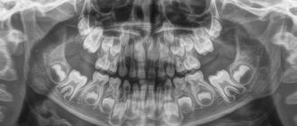
Baby teeth
Their formation in a child begins to occur in the womb at the twelfth week . As a rule, the first to appear in a child are the incisors and canines, and only at the very end the molars.
The timing of this process is purely individual and can vary, but in most cases, the formation of the primary bite begins to occur at the age of seven months and ends at three to four years. By this time, the child should have twenty baby teeth.
Compared to permanent teeth, milk teeth have their own characteristics:
- smaller sizes;
- fewer chewing tubercles;
- the roots spread out to the sides.
Despite this, primary and permanent teeth have the same number of roots.
The deciduous row in the jaw consists of ten teeth: four molars, four incisors and two canines. At the age of six or seven years, baby teeth begin to fall out and are replaced by permanent ones.
First of all, the large molar is replaced, and the final formation of the row ends by the age of twelve to fourteen, with the exception of the third molar.
If you find an error, please select a piece of text and press Ctrl+Enter.
Tags: tooth structure
Did you like the article? stay tuned
Previous article
Photos of the smiles of stars and ordinary patients BEFORE and AFTER installation of veneers
Next article
What to do if a wisdom tooth grows into the cheek?
Name of human teeth
Depending on the location and structure, dental units have their own functional characteristics and are called differently.
- Incisors.
On both jaws there are four front teeth in humans - medial and lateral incisors, which are used for biting food. - Fangs.
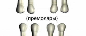
Sharp teeth designed for chewing hard foods. - Premolars.
"Fours" and "fives" on the left and right sides of each jaw arch grind soft or small pieces of food. - Molars.
Three large outer teeth in each row are aimed at grinding coarse substances. - The canines
and incisors are part of the anterior group, or the “smile zone,” and the human molars are part of the chewing segment.
In addition, teeth are divided into temporary and permanent. In the first case, we are talking about dairy products that appear in children from the fifth month of life to three years. The second refers to the final bite, which is formed between six and thirteen years of age. Milk teeth differ from permanent teeth only in size, but in structure they are identical.
Incisors and canines
Dentists divide human front teeth into canines and incisors.
Incisors
The incisors include two teeth located in the upper and lower jaw arches. The crown has a narrow, flattened shape with a sharp edge, as it is intended for cutting pieces of food, which are subsequently chewed by molars and premolars.
The incisors of the upper jaw are much wider and more massive, while the lower ones are almost half as large. The roots are single and flat, especially for the incisors located below. The upper part of the roots deviates to the side.
Fangs
The canines are located directly behind the incisors in the upper and lower jaw arches. Their distinctive feature is that both cutting edges converge at an angle at one point, forming such a recognizable shape. The canines have one long root with grooves on the side.
The upper canine is larger and more massive, while the lower one is less pronounced. The fangs located below have a shorter and smoother cutting edge and narrow longitudinal ridges. The roots are noticeably shorter than the upper ones and have pronounced grooves.
How many teeth does a person have?
The number of teeth a person has depends on age and anatomical features. The child has a set of 20 primary teeth, which are replaced by a permanent bite of 28 teeth. Third molars erupt, as a rule, after twenty years or do not grow at all, which is not a pathology.
In dentistry, a single numbering of human teeth is adopted. Doctors classify teeth as lower and upper and distinguish the right and left segments of the jaws. Each of them includes two incisors, a canine, two premolars and three molars. The countdown starts from the first front tooth and ends, accordingly, with a figure eight. Sometimes a number is added to the serial number indicating the location zone. For example, the right canine of the top row is numbered 13. This order in the schematic representation is called the formula of human teeth.
Polyodontia
In rare cases, an anomaly such as polyodontia is observed - supernumerary, or extra teeth in a person. Dental units can appear in the primary and permanent dentition anywhere in the jaw, separate from or fused with the main teeth. The defect affects not only the aesthetics of the smile, but also leads to the formation of incorrect occlusion, impairs the quality of chewing food and diction. Most often, supernumerary teeth are removed in childhood or built into the dentition.
Edentia
There is also a deviation of the opposite meaning called edentia - congenital or acquired absence of dental units. The causes of the phenomenon include heredity or improper development of the embryo in the womb. People without teeth cannot fully eat and speak, have a deformed facial contour and weakened immunity.
Useful information for patients
The jaws are the basis of the facial skeleton. Not only the beauty of the profile depends on their anatomical structure, but also functional capabilities that are important for life - chewing, swallowing, breathing, speech, the formation of cavities for the sensory organs and much more.
SYMPTOMS OF CORRECT BITE:
- There are no gaps between teeth.
- Chewing is not difficult.
- There is clear pronunciation.
- There are no cracks, chips or stains on the enamel.
- The teeth have the correct shape.
- There is no crowding of teeth.
- There is no pronounced inclination of the teeth in or out.
- The center of the incisors is located right in the middle of the face.
With improper development of bone tissue and the alveolar process, deformations such as microgenia, prognathia, overbite and others occur. In adults, the shape can also change, and this is mainly due to factors such as:
- Severe mechanical injuries.
- Consequence of surgical interventions.
- Various pathological processes (osteomyelitis, ankylosis, purulent inflammation and others).
- Incorrect orthodontic treatment or relapses after it.
- Partial or complete adentia (senile jaw).
Changes in structure, various inflammatory and tumor processes lead to severe diseases of the jaw. The most common pathologies are osteitis, periostitis, osteomyelitis. At the first signs, such as swelling, redness, pain, fever, you should contact a dentist or surgeon.
ANATOMY OF THE JAW
UPPER JAW
The upper jaw is a fixed paired bone of the facial skeleton. It has an air cavity (sinus), 4 surfaces (orbital, anterior, posterior, internal), as well as 4 processes (frontal, zygomatic, palatine and alveolar).
The dentition of the upper jaw is deviated slightly forward and outward. The crowns of the teeth increase in size from the incisor to the first molar, but the second molar is smaller than the first, the third is smaller than the second. The height of the crowns decreases successively from the incisor to the third molar, with the exception of the canines. The large roots of the teeth provide significant stability to the dentition of the upper jaw and to each tooth individually.
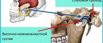
LOWER JAW
The lower jaw is a movable horseshoe-shaped bone of the facial skeleton. A large number of muscles are attached to it. The dentition of the lower jaw has a parabolic shape.
The teeth of the lower jaw are characterized by the fact that the incisors and canines are located perpendicular to the alveolar process, the chewing teeth are slightly inclined towards the tongue. The pattern of changes in the size and height of tooth crowns on the lower jaw is the same as on the upper jaw.
JAW DEVELOPMENT
The growth and formation of the jaw is closely related to the development of tooth germs and alveolar process. This process begins as early as the 7th week of intrauterine life. It is influenced by various endogenous factors (maternal toxicosis, hormonal imbalances, levels of vitamins and minerals), and after the birth of a child - also exogenous (external).
The jaw of a newborn is not yet completely ossified and fused; the lower part is shifted back in relation to the upper.
When a permanent dentition is formed (from the age of 6), intensive growth occurs due to the eruption of molars and incisors. Also, growth spurts are observed at 11–13 years of age; in boys, this usually occurs later. By the age of 18, the formation of bone tissue is completely completed.
CHANGES IN THE SHAPE OF THE LOWER JAW WITH AGE
| Jaw of a newborn |
| Jaw of a 4 year old child |
| Jaw of a 6 year old child |
| Jaw of an adult |
| Elderly man's jaw |
For patients who want to get a free consultation with a dentist in Krasnodar , we inform you that pre-registration for an appointment is being made at Dentistry on Gagarin. Call the numbers indicated on the website and sign up for a free consultation with a dentist with a diagnosis (consultation duration up to 15 minutes).
Dimensions of human teeth
The upper central incisors are twice as wide as their antagonists. The remaining dental units of the same name have approximately equal parameters. The size is determined using special tables with the optimal size and permissible deviations. Experienced doctors calculate proportions by dividing the length of a person’s teeth by the width. A result of about 0.75 millimeters is considered close to ideal. For more detailed diagnostics, other professional formulas and techniques are used.
Size deviations from the norm occur due to improper formation of the jaw, fusion of tooth buds, or genetic predisposition. Teeth that are too large are called macrodentia, and abnormally small teeth are called microdentia. Pathologies are accompanied by problems with bite and chewing functions, but can be successfully corrected by a dentist.
Interesting fact!
The longest tooth in the world belongs to an Indian teenager. The size of its crown is almost four centimeters. About a year ago, the tooth was removed, and the young man was included in the Guinness Book of Records.
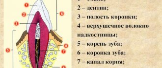
The structure of the human tooth
Anatomy
From an anatomical point of view, a human tooth consists of three parts.
- Crown.
The visible part protruding above the gum. It has four sides: the occlusal, or cutting edge, in contact with the antagonist teeth; contact wall adjacent to adjacent dental units; vestibular and lingual surfaces facing the lips and tongue, respectively. - Root.
Fixed in the socket by connective tissue, located in the recess of the jaw. As a rule, premolars have two roots, and molars have three, four or even five. The remaining dental units have one root canal. - Neck.
It is located between the coronal part and the root of a human tooth, surrounded by periodontium.
Histology
What are human teeth made of? Let's look at the cross-section of the structure of a human tooth.
- Enamel.
A transparent protective coating of the crown, almost entirely consisting of inorganic microelements. - Dentine.
The hard base of the tooth, containing 80% mineral components and 20% organic substances. The shade of dentin is responsible for the color of dental units, as it shines through the enamel. - Cement.
The bone tissue covering the tooth root. Plays the role of a fastening element connecting the tooth to the alveolus. - Pulp.
Soft tissue filled with bundles of nerves and capillaries. Painful sensations during caries are explained precisely by the presence of nerve endings.
Upper jaw
The upper jaw (in Latin - maxilla) occupies a central place among the bones of the facial part of the human skull. This bone structure has a complex structure and performs a number of vital functions.
INTERESTING : As their work activity developed, ancient people transferred some of the grasping functions from the jaw to their hands. As a result, the size of this bone structure has decreased significantly
Functions and purpose
The upper jaw bone performs a number of important functions. Below is a description of some of them:
- Shape-forming. Forms the nasal and eye cavities, the partition between the mouth and nose.
- Aesthetic. The size and shape of this bone will determine the oval of the face, the setting of the cheekbones, and the external attractiveness of a person.
- Respiratory. Forms an extensive maxillary sinus, in which the inhaled air is moistened and heated.
- Chewable . The teeth located on the jaw ensure chewing of the food consumed.
- Swallowing . The muscles and ligaments involved in the process of swallowing food (including the tongue) are attached here.
- Sound-forming. Together with the lower jaw and air sinuses, it takes part in the formation of various sounds. When this bone structure is damaged, a person’s diction is impaired.
IMPORTANT ! During the day, a person makes about 1.4 thousand chewing movements. When chewing bread, the jaw experiences a pressure of 15 kg, fried meat - 25 kg, maximum pressure - 72 kg
Structural features
The upper jaw bone has a complex structure. It consists of several segments and processes, shown in the following picture.
Below we will consider how the body of the jaw bone is structured and how many interconnected surfaces it consists of.
Jaw body
The anterior surface , located under the infraorbital margin, has a slightly curved shape. On it you can see the infraorbital foramen and the canine fossa.
The posterior surface consists of a tubercle and several alveolar openings for nerves and vessels. Next to the tubercle is the palatine groove.
The orbital surface consists of the lacrimal notch and the infraorbital groove, which passes into the infraorbital canal.
The nasal surface and the anterior surface are isolated from each other by the nasal notch. The main part of the nasal surface consists of the maxillary cleft.
REFERENCE : The fixed upper jaw bone is stronger than the movable lower one. Together with other bone structures of the skull, it protects the brain from injury and bruises.
Processes
The palatine process occupies a significant area of the hard tissues of the palate. It is connected to the second process, located on the opposite side, using a median suture.
The frontal process with its upper side is attached to the nasal region of the frontal bone, its anterior side to the new bone, and its posterior side to the lacrimal bone. The lower edge of the process connects to the body of the jaw. The process has a lacrimal groove and a ethmoidal ridge.
The zygomatic process begins at the outer upper corner of the body and has a lateral location. The upper part of the zygomatic process is adjacent to the frontal bone.
The alveolar process is a bone formation with a complex structure. It includes walls, dental alveoli, interdental and interradicular bone septa.
Mounds
The infratemporal part of the jaw has a convex shape. Its most prominent area is called the “maxillary tubercle” (in Latin - tuber maxillae). At the base of the tubercle there are alveolar openings for blood vessels and nerves. The oblique head of the pterygoid lateralis muscle is attached to the maxillary tubercle.
In international practice, the following abbreviations are used to designate tubercles: PNA (according to French nomenclature), BNA (according to Basel nomenclature) and JNA (according to Jena nomenclature).
Features of blood supply
The maxillary internal artery, or rather its four branches, is responsible for the blood supply:
- posterior superior alveolar;
- infraorbital;
- descending palatine;
- nasopalatine (see the following diagram).
The following table shows which areas the listed vessels supply blood to.
Blood supply to the maxillary bone
| Arteries | Areas to which blood is supplied |
| Posterior superior alveolar | Gums, teeth, alveolar process, mucous membrane of the maxillary cavity, bone walls (posterior, lateral) |
| Infraorbital | Inferior orbital canal, orbit, anterior gums, teeth |
| Descending palatine | Hard palate, pterygopalatine canal, soft palate and surrounding tissues |
| Nasopalatine | Nasal cavity, incisive canal, posterior parts of the nasal septum |
The venous network, which is responsible for the outflow of blood, does not always follow the pattern of the supply vessels. It is represented by parallel veins and venous plexuses. From the pterygopalatine ganglion, blood flows into the maxillary vein, and from there into the external jugular vein. From the plexus of the alveolar process it enters the facial vein, and then into the internal jugular vein.
Teeth
When studying the anatomy of the human upper jaw, one should dwell in more detail on the structure of the teeth. This bone structure contains incisors, canines, premolars and molars.
Below is a brief description of the structure of the teeth of a normal, healthy human upper jaw.
Teeth located on the human upper jaw
| Tooth name | Tooth shape | Number of tubercles | Root structure |
| Central incisor | Chisel-shaped | 3 | Single, cone-shaped |
| Lateral incisor | Chisel-shaped | 3 | Flattened from center to edge |
| Fang | Pointed | 1 | Single, powerful |
| First premolar | Prismatic | 2 | How many tubercles, so many roots |
| Second premolar | Prismatic | 2 | Cone-shaped, compressed front and back |
| First molar | Rectangular | 4 | With three branches |
| Second molar | Cubic | 4 | With three branches |
| Third molar | Cubic | 4 | Short, powerful |
Despite the fact that teeth differ in types (types) and shapes of crowns and roots, their internal structure is the same.
Diseases and pathologies of the upper jaw
Inflammatory processes in the oral cavity can provoke the appearance of cysts on the human jaw - hollow tumors filled with fluid. Cysts are treated in several ways, but surgery is considered the most successful. More information about the treatment of cysts can be found in the article “Treatment of jaw cysts: radicular, follicular, odontogenic and others.” Bone inflammation can lead to osteitis, periostitis or osteomyelitis, the characteristics of which are presented in the following table.
Inflammatory diseases of the human maxillary bone
| Name | a brief description of |
| Osteitis | Bone damage |
| Periostitis | Inflammation of the periosteum |
| Osteomylitis | Bone marrow inflammation |
Periostitis can occur in fibrous, purulent or serous forms, and osteomyelitis - in acute or chronic forms. The listed diseases can cause odontogenic sinusitis - a disease associated with the penetration of infection into the maxillary sinuses.
Among malignant formations of this bone structure, tumors of epithelial origin predominate.
Human wisdom teeth
A “wisdom tooth” is the third outer molar with three to five roots. In structure it is no different from its “neighbors”. To the question “How many wisdom teeth does a person have?” cannot be answered unambiguously. They erupt around the age of twenty, one on each side of both jaws. However, there are people without wisdom teeth. This is a variant of the norm, since in the process of human evolution the need for the “eight” disappeared, and the structure of the jaws underwent corresponding changes. Today, third molars are considered a vestigial organ.