A targeted dental photograph is a popular option for diagnosing most problems in the oral cavity. The method is based on the use of X-rays and a patch receiver for these rays (radiovisiograph), which makes it possible to obtain data on the internal tissues of the tooth, as well as the tissues surrounding the root. Taking into account the local direction, X-rays can be taken for 1-3 teeth. The purpose of the study is to supplement visual examination data, simplify the diagnosis of a number of diseases, and also control the quality of treatment provided by a specialist.
| For example, the image allows you to check the quality of cleaning and filling of the canals. If endodontic treatment has been carried out, a photograph of the teeth will allow you to find out how thoroughly the foci of infection were removed and how tightly the filling fits. |
Indications for prescribing a targeted photograph of a tooth
A modern targeted X-ray of the tooth (radiovisiography) should be performed before treating:
- caries. In the image, the doctor will be able to detect carious lesions under a filling or prosthesis;
- pulpitis, periodontitis. The doctor will be able to analyze the condition of the tooth roots, see the number of canals, and correctly carry out endodontic treatment;
- dental granuloma, cyst; The dentist must make sure that there are medical indications for tooth extraction. Otherwise, a “program to save” the tooth will be offered.
- pericoronitis. For example, a photograph of a wisdom tooth (“eight”) that has not yet erupted will allow you to clarify its position. In general, radiovisiography is rarely prescribed for wisdom teeth due to the difficulty of positioning the radiovisiograph sensor in the patient’s oral cavity. If radiovisiography is not possible, an OPTG (orthopantomogram) is prescribed.
- incorrect position of a growing tooth.
As for contraindications, traditionally, dental x-rays are not recommended in the first trimester of pregnancy. However, modern diagnostic equipment is extremely safe for the body. Therefore, if a correct diagnosis is needed for medical reasons, it is not prohibited to take a photo of the teeth while expecting a child.
Summary: important points
As a practicing dentist who knows the system from the inside, I want to draw your attention to the following points that are important for your safety. If the clinic has an X-ray machine, then a license must be obtained for it, the issuance of which presupposes the mandatory presence of a certified radiologist on the staff of the dental clinic. However, in reality, even in large clinics and public clinics, it is not always the case that x-rays will be taken by a trained specialist.
Even if he is, he may go on vacation or get sick, and a regular nurse (dental assistant) will take the pictures instead. This is a gross violation that leads to both the production of low-quality images and an increase in the radiation dose. In small clinics, the risks of receiving a poor-quality x-ray examination are much higher, and the first thing that makes you suspect a forgery is if the picture is taken not by a special employee, but by a nurse from the dentist you came to see.
The second very important point: if you see that the x-ray is not done in a special room, but the x-ray machine is located right next to the dentist’s chair, then you should change the clinic and the doctor. The fact is that all objects in this office (including the dental chair and doctor’s instruments) will have an increased background radiation, and this is no longer safe for health. We hope that our article on the topic: X-ray of the jaw and teeth was useful to you!
Types of targeted dental imaging
To take a single photograph of teeth, dental clinics use two types of equipment:
- an outdated analog X-ray machine: it can be used to take pictures on X-ray film. The process is not always comfortable, printing pictures takes time, and after 2 years the resulting image fades;
- modern digital radiovisiograph: a device that creates high-resolution electronic images. If necessary, the image can be enlarged and detailed information can be obtained. The images are permanently stored in the dental clinic’s database, and you can return to them at any time – even after several years. Plus, electronic equipment has 2-10 times less radiation exposure compared to analogue equipment.
Technology
The procedure takes place in an x-ray room. The patient wears a protective lead apron to reduce exposure. Next, install the sensor or film to the desired tooth. After this, a targeted photo is taken. It lasts no more than a couple of seconds. The sensor is taken out and the information is displayed on the screen. If the image is taken on film, it is first developed in a dark room in a special solution, after which it is given to the patient. Film radiography is slower than digital radiography; image clarity is affected by the quality of the film and the technique of working with it, so it is becoming a thing of the past.
Advantages of a targeted tooth image
A dental image taken on a digital radiovisiograph has several advantages:
- a clear and detailed image of the tooth, as well as the tissues that surround it;
- safety of the study for the body and absence of harm to the body;
- you can prescribe repeated images in the required quantity without health consequences;
- organized and secure storage of images, the ability to return to previous results;
- results can be saved on digital media or sent by email;
- you can enlarge a certain area of the image, obtaining more information;
- There is no need to wait for the film to be developed - the results are available immediately.
The key advantage is high accuracy and information content, which makes it possible to detect problems that are invisible during visual examination, as well as pathologies and diseases in the early stages.
General overview
From a technical point of view, the procedure is an X-ray analysis of the state of one or more adjacent units, characterized by the speed of obtaining complex data and ease of execution. The resulting image allows the dentist to:
- To formulate a correct diagnosis, the formulation of which on the basis of visual examination data alone may be impossible.
- Determine and prescribe an effective course of treatment, as well as control its pace.
- Monitor the intensity of the spread of pathology formed in the oral cavity.
- To study the condition of the structure of bone tissue and root canals, dentin and blood vessels.
- Identify hidden anomalies and pathological processes, determining their localization.
To obtain targeted dental images, digital equipment is used - a radiovisiograph, one of the functional advantages of which is the minimum radiation dose to the patient during the shooting process. The directed beam flow maintains focus, affecting only a selected area of the jaw row, which makes the diagnostic procedure safe for the patient’s health.
General description of the sighting shot
A targeted X-ray is a familiar type of x-ray, in which one tooth or a row of three adjacent units is illuminated. The format of the captured image is a 2D image.
Possibilities for reviewing problem areas include:
- dentin with its cavity;
- root canal system;
- vessels close to the diseased tooth.
Instructions for prescribing a targeted photo
The patient is sent for an x-ray if necessary:
- examine the condition of the gums for the presence of periodontal disease;
- establish the causes of acute toothache;
- confirm caries or pulpitis discovered during sanitation;
- examine the integrity of bone tissue after injury;
- check the filling of the canals after depulpation;
- monitor the development of wisdom teeth.
Types of targeted shots
List of existing X-ray images:
- Standard shot. It is produced using analog X-ray equipment to ultimately obtain a planar image. The main disadvantage of the outdated methodology is the short period of preservation of the clarity of the basic result.
- Digital photo. A more modern method of performing x-rays involves the use of a radiovisiograph. The device digitally records the area of the problem area, which can then be saved on your personal PC resources.
- Interproximal image. Such an image is prescribed, if necessary, to examine the adjacent surfaces of teeth for the possibility of hidden carious cavities between them.
- Periapical image. It is done to obtain data on the condition of the tooth root and the adjacent soft tissues.
- Intraoral image. This type of x-ray is the best way to check the integrity of dentin and diagnose the initial stages of caries.
The voiced types of targeted photographs are not suitable if we are talking about significant problems with teeth. In this case, a panoramic x-ray is prescribed, which has much greater diagnostic capabilities.
Advantages of X-ray
- safety of diagnostics at permissible radiation dosage;
- the ability to take several pictures (up to 3-5 pieces) at one time;
- affordable cost of this service (350-500 rubles);
- the reality of taking a targeted photo in almost all clinics.
Cons of X-ray
- limited viewing range of problem areas;
- presentation of the image in a single-plane perspective;
- ineffectiveness when wanting to plan corrective actions for malocclusion;
- the inability to detect literally all cracks in the tooth root and diagnose nerve inflammation;
- the minimum term for the preservation of a photograph when it fades.
Sight Shot Limitations
The prohibitions do not apply to pregnant women in the 2nd - 3rd trimester, whose dental condition may deteriorate during pregnancy. For breastfeeding mothers, problems arise not from the procedure itself, but from increased sensitivity to myths about the dangers of radiation for milk. As a result, they lose teeth and then have to be removed.
If unwanted spots on the enamel were discovered during sanitation in a child, then he will definitely be prescribed a targeted scan. If the clinic client’s age is too young, the procedure is prescribed only for serious damage to the baby’s jaw (fall, difficult birth of the mother).
However, there is a list of patients who are prohibited from diagnosing using X-rays:
- children under 2 years of age, if it is possible to avoid the procedure;
- pregnant women in the first trimester;
- people during a serious illness and in the postoperative period;
- clinic clients with reduced immunity and severe anemia;
- patients with bleeding of any etiology and intensity.
Execution steps
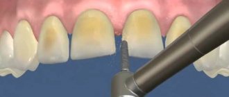
Interproximal radiography is one of the types of intraoral examination that allows you to obtain a picture of 1 section of the oral cavity with an image of the upper and lower dentition. To obtain the image, a special holder is secured between the closed teeth. It allows you to identify interdental caries and various changes in bone tissue due to gum disease. And also check the correct installation of crowns, dentures or fillings.
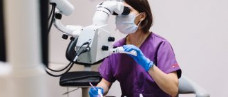
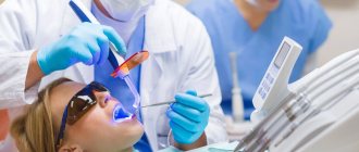
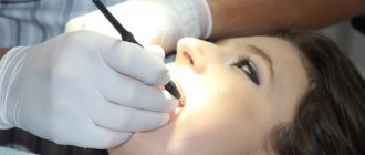
An X-ray of the tooth is taken by a doctor in an office specially equipped for this purpose. Before taking an X-ray of a tooth, the doctor gets acquainted with the problem and studies its location.
To obtain a clear image of the problem area, the patient's head is fixed in the required position. The process of visiography takes a matter of seconds, but during this time the patient is required to remain completely still. The video in this article shows how to take targeted photographs of teeth so that the image is as informative as possible.
Using a digital sensor, the radiologist directs the beam of rays to the desired area. The photograph is taken either from the inside of the mouth or from the face. During the manipulation, the patient does not feel any pain or discomfort.
Features of the procedure in children
Contact intraoral radiography can be prescribed for children in cases where tooth damage cannot be examined in any other way. The technique allows early detection of disturbances in the process of teething, bone diseases, and prescribing effective treatment.
In addition, this method allows you to control the implementation of orthodontic manipulations if the child has problems with the formation of the jaw.
The study is carried out in the same way as in adult patients. Children under 2 years of age are recommended to undergo x-rays only in case of urgent need. For example, in case of injury during childbirth, to monitor the development of the jaw, or after a fall from a height, to assess the integrity of the teeth.