How is dental implantation done, what types of implants are there, how are they installed, and what are the differences between the methods of implanting them? Dental clinics use various methods. The choice depends on the clinical picture, the scale of the problem, the patient’s financial capabilities and other factors. There is a list of absolute and relative contraindications when such intervention is prohibited.
The procedure is associated with a high risk of injury, soft tissue infection, and rejection of the implanted material. To avoid negative consequences, you need to carefully consider treatment tactics and adhere to all doctor’s recommendations.
What is implant installation?
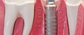
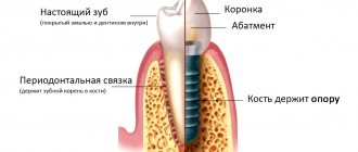
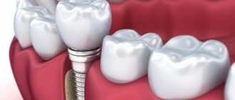
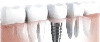
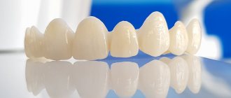
This is the process of implanting metal screws and plates (usually titanium) into bone tissue. They come in various forms, the choice depends on the type of intervention. Most often, pins are implanted into the gum bones, and then artificial crowns and bridges are installed on it. High-strength hypoallergenic materials are used in production.
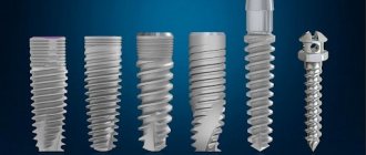
Implanted screws come in conical and cylindrical shapes. They include a stem, a neck and a support head for the abutment. The cost and quality of treatment depends on the origin. Models made in Germany are considered the most popular in the modern dental market.
Implant shape
The shape of the implant does not affect the size, but it determines how natural the breast will look after mammoplasty. There are two types.
Round (hemispherical) implants
The peculiarity of such an implant is that the highest point is located in the center of the endoprosthesis. After the operation, the volume of the upper pole of the gland becomes filled, a push-up effect occurs - raised breasts in the décolleté area.
Piezo devices are distinguished by their compact size and adjustable power. Such implants look natural when correcting ptosis, and if a woman has a layer of subcutaneous fat in the décolleté area. Even if immediately after surgery the breast does not look as natural as possible, over time, under the influence of gravity, the silicone implant lowers slightly, creating a harmonious shape. The second point in choosing such an endoprosthesis is the correct selection of access.
Anatomical implants
They have a teardrop shape, repeating the natural shape of the mammary glands. The anatomical endoprosthesis immediately imitates the effect of gravity, maintains proportions in which the upper pole of the chest is less filled in comparison with the lower pole. This shape is maintained in the supine position. Such implants have only one drawback: compared to the hemispherical shape, they have a higher probability of displacement in the capsule.
It is impossible to say unequivocally which form is better - it all depends on the anatomical features of the patient’s mammary glands.
Basic methods of implant installation
There are several methods that are most in demand. The choice of one technique or another depends on the type of structure, the length of time teeth are missing in a row, the financial capabilities of the patient, the location of the problem and other factors.
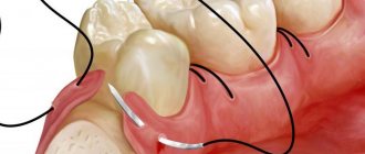
Patchwork method
This is the most common intervention option. Main stages (after all preparatory activities):
- anesthesia (administration of a drug for anesthesia);
- treating gums with antiseptics;
- incision of gum tissue to form a flap;
- creating a hole (bed) for the exact dimensions of the structure being mounted;
- screwing the screw into the bone and installing a plug;
- suturing.
The main disadvantage is the long healing time. This is very traumatic, recovery lasts up to 2 weeks. The risk of complications increases, such as bleeding, pain, ingress of harmful microorganisms and, as a result, an inflammatory process and rejection.
Minimally invasive surgery (transgingival implantation)
This method is used primarily for express implantation using one-component models. The pins are installed very quickly and with minimal trauma.
The doctor does not make massive incisions. In this case, the screw is screwed into the bone through the gum. No flaps are formed, no bed is created. The main advantages of the procedure:
- speed of manipulation;
- relief of symptoms during the recovery period;
- reducing the duration of rehabilitation.
Many people pay attention to the fact that after 2-3 days the discomfort completely disappears. The risk of infection is reduced, the pins are not rejected.
Using laser
This is not an independent technique, but an additional method of intervention that helps reduce the likelihood of injury. The laser system is most often used with the patchwork method, when it is necessary to peel off soft tissues. The beam is an alternative to a scalpel. Very thin incisions are formed, and the gums are minimally injured.
During the manipulations, disinfection occurs, capillaries are cauterized, which means there is no bleeding. Healing occurs several times faster.
Doctors carry out antibacterial treatment in this way. The tone increases, which means the period of restoration of cellular structures is reduced.
Lateral method
This method is associated with many risks and increased trauma. This is usually how extensive disk and plate structures are installed, which are integral with the support. During the operation, large flaps are formed, including from the side. The dentist uses a special tool to cut down the bone area intended for the screw.
When installing a dental implant, the bed must be identical in size to it. A doctor’s work is practically a piece of jewelry; it is important to prevent even the slightest deviations. To avoid loosening, a prosthesis is placed immediately.
This option is now practically not used. This is associated with a high risk of rejection, injury, and inflammation.
One-step method
Implantation is possible not only in case of long-term absence of a tooth. For example, the structure can be fixed immediately after a unit is pulled out or falls out. Thus, the pin is screwed into the fresh hole formed after extraction. At the same time, it is allowed to replace as many removed incisors, canines and molars as desired. However, the main condition remains the mandatory preparatory stage. That is, such operations are not performed urgently; treatment must be planned in advance.
Which screw will be installed depends on the readings. Both one-piece and two-piece elements are used.
Types of accesses in mammoplasty when installing breast implants
Endoprosthesis replacement is performed in three access options. After consultation and examination, the plastic surgeon decides which way is best to insert endoprostheses.
Submammary (submammary) access
This is the first and well-developed type of plastic surgery, which is now operated using new techniques and technologies. The incision is made along the natural fold located under the breast. A pocket is formed under the breast or muscle into which the implant is placed. The endoprosthesis looks more natural under the muscle.
The advantages of the inframammary approach include:
- correction techniques brought to perfection;
- easy recovery period;
- possibility of prosthetic implants of large volumes;
- universality of the method;
- maintaining the integrity of the mammary gland.
This method also has disadvantages. Due to the need to transfer the own submammary fold, there is a possibility of implant prolapse.
Plastic surgeons recommend this method to all women who have a pronounced submammary fold under the breast. It is not done for girls with small and high breasts, which after plastic surgery will not hide the postoperative scar.
Periareolar access
This is a method of installing an implant through the areola. To do this, at the border with the skin, along the lower edge of the areola, a small incision is made into which the implant is inserted. It can be installed behind a muscle or gland.
From an aesthetic point of view, this is the most acceptable method.
With mammoplasty with periareolar access, you can not only increase the size, but also correct other breast defects:
- reduce the size of the areola,
- restore the symmetry of the mammary glands;
- pull up the nipples;
- perform a mini gland lift;
- move the nipples to the periphery or closer to the center.
This access also has disadvantages, and quite significant ones:
- the possibility of the formation of a keloid scar cannot be excluded;
- discomfort during the rehabilitation period when installing an implant under the muscle;
- possible loss of sensation in the nipple;
- there is a possibility of the formation of pericapsular contracture - excessive formation of fibrous tissue around the endoprosthesis and deformation of the mammary glands;
- there is a possibility of retraction of the scar under the areola.
Surgeons do not recommend this method for women planning to breastfeed for a year after plastic surgery. It is not suitable for patients with light-colored nipple areolas, as well as those who have a tendency to form a keloid scar.
Transaxillary access
This is an endoscopic method of installing an implant through the armpits. An incision is made along the posterior edge of the pectoralis major muscle, in the armpit area. It can be vertical or horizontal. Under the control of endoscopes, surgeons form a cavity, place an endoprosthesis, quickly eliminate bleeding, and control all risks. At the same time, the doctor, using a video camera, sees the correct installation of the implant.
In some clinics, this operation is performed “blindly”, but this method is too traumatic and there is a high risk of bleeding under the muscle. Now it is practically not used. Optics reduced the risk of hematomas, massive bleeding and other complications.
The endoprosthesis is inserted without injuring the mammary gland, since its tissue is not affected during surgery. This is important for women who want to breastfeed their baby. This access is one of the most aesthetic methods, in which it is easiest to hide traces of surgical intervention.
This method has other advantages:
- recovery time after surgery does not exceed two weeks;
- the scar completely resolves within five months and does not form a keloid;
- these methods provide the possibility of fixing the implant and reduce the risk of displacement after correction;
- The duration of the surgical intervention does not exceed 40 minutes.
The disadvantages of the method include the impossibility of plastic breast correction during surgery. This type of access is not used when it is necessary to eliminate the asymmetry of the glands and shift the inframammary fold. The method does not exclude the possibility of the formation of fibrous contractures, in which excessively overgrown fibrous tissue deforms the endoprosthesis and the breast.
After correction with axillary access, scars do not require special care. Physical activity is not excluded, you can raise your arms.
According to plastic surgeons, this method of access gives excellent results on a “boyish figure” with small breasts.
Vertical and inclined installation
Most structures are installed this way. If several pins are installed simultaneously, the doctor must observe the principle of parallelism. In some cases, the material is screwed into the bone tissue at an angle of 45 degrees. This is indicated when the base thickness is insufficient or its density is low due to atrophy and inflammation.
Inclined placement allows for large areas of solids to be brought into contact with the implant surface. In addition, this way you can bypass atrophied areas, anatomical structures, and prevent possible plastic surgeries. However, not all models are suitable for carrying out such manipulations.
Installation options depending on bone type
Often screws are implanted not only in a vertical position, but also in the central spongy tissue. It is this area that is most susceptible to atrophic processes and resorption after extraction. It is not always possible to use this area during therapy; some dentists resort to extensions.
Manufacturers have developed models designed for attachment to other types of bone structures:
- Basal and compression. They are located in the deepest layers that are not prone to atrophy. A well-known, time-tested brand is Oneway Biomed.
- Zygomatic Elongated, mounted at an angle exclusively on the upper jaw. Such structures are characterized by reliable fixation without deformation.
Alternative Methods
All options considered involve the implantation of screws into the bones. Nowadays all implantation materials are screwed in this way. An artificial metal root completely replaces the natural one. The implant has a similar appearance and is placed in almost the same area. Additional segments may be affected at greater depth.
Intramucosal technology for installing dental implants
The pin is not screwed into the bone structures, but directly into the gums and a little deeper. This way the installed structure will not fall out. Outwardly, it resembles buttons, and not an imitation of the root part of the unit.
The technique is gradually becoming a thing of the past due to its fragility. There is a high probability of complications (injury, inflammatory processes).
Endodontic-endosseous method
This option is also outdated. A device in the form of a small awl is inserted into the root canal, then it is moved slightly beyond its limits and fixed in hard tissues. It was used mainly in the presence of a mobile or broken incisor, canine, or molar. Such fixation is difficult to implement and becomes loose over time. The technology is not suitable for long-term restoration and is indicated for patients with narrow-shaped canals.
Mini implantation
How dental implants are inserted: tooth installation technology involves shallow immersion into bone structures. Over time, the pins become loose, the fastenings wear out, which means the prosthesis becomes mobile and causes severe discomfort.
This method is welcomed only as a short-term method while the patient is undergoing long-term therapy. For example, this happens when restrictions are discovered on the use of more “long-lasting” techniques. The treatment period is up to 5 days. The prosthetics are removable; screws can be inserted even in cases of complete edentia.
Stages of implantation, technology for installing dental implants
A whole team of experienced doctors goes through several successive steps. Each of them has its own characteristics.
Survey
Before performing surgical procedures, it is imperative to conduct a high-quality diagnosis. The patient is sent for laboratory tests, x-rays, and CT scans of the jaw. Thanks to a thorough examination, the structural features of the maxillofacial apparatus, the location of nerve endings are revealed, and the thickness of the bones is determined.
In addition, a visual examination of the oral cavity is mandatory. The specialist fills out a card indicating the presence of chronic pathologies.
Implantation plan
Depending on the results of diagnostic procedures, a therapeutic regimen is developed. The dentist selects the optimal method of intervention, advises the most suitable designs, and calculates the number of units used. A 3D model of the jaw is created using a computer. Online you can “try on” different types of implants, as well as depict their angle of inclination. A prosthesis is added to the image. In this way, the doctor is able to assess the resistance of the pins to chewing load.
Surgical stage
This is the direct implantation of a screw into hard and soft tissues. Anesthesia is indicated before surgery. Anesthesia in such cases can be either local or general. Sedation (medicated sleep) is often used.
All doctors’ manipulations take about 40 minutes on average (if only one unit is missing). When entire rows need to be restored, it will take several hours.
Rehabilitation
Regardless of the chosen technique for installing a dental implant, the patient goes home immediately after the operation. Prosthetics are rarely performed on the same day as surgical procedures. It is important for gingival and bone tissues to recover after serious mechanical impact.
Medical recommendations:
- to prevent damage to the integrity of the suture material, do not rinse the oral cavity for several days; baths are encouraged;
- careful hygiene (it is important to take care of the health of the remaining incisors, canines and molars);
- follow advice on proper nutrition and a healthy lifestyle;
- do not neglect scheduled visits to the doctor even in the absence of adverse reactions.
Installation of prostheses
Prosthetics are of the following types:
- Immediate. The structure is installed 4-5 days after successful implantation. The technique is more often used for large volumes of work on the entire jaw.
- Deferred. It is necessary to wait for complete healing. Usually it lasts up to six months. This method is indicated in the absence of 1-2 teeth.
- Early. An intermediate option between the two discussed above. This means that the process will not be completed in a couple of days, but at the same time the patient will not have to wait 6-8 months. If the device model is modern and progressive, 90 days or even less is enough. Rapid integration with bone tissue is achieved due to special surface characteristics.
Preparatory stage
Goal: identifying contraindications to implantation, detecting hidden dental diseases, obtaining a detailed clinical picture, planning an operation with a predictable result.
As a rule, preparation for dental implantation lasts no more than 1 day. But if the patient has chronic diseases and possible contraindications to the installation of dental implants, the preparatory stage may drag on for a week.
Visual examination of the oral cavity
The doctor receives basic information about the health of teeth and gums, the presence of stone, etc.
Duration: 30 minutes.
Taking tests
Only if the patient is suspected of having some contraindications for dental implantation, he is prescribed a series of tests:
- clinical blood test with leukocyte formula + ESR;
- coagulogram (fibrinogen level, prothrombin and thrombin time);
- biochemistry (total protein, protein fractions, urea, creatinine, bilirubin, etc.);
- blood glucose levels;
- detection of antibodies to HIV and hepatitis.
It is important to know that pre-implant laboratory test results are only valid for 14 days.
Duration: up to 7 days.
Hardware diagnostics
To determine the anatomical features of the bone, assess its quality and determine the location for installation of implants, a panoramic image of the jaw (OPTG) and cone beam computed tomography (CBCT) are performed.
At this stage the doctor:
- assesses the condition of the jaw bones and bone density in areas of missing teeth;
- identifies pathologies in the periodontium;
- records existing tooth movement;
- calculates the distance of the future implant from the mandibular nerve;
- determines the exact number of root canals and analyzes the features of their structure;
- checks for cracks in the roots of teeth, unerupted eights, malocclusions, and neoplasms in the maxillofacial area.
Duration: up to 1 hour.
Sanitation of the oral cavity
Identified dental diseases are treated: caries, gingivitis, pulpitis, etc. If necessary, previously installed dentures, crowns, and veneers are replaced. Teeth that cannot be treated are removed.
Duration: up to 3 days (depending on the amount of work).
Bone grafting
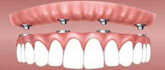
The All-on-4 (all on four) and All-on-6 (all on six) dental implant concepts make it possible to restore the dentition without bone grafting in most clinical situations.
However, if increasing the volume of spongy tissue is indicated, one of the following methods is used:
- sinus lift (only for dental implantation in the upper jaw) - through an incision in the gum and bone, the bottom of the maxillary sinus is raised and filled with artificial bone material;
- osteoplasty - replanting natural or artificial material from a donor (human or animal) under the gum or into the alveolar ridge;
- autogenous transplantation - replanting a block of your own bone to increase the volume in height and width.
Duration: from 3 months to 1 year.
Contraindications to inserting dental implants
Implantation is not permitted in all cases. There is a list of conditions when medical manipulation is absolutely contraindicated:
- Problems with blood clotting. In this case, any surgical intervention is prohibited.
- Epilepsy. During the procedure, the risk of an epileptic seizure increases sharply.
- Exacerbation of mental illness.
- Fall of immunity. The implanted pin may be rejected.
- Connective tissue diseases. The regeneration process will take a long time.
- Syphilis, tuberculosis. The bones are affected and the screw is rejected.
- Deep periodontal disease, periodontitis.
- Severe forms of rheumatism.
- Malignant tumors. Relapse is possible.
- Allergic reactions to the materials used and medicinal formulations.
- Cardiovascular pathologies in advanced cases. Seizures are possible that can cost the patient his life.
Relative contraindications, when surgery to install a dental implant instead of a tooth is undesirable, but under certain conditions is sometimes allowed:
- Diabetes. Metabolism is disrupted, and wounds take a very long time to heal. Treatment is carried out at the compensation stage in compliance with special rules.
- Pregnancy. Accompanied by an increased risk of miscarriage.
- Exacerbation of the inflammatory process. Therapy is carried out only after complete recovery.
Possible complications
One of the most serious consequences of medical intervention is the rejection of material implanted into bone tissue. The screw is rejected when the patient’s immune status is elevated and allergic reactions occur. Fortunately, this phenomenon is rare.
Often the gums respond with an inflammatory process to a foreign object, pain, swelling, and redness appear. In severe cases, surgical removal of the pin cannot be avoided. After this, rinsing and disinfection are performed, and a course of antibacterial and anti-inflammatory drugs is prescribed.
If the doctor does not follow the rules of asepsis, pathogenic microorganisms may enter the wound. As a result, acute infections develop, which are accompanied by dangerous symptoms, including suppuration. Also, treatment can be complicated by otitis media, neuritis of the facial nerve, and abscess. In such situations, intragingival injections and antimicrobial therapy using a dropper are indicated.
The procedure for installing dental implants, placement and treatment methods depend on the clinical picture, the degree of damage to the dentition, the presence of contraindications, financial capabilities, the age of the patients and other factors. The older a person is, the higher the likelihood of encountering such a problem. Many people are afraid that a smile looks unattractive to their interlocutors, although the real danger from missing elements in a row is more serious complications.
How does enlargement occur through the areola?
Mammoplasty through the areola is performed under general anesthesia and lasts about an hour. First of all, the surgeon makes a 3.5-4 cm incision to install the implant, along the pigmented edge of the areola, which differs from the skin in structure. Thanks to this, the scar is completely invisible in the postoperative period. Then Philip Nikolaevich creates a subcutaneous tunnel under the base of the mammary gland or under the pectoralis major muscle, without affecting the breast tissue. In this place there will be a so-called “pocket” for placing the implant. The implant is inserted into the pocket. After surgery, dissolvable sutures are placed in the skin and the patient is placed in a compression bra.