Author: Salatsky Dmitry Nikolaevich Chief physician, orthopedic dentist, gnathologist, maxillofacial prosthetist When treating most dental diseases, doctors do not limit themselves to simply removing the affected necrotic tissue, cleaning out the carious cavity. Very often, the dentist’s task becomes the need to remove the pulp, the neurovascular bundle located inside the tooth, and clean it with further filling of the tooth canals (endodontic treatment). In this case, the dentist must go through all the canals in the tooth without exception, since the anatomical space inside the dental unit remaining uncleaned and unsealed can lead to irreversible consequences due to which it will have to be removed.
What are root canals
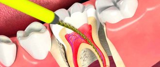
The dental nerve, or pulp, is located not only inside the tooth, its crown, but also inside the roots. Root canals are elongated cavities located inside the roots of teeth. This is where the dental nerve passes within the root. The number of root canals in one root may vary. There may be two or more channels in one root. The root canal can be very thin, thinner than a human hair, so high-quality treatment can take quite a long time. The difficulty is that even with the use of modern microscopes, the doctor cannot see absolutely all parts of the root canal. Work in the deepest part of the root is carried out almost blindly, which requires experience and skill from the doctor.
Signs of canal infection
In some cases, patients can independently determine the presence of problems with a previously treated tooth. The main signs of canal infection are:
- pain that did not go away after treatment or occurred some time after it;
- a feeling of fullness in the gums, discomfort when pressing on the tooth;
- the presence of swelling, swelling on the gums, as well as the formation of a fistulous tract (cyst breakthrough);
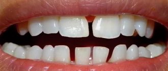
- changing the shade of the dental crown from light to gray. This may indicate dentin destruction;
- persistent unpleasant odor.
If you have the above symptoms, you should not waste time by self-medicating. At the IMEZA clinic, the patient can count on a quick, accurate diagnosis and adequate treatment.
Root canal treatment
Root canal treatment takes place in several stages. Before treatment, X-rays or computed tomography must be taken. Using CT or X-ray images, the doctor will determine the approximate length of the root canals, shape, and the presence of obstacles to their cleaning.
Preparation for root canal treatment involves administering anesthesia and isolating the tooth from saliva using a rubber dam. If a tooth is severely damaged, the doctor can first restore its walls using temporary filling material.
Clinic doctors
Often during endodontic dental treatment, dentists encounter root canal obstruction (obstruction of the root canals of the teeth). But in most cases, this problem can be solved thanks to the high professionalism of specialists and the excellent equipment of the DentaBravo clinic.
Why does tooth root canal obstruction occur?
There are several causes of root canal obstruction:
- Anatomical structure of the root.
The canals may be flattened, curved, with small branches or transverse bridges.
- Inflammatory phenomena in the pulp. Chronic caries, subacute and chronic pulpitis, mechanical overload of the tooth provoke overgrowth of the canals.
- Age-related changes. Over the years, deposits of dentin or dentin-like tissue (denticles) accumulate on the walls of the canals. They narrow the lumen and often contribute to root canal obstruction.
- Treatment using phosphate cement. This is a dense material that cannot be removed by ultrasound, solvents, or mechanical methods.
How to determine tooth canal obstruction
Diagnosis of root canal obstruction is very important, because quality treatment requires careful preparation. And in order to freely manipulate during cleaning and expansion, the doctor must determine the working length.
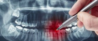
The most common research method is radiography. A photograph of the root of a tooth with an instrument inserted into it makes it possible to see:
- tooth length;
- direction of movement of the endodontic instrument;
- obstruction of the root canals of the tooth;
- canal curvature;
- presence of perforation;
- periodontal condition, etc.
If there are symptoms of root canal obstruction, they need to be opened to the apical narrowing so that endodontic treatment can be carried out.
How is tooth canal obstruction treated?
Usually, when treating pulpitis or periodontitis, the doctor works with a pulp extractor. If there are no problems with the channels, then difficulties usually do not arise. But if the canal is curved, narrowed, closed, or adjacent to an adjacent root, a special approach is needed.
Treatment of root canal obstruction requires the use of special chemicals and a stronger extractor to avoid metal fatigue and instrument fracture. For example, nickel-titanium files (titanium needles) are used - rotating extractors, which make it possible to pass root canals without preparation and reduce the risk of pinching or breaking the instrument.
Also, in case of obstruction of the tooth canal, thin drill burs are used - instruments with spiral blades designed to eliminate old filling material (pastes, cements, gutta-percha).
Unsealing is also done using solvents that work well with soft and flexible materials. Difficulties arise with channels filled with resorcinol-formalin paste. When it hardens, it becomes very hard, and to eliminate the obstruction of the tooth canal it is necessary to use strong substances, drilling with a bur and nozzles with ultrasound. Unsealing a resorcinated canal is a long and labor-intensive procedure associated with the risk of tooth perforation. And if the patient has a phosphate-cement filling, the root is generally amputated or its apex is cut off.
Treatment methods for dental canal obstruction are selected individually. For consultation and diagnostics, sign up at the DentaBravo clinic!
Cleaning and expansion of root canals
Root canal cleaning begins with measuring its length. A special instrument that resembles a needle in appearance is inserted into the root canal. A special apparatus is attached to this instrument - an apex locator. When the root apex is reached, the device beeps and the distance to the apex appears on its display.
After measuring the length, the doctor begins to clean the canal from the remnants of the nerve and expands it. Root canals can be expanded using hand instruments that the doctor holds in his hands or using machine or rotary instruments. Rotary instruments are inserted into a special tip - an endomotor. Development with machine tools is more modern and more expensive. It speeds up the process and provides higher quality processing. In order for a doctor to achieve a similar effect using hand instruments, the doctor will need much more time. After each insertion of each instrument, the doctor rinses the root canal with a chlorine-containing antiseptic.
After processing and drying the root canal, it is filled.
Conservative treatment method
This method allows you to keep the nerve alive. This is a more preferable option, which is possible in case of a minor inflammatory process, or in the presence of a deep carious lesion, in which the lesion is close to the root, but has not yet had time to fully move to the pulp. In addition, complete and rapid nerve regeneration is possible mainly in young patients under 25-30 years of age due to the physiological characteristics of the structure and functioning of the pulp.
During the conservative treatment method, carious tissue is removed, after which a special medication is applied on top of the nerve. A temporary filling is fixed, which will reliably cover the area from bacteria and plaque. Such treatment can be repeated - the doctor assesses the condition of the dental nerve using X-rays and if the pulp regeneration is insufficient, the medicine can be prescribed for a few more days. After therapeutic treatment, the temporary filling is removed and replaced with a permanent one.
Surgical method of treatment
The surgical method is more common in the treatment of pulpitis, since the pulp most often cannot be cured and it is necessary to remove it so that the inflammatory process does not spread to the tissues around the tooth. This procedure is called depulpation or endodontic treatment.
Measles canal filling
The procedure for filling root canals is carried out in various ways, for example: by the method of cold lateral (side) condensation of gutta-percha pins (reliable and affordable). Pins that resemble gutta-percha sticks in appearance are inserted into the root canal. After insertion, these sticks are compacted against one of the walls of the root canal. Between the gutta-percha there is a special paste for filling the canals.
The most modern method is filling the root canals using the vertical condensation method of hot gutta-percha. With this method, the doctor seals the top of the root canal using a heated gutta-percha pin. After this, the remaining volume is filled with hot liquid gutta-percha. This method is more expensive.
After the filling, if everything is done well, the doctor removes excess gutta-percha and sealer, and then installs a temporary filling. X-ray control is required. Permanent tooth filling in one visit is carried out only in exceptional cases, since it is necessary for the material to cool completely.
After root canal filling, discomfort may persist for some time. The tooth may be sensitive when biting. Within a few days the pain goes away.
Causes of obstruction of dental canals
The root canals of teeth can become obstructed for a variety of reasons. The most common include:
- features of the anatomical structure of the tooth - highly curved roots with small branches, flattened areas and transverse bridges;
- inflammatory processes in the neurovascular bundle - with dental injuries and chronic pulpitis with a subacute course, the root canals can become overgrown;
- age-related changes in tissues - over time, dentin deposits accumulate on the walls of the root canals, which lead to a narrowing of the lumen;
- dental procedures using phosphate cement - this material has a high density and cannot be destroyed by ultrasonic waves, instruments and solvents.
Before starting endodontic treatment, it is important to accurately determine whether the canals are patent.
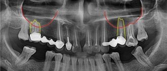
How to understand whether the root canal is filled properly
Signs of a properly treated canal are visible on x-ray: filling to the tops of the root, dense filling of the canal without any excess or voids. It is not desirable to have material residues outside the root canal. If the paste does not dissolve, an inflammatory process may develop in the future. But the quality of work depends not only on the attending physician: the patient himself also plays an important role. By following all the dentist’s recommendations, observing the rules of oral hygiene, as well as a careful approach to the health of your teeth, this is the only way you can achieve the desired results.
Root canal treatment and filling should always be trusted only to experienced specialists. An experienced dentist in Minsk is waiting for you at the Family Dentistry Center. Comfortable treatment, modern equipment of European quality, highly qualified specialists and an individual approach to each patient. Thanks to the use of drug sedation, all procedures can be performed without stress and pain, and children's fears of the dental office will go away on their own.
What is the essence of root canal treatment?
Treatment consists of removing the pulp from the crown of the tooth and directly from the root, followed by mechanical and medicinal treatment of the canals. The final touch is filling.
Endodontic treatment is always carried out under anesthesia, as it is a painful procedure.
How is the dental canal treatment procedure carried out step by step?
The first thing a qualified doctor should do is send the patient for an x-ray examination. You can take a targeted photo if the source of pain is identified. Or you can send the patient for a panoramic photograph, because it shows the condition of the entire dentition as a whole.
After studying the image, the dentist examines the oral cavity, listens to the patient’s complaints, and performs probing, percussion and palpation of questionable teeth.
After the causative incisor is accurately installed, anesthesia is administered. Today, the main goal of dental treatment is painlessness. Anesthesia takes effect within 5-10 minutes, depending on the physiological abilities of the body.
While the anesthesia is in effect, the doctor begins to isolate the surgical field. Today, a very popular dental accessory designed for isolation is the cofferdam.
Once the anesthesia has completely taken effect, the dentist begins to open the tooth cavity and clean the canals. A very important point at this stage is processing to the required depth. It is impossible to go beyond the apex of the incisor so as not to widen the apical foramen of the root.
To determine the required depth, a special device is used - an apex locator. If the doctor accidentally goes beyond the top, he will sound an alarm. If there is no apex locator, then you can use the intermediate radiography method. To do this, an endodontic instrument is left at the root at the time of the photograph. This method shows not only at what depth the endodontic instrument is located, but also whether the canal is expanding correctly and whether the root walls are thinned.
Mechanical treatment is periodically alternated with medicinal treatment with antiseptic solutions.
After it is fully expanded, it is dried using special paper points and filling begins.
After filling, it is necessary to take a control x-ray to see whether the entire length of the canal is sealed and whether the filling material is evenly distributed in it. If it is not sealed well enough, it must be treated, otherwise the inflammatory process will spread to the periapical tissues.
The most common method of filling dental canals is described above, but there are others:
- Devital method. It is characterized by the fact that a devitalizing (pulp-killing) paste is placed on the pulp horn for 2 weeks. After 2 weeks, it is processed in the same way as in the first case, only without anesthesia.
- Delayed filling. This method is indicated when, after opening the tooth, pus is detected from the canal. In this case, the tooth is left open for 3 days. The patient is recommended to rinse the mouth with a soda-saline solution or a solution of Chlorhexidine Bigluconate. A course of non-steroidal anti-inflammatory drugs is prescribed. While eating, such open teeth must be covered with a ball made of cotton wool so that food debris does not get into it and does not aggravate the pathological process. Three days later, an X-ray is taken, if there are no pathological elements in the area of the apex of the tooth and if the canal is free of purulent contents, it is filled.
Cost of treatment
Dentistry Dentpremium is a center of expert medicine that provides professional services at affordable prices. For dental canal treatment, the cost is calculated taking into account several factors: the number of root canals, the method and materials used, the total volume and complexity of the specialist’s work.
On the clinic’s website you can see the price list, which indicates the average cost of root canal treatment. After an examination in the dentist’s office and receipt of diagnostic results, the attending physician will be able to announce what the final cost for root canal treatment will be.
On the website of the Dentpremium clinic you can find all the information you are interested in, make an appointment or a free consultation, and also read reviews about root canal treatment in our center. If you have any questions, you can contact us by phone.
If your tooth hurts after root canal cleaning
Toothache is an acceptable consequence of endodontic treatment; provided that all doctor’s instructions are carefully followed, the pain should gradually go away within three days.
If the pain does not go away within three days (and even earlier, if the intensity of the pain increases), a repeat visit to the clinic is necessary. This is possible, for example, with periodontitis, when even high-quality canal treatment is not enough and further treatment is necessary.
Important! Only a doctor can prescribe adequate treatment (medical or surgical) after diagnosis; self-medication is extremely dangerous!
In conclusion, here are some tips for choosing a clinic for endodontic treatment. When choosing dental services, pay attention to the following points:
- Quality of the initial consultation: this should be a thorough examination of the oral cavity and the most detailed interview of the patient, who should be given the opportunity to learn about the state of health. Already at this stage, the doctor must demonstrate his ability to create a comfortable environment and win over the patient.
- The quality of the proposed diagnosis: the patient should be offered an X-ray examination; canal treatment should be preceded by a computed tomography scan, which allows one to accurately determine the number of canals to be treated in the tooth.
- Use of latex plates in practice (read about the purpose of rubber dam above).