Bone exostosis or osteochondroma is the most common skeletal tumor. It accounts for about 20% of all cases of the formation of bone tumors and almost 40% of the development of all benign bone tumors. Most often it is detected before the age of 20, although it is usually asymptomatic. Only in isolated cases does pain and other disorders occur. Nevertheless, the formation of a growth on the bone necessarily requires consultation with a doctor, since malignancy or malignancy of osteochondroma is rare, but still possible.
What is bone exostosis
Osteochondral exostosis is a benign osteochondral formation on the outer surface of the bone. Most often, it is detected at a young age before the end of skeletal growth, including in children.
The formation of osteochondroma provokes a disruption of the natural processes of resorption (resorption) and the formation of new bone tissue (remodeling).
Exostosis is a growth formed by spongy bone and having a cortical layer (a strong, hard “shell”). It can grow on a stalk or be attached to a bone with a wide base. On the outside, the bone growth is covered with cartilage tissue, which has much in common with articular cartilage. Its thickness does not exceed 1 cm. The top layer of exostosis is also called a cartilaginous cap.
Osteochondroma is a consequence of impaired bone growth in the area of the epiphyseal plates, i.e., their displacement from the anatomically correct position with the “throwout” of cartilage tissue to the side. Subsequently, just like the metaphysis, it ossifies in the direction from the base to the apex, which leads to the formation of bone exostosis with a cartilaginous cap. At the same time, it continues to increase in size until the growth zones close, and can appear in absolutely any bone. The bony part of osteochondroma is formed from unevenly distributed bone beams. Between them there is adipose tissue with islets of hematopoiesis. Bone septa are formed due to enchondral ossification of cartilage. This raises the possibility of the presence of calcifications in the cancellous bone.
The metaphyseal areas of long tubular bones are most often affected:
- femoral (30%);
- shoulder (10-20%);
- tibial (15-20%).
Metaphysis (sometimes neck) is a small part of a tubular bone that is located between its epiphysis (head) and diaphysis (body). At 18-25 years of age, it stops growing and ossifies, which indicates the completion of bone formation.
Thus, articular exostosis is often observed - the formation of a growth on the upper (proximal) part of the tibia or lower (distal) part of the femur, i.e. the tumor is localized just below or just above the knee. Damage to flat bones, i.e. exostosis of the rib, scapula, pelvic bones, are less common. Osteochondromas of the bones of the fingers, toes, and clavicle are diagnosed even less frequently. The most rare occurrence is the formation of bone growths in the spine.
Also, bone exostosis can form on the lingual or buccal surface of the jaw. At the same time, it can take the form of a ridge, protrusion, mound, or take on bizarre shapes. In such cases, patients are advised to consult a dentist.
What it is
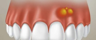
Epulis is a tumor-like neoplasm that develops on the alveolar process of the jaw. It also has other names - epulid, supragingival, giant cell granuloma. More often it is localized in the area of incisors, canines and small molars of the upper jaw, less often the tumor is detected in the lower jaw. The growth has a round or irregular shape and a wide stalk; the size of the formation varies from 3 mm to 5 cm.
Despite the fact that such a tumor looks frightening, it practically does not cause concern to the patient, unlike, for example, a burn, with the exception of a violation of aesthetics and the sensation of a foreign body in the oral cavity. Large tumors may cause difficulty chewing and swallowing food.
If epulis is accompanied by bleeding, infection may occur, which will lead to complications.
The disease mainly affects adults, but the neoplasm can also appear in children; in the latter, it occurs during teething. Studies have shown that women are more susceptible to developing epulis than men.
Exostosis in a child
It is in children that osteochondroma is most often first diagnosed, which is due to its formation from cells of the epiphyseal plate that is present only until the end of the growth period, which is adjacent to the metaphysis. It is also called the bone growth zone, since it is a hyaline cartilage whose cells are in constant miotic division. As a result, new chondrocytes (cartilaginous tissue cells) are formed, forming the epiphyseal plate, and the old ones are shifted to the metaphysis and subsequently replaced by osteoblasts (bone tissue cells).
In infants, violation of the rules for the prevention of rickets, in particular excessive use of vitamin D preparations, increases the likelihood of the formation of exostoses.
After puberty, the growth plates gradually close and are replaced by bone tissue, transforming into a thin epiphyseal line. If hormonal imbalances occur during this period, it is possible that the growth zones may remain open, which creates the preconditions for the formation of osteochondromas.
Usually, until the age of 7-8 years, bone exostosis does not manifest itself in any way in a child and makes itself felt only during the period of intensive growth, i.e., at 8-16 years, since it also begins to grow actively. In young children, such growths are present in the metaphysis area immediately near the epiphyseal plate, but subsequently move away from it and approach the diaphysis. Therefore, by how far the bone exostosis in children is from the epiphysis, the time of its formation is determined.
The growth of the neoplasm continues until the end of the growth period.
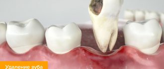
Why does exostosis occur on the gums after tooth extraction?
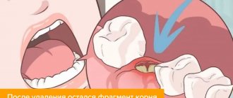
The cause of the growth is excessive growth of bone tissue. This mechanism is triggered when they are damaged, which occurs during the extraction of teeth or a jaw fracture. Usually bones are injured during complex operations to extract molars. After wisdom tooth removal, exostosis is diagnosed more often than other cases.
Factors that provoke pathological bone growth:
- hereditary predisposition;
- infections, inflammation in the oral cavity;
- injuries during dentofacial operations;
- endocrine disorders.
After tooth extraction, the patient may feel small nodules with his tongue. Over time, they increase in size and cause concern.
Types and stages of development
Exostosis can be present only on one bone, i.e., be solitary, or affect all metaphyses of the bones of the skeleton. In the second case, they talk about a generalized form of the disease, which is more common in men and is mainly hereditary. It is also called multiple exostotic chondrodysplasia. In this case, several bones or the vast majority of them are affected, but the formations have different shapes and sizes.
Depending on the shape and direction of growth, there are:
- hilly;
- linear;
- spherical exostoses.
Single exostoses have a narrow or wide base, while multiple ones are usually spherical or oval growths measuring 2-12 cm, sometimes more.
In its development, any osteochondroma goes through 3 successive stages:
- the formation of cartilaginous exostosis, which is not determined by palpation (palpation);
- ossification and active growth of growth;
- stopping the growth of the bone part of the neoplasm while maintaining the possibility of increasing the size of the cartilaginous cap.
Bone exostoses can already be felt during examination and can lead to discomfort, pain during physical activity and other symptoms.
Based on the nature of development and clinical picture, osteochondromas are divided into the following types:
- With normal growth rate. The osteochondral formation grows slowly, and there is a correspondence between the growth rate of the cartilage and the affected bone. Such osteochondromas have the most favorable prognosis, since after the end of the growth period they do not tend to continue to grow and almost never become malignant.
- With a high growth rate. An increase in the size of the growth occurs due to the proliferation of cartilage tissue and can also continue after the completion of the formation of the skeleton due to the preservation of the growth zone. In such cases, removal of exostosis is indicated, since there is a fairly high risk of malignancy.
- Malignant exostosis has the most unfavorable prognosis. Most often, osteochondral neoplasms of the ribs, pelvic bones, scapula and spine degenerate into chondrosarcoma or osteogenic sarcoma.
Gum cancer
This dangerous neoplasm is worth highlighting separately. Mutation cells begin to divide uncontrollably, which is why a red tumor forms on the gum. The disease progresses quickly and over time can develop into jaw cancer.
What are the symptoms?
- General weakness;
- increased body temperature (37 C°);
- loss of appetite;
- constant drowsiness.
The doctor makes an accurate diagnosis after a detailed examination. As a rule, treatment is long and painstaking. With timely medical intervention, the outcome is favorable.
Causes of exostosis
There is still no consensus on the reasons for the appearance of exostoses. A number of authors believe that they are of a tumor nature. But most agree that they are a consequence of disorders of enchondral ossification processes that arise as a result of dysembryogenesis (disorders of embryonic development). Therefore, today the main theory of the formation of bone exostoses is the displacement of the epiphyseal plate during intrauterine development of the fetus. It is a bone growth zone located directly below the epiphysis and is responsible for its lengthening. Therefore, the disease is considered congenital, and its development continues throughout the entire period of growth.
The following can increase the risk of osteochondroma:
- ionizing radiation (in 10% of cases the disease develops in patients who underwent radiation therapy in childhood);
- disturbances in the functioning of the endocrine system, taking hormonal drugs;
- smoking and alcohol abuse of parents.
The disease can also be hereditary and transmitted to the child from one of the parents. Especially often, multiple bone lesions due to exostoses are genetically determined.
The acquired nature of the disease cannot be ruled out. The following can provoke the appearance of neoplasms:
- bone injuries;
- microtraumas caused by excessive physical activity, which is typical for professional sports;
- infectious diseases and chronic inflammatory processes of any localization (for example, a heel spur often develops against the background of plantar or plantar fasciitis);
- pathologies of the periosteum, degenerative-dystrophic diseases of cartilage, ankylosing spondylitis;
- microcirculation disorders in soft tissues;
- muscle dystrophy;
- obesity;
- severe forms of allergic diseases;
- compression of the limbs, including an incorrectly applied plaster cast or tourniquet.
Wearing improperly selected clothing and shoes, in particular frequent hypothermia of exposed areas of the body, increases the risk of developing bone tumors.
Of particular importance in the development of osteochondroma is hormonal imbalance in children during puberty. Often the formation of bone exostoses is observed against the background of increased synthesis of sex hormones in adolescents. This can provoke the growth zones to remain open, which can subsequently cause gigantism. Also, an increase in the size of osteochondral exostoses can continue after the closure of growth plates in women as a result of hormonal changes.
Causes
The question of the etiology of osteochondromas remains open to this day. A number of authors do not recognize the tumor nature of osteochondromas, but consider it as a violation of the process of enchondral ossification (as a result of dysembryogenesis ). Many authors are inclined to believe that the appearance and development of osteochondroma is associated with a congenital pathology, the development of which continues throughout the entire period of bone growth (that is, the epiphyseal plate is displaced during the formation of the child’s bones even during the intrauterine formation of the skeleton).
Factors that increase the risk of its development include:
- Ionizing radiation.
- Severe bone bruises and fractures.
- Disruptions in the endocrine system.
- Infectious diseases.
Symptoms
The clinical picture depends on the form of the disease, location, size, shape of exostoses, and the degree of their influence on surrounding tissues. Single neoplasms are usually immobile and their growth is slow. Therefore, in most cases they are asymptomatic and there is no pain. The condition of the skin with solitary exostoses is usually not changed.
But in some cases, there is active growth and acquisition of large osteochondromas. They can mechanically compress blood vessels, nerves, and joints passing near them and provoke the development of reactive bursitis and myositis. This is accompanied by the appearance of pain of varying severity, swelling, redness of the tissues, and sometimes a feeling of numbness. If joint exostosis is present, the range of motion may be limited.
What symptoms exostosis will have depends largely on its location. With multiple exostoses, often the first signs of the disease are stunted growth, valgus deformity of the knee joints and clubhandedness. Fractures of the osteochondroma pedicle may also occur.
Exostosis of the knee joint
It may be caused by the formation of a growth at the end of the tibia and or femur. Exostosis of the tibia provokes severe deformation of the knee joint and is easily detected with the naked eye. Upon palpation, it is possible to detect a dense but painless formation under the skin, which can be either smooth or rough. Reaching large sizes, it provokes pain in the knee when walking (especially often in women who tend to wear high-heeled shoes). In this case, exostosis of the joint tends to injure the adjacent soft tissues, which provokes the development of tendonitis (inflammation of the tendons) and bursitis (inflammation of the synovial bursa).
Damage to the femur in the early stages of development is asymptomatic. But when the tumor reaches a large size, pain in the thigh and dysfunction of the affected limb may occur. Often there are many areas of deformation up to the covering of its entire surface. Since the femur is located deep in the soft tissues, it is difficult to detect exostoses during palpation.
Classification
The classification is based on several characteristics. Based on quantitative characteristics, solitary (single) and multiple (generalized) forms are distinguished.
According to the stages of osteochondroma formation:
- The neoplasm is formed from cartilaginous tissue and cannot be detected by palpation.
- Ossification of the growth and its increase. The bone tissue is covered with cartilage, and active growth continues.
- The growth of the bone part of the tumor stops, but the growth of the cartilaginous “cap” of the tumor may continue. Determined by palpation examination. During physical activity, it can manifest as pain and impaired limb function.
Based on the shape and direction of tumor growth, there are hill-shaped, linear and spherical exostoses.
Exostosis of the calcaneus (calcaneal spur)
This is a well-known type of osteochondroma. The growth can take on different shapes, which determines the characteristics of the symptoms that arise. With a spherical or mushroom-shaped neoplasm, after it reaches a size of 3-4 cm, pain appears when walking and the inability to fully step on the foot. Since the growth is located in an area that is constantly subject to friction from shoes, the skin in its projection gradually becomes coarser and thickens. A characteristic symptom is an increase in pain in the morning and a gradual decrease in its severity during the day. In the evening, swelling of the lower extremities is often observed.
Spinal exostosis
The lesion can occur at any level, but the thoracic region is most often affected. The neoplasm is located in the area of the arches or processes of the vertebrae. It can grow towards the vertebral or spinal canal or in an anterior direction. The first option is more dangerous, since it is capable of mechanically compressing the spinal cord and the nerve roots extending from it. This provokes the development of the so-called radicular syndrome with sharp, severe pain, radiating in accordance with the level of damage to the body part, impaired sensitivity and mobility.
Exostoses of the ribs
The formation of growths on the ribs is more often observed with curvatures of the spine, which also lead to deformation of the chest. Their growth causes fixation of the pathological position of the chest and limitation of the vital volume of the lungs. As a result, respiratory function disorders may occur, which provokes the appearance of signs of hypoxia (oxygen starvation). To a greater extent, the lack of oxygen affects the brain, which can be accompanied by dizziness, headaches and other disorders. The heart, liver, spleen, and other internal organs are also sensitive to changes in the gas composition of the blood.
Preventive actions
Monitoring the condition of the teeth and oral cavity will reduce the likelihood of tumors.
- Timely sanitation of the oral cavity. Visiting the dentist for a preventive examination and professional hygiene twice a year will prevent the growth of caries, the appearance of defects in fillings and the formation of tartar, leading to gum injury and the appearance of epulis.
- Prevention of gum injury. If systematic injury to soft tissue occurs as a result of poorly fitted orthopedic or orthodontic structures, consult your doctor about this problem. He will adjust the crowns or dentures.
If these preventive measures are followed, the prognosis is favorable and epulis does not recur.
We hope that our article about epulis will be for informational purposes only. And if you want to soothe your gums, make them strong and strong, try the unique two-component mouth rinse ASEPTA ACTIVE.
This is the only rinse with a combination of chlorhexidine + benzydamine for the treatment of inflammatory periodontal diseases. The product has a combined effect: antimicrobial, anti-inflammatory and analgesic. Instant anesthetic effect allows you to quickly reduce pain.
Exostosis of the humerus
The humerus is one of the most common sites of osteochondroma. During its formation, the following may be observed:
- dull pain in the shoulder area that occurs when moving the arm with a large amplitude;
- a feeling of stiffness in movements, present mainly in the morning, until you manage to warm up;
- limited mobility of the upper limb in the shoulder;
- dystrophy of the muscles of the shoulder and forearm, which in difficult cases can be seen with the naked eye.
Complications
Cartilaginous exostosis in children has a dangerous negative impact on the condition of the growth plates, which can result in shortening of the lower leg, thigh, forearm, shoulder, etc. This can also cause curvature of the arms and legs, increase the risk of fractures and cause disability.
Patients with severe forms of the disease feel inferior, which negatively affects their mental and emotional state.
The most dangerous complication of osteochondroma is its malignancy, i.e. transformation into a malignant tumor - chondrosarcoma. The highest risk of malignancy is with multiple exostoses. In this case, growths localized in the pelvic bones are more likely to undergo malignancy. Less commonly, exostoses of the ribs, scapula, and spine turn into malignant neoplasms. Single osteochondromas transform into a malignant tumor in no more than 1% of cases.
Atypical growth can be observed in any part of the exostosis: the cartilaginous cap, at the base or in the middle part.
Reasons for the formation of bumps on the gums
To find out why a lump appeared on the gum, you need to make a correct diagnosis; only a specialist can do this. There are several reasons why swelling appears, but none of them are harmless. When diagnosing, the nature of the formation, color, density, presence or absence of pain, purulent exudate is taken into account, and the nature of the disease is clarified - infectious or other.
Important! Basically, most dental problems are associated with poor oral care. As a result, mucosal pathology develops with its own symptoms.
The reasons why a lump appears between the gum and cheek may be the following:
- irregular and illiterate oral care, resulting in infection affecting periodontal tissues;
- complications of untreated caries - infectious lesions of the roots (fistula, periostitis);
- periodontitis;
- complications resulting from dental implantation;
- improper wearing or manufacturing of orthopedic structures;
- cystic formations;
- gingivitis;
- periodontal trauma (hematoma);
- benign neoplasms (epulis, fibropapilloma);
- malignant tumors.
There is another reason for the occurrence of periodontal compaction. If a child has a white bump on his gum, this may indicate the eruption of a new tooth. Short-term swelling is not terrible, but if after a long time the tooth does not appear, you should consult a surgeon.
Attention!
Many are in no hurry to go to the doctor, since the growth does not hurt. The absence of discomfort does not indicate the absence of disease. Remember: if a lump appears on your gum, only a doctor will determine what to do and how serious the consequences will be!
Diagnostics
Diagnosis of bone exostoses is not difficult. If these symptoms occur, you should consult an orthopedist. Based on the patient’s complaints, medical history and examination results, the doctor can already assume the presence of a neoplasm.
To clarify its size and other features, radiography is prescribed. It is carried out in 2 projections. Osteochondroma on x-rays is visualized as a shadow with smooth, clear contours and a pedicle or wide base connected to the bone. If there is no calcification in the cartilaginous layer, it will not be visible on x-ray.
To obtain additional data, the following may be assigned:
- CT scan, which makes it possible to detect the presence of bone marrow contents in exostosis and trace its connection with the medullary canal of the affected bone;
- MRI, which provides accurate data on the thickness of the cartilage cap and thereby clarifies the nature of the neoplasm (malignant tumors are characterized by a thickness of the cartilage tissue covering them of more than 2 cm).
If there is a suspicion of the development of oncology, patients are advised to undergo a biopsy - taking a fragment of exostosis for histological examination. In this way, benign osteochondroma is differentiated from malignant neoplasms.
If exostoses are detected in childhood and adolescence, it is imperative to conduct a full endocrine examination and determine the content of all significant hormones in the blood.
A growth has appeared on the gum: what is the reason?
A lump or growth is a compaction on the gum caused by damage to periodontal tissue, which may appear without any previous signs. Growths appear in people of any age, but mainly in young children, as they unknowingly introduce infection into their mouths. The first thing you need to do when you detect formations in your mouth is to determine the nature of their origin (only a doctor can do this). It comes in two types:
- Infectious. The appearance of cones is caused by the activity of bacteria.
- Non-infectious. Bumps and growths as a result of injury, mechanical or chemical damage.
If a lump has formed in your mouth, you should consult a doctor to rule out possible complications. The doctor will help diagnose the problem and prescribe appropriate treatment.
Treatment of exostoses
When diagnosing exostosis, treatment is prescribed only when symptoms appear. Otherwise, dynamic monitoring of the tumor is sufficient. If the tumor bothers the patient, depending on its type, the nature of the manifestation of osteochondroma, conservative treatment is selected or surgical intervention is prescribed. But the main method of treating exostosis is surgery.
Conservative, i.e., non-surgical, therapy is indicated mainly for exostosis of the calcaneus and rib, but only in the early stages of development. First of all, the use of special orthopedic insoles, made individually, and the replacement of regular shoes with models with offset edges are prescribed. This helps eliminate mechanical pressure on the tumor, reducing the load on the Achilles tendon and calcaneal tubercle.
If severe pain and inflammation occur, drug therapy is prescribed, including:
- NSAIDs in the form of topical agents (Nimid, Dolaren, Ketoprofen, Voltaren);
- drug blockades with the introduction of a mixture of anesthetics and corticosteroids (performed for severe pain that cannot be eliminated by other means).
Courses of physiotherapeutic procedures are also indicated. Shock wave therapy (SWT) is the most effective. It involves the impact of infrasonic acoustic waves on the affected area. The mechanism of action of the method is based on the cavitation effect that occurs at the interface between media. The acoustic resistance of soft tissue is less than that of bone. Therefore, sound waves penetrate through them and affect bones and cartilage. This provokes an improvement in blood supply, restoration of normal cartilage tissue and bones, and a reduction in the size of tumors.
Additionally, we may recommend:
- electrophoresis with the introduction of anesthetics;
- ultrasound therapy;
- laser therapy;
- Ural Federal District;
- magnetotherapy.
PREPARATION
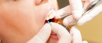
Surgical intervention is the only option for removing a growth of bone or cartilage tissue from the surface of the jaw row, and therefore requires careful preparation to ensure a favorable outcome.
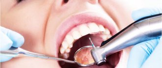
During the patient’s initial appointment, the dentist clarifies his complaints and conducts a visual examination of the oral cavity in order to identify visible pathologies. At this stage, a number of concomitant diseases are often identified: caries, gum or periodontal inflammation, which require treatment.
To determine the exact location of the growth, its shape, size and nature, the specialist sends the patient for an x-ray examination. The completed image is the basis for making an accurate diagnosis and drawing up a treatment plan.
At the stage of preparation for surgery, the dentist directs the patient to undergo tests, which can be used to identify additional diseases that impede the procedure. Thus, mandatory steps include checking blood for clotting and sugar levels, assessing the balance of hormones and the functioning of the adrenal glands.
At the preparatory stage, the specialist also decides on the option of administering an anesthetic drug.
Surgery to remove exostosis is often performed using local anesthesia, but in some situations general anesthesia may be required.
Before administering it, the specialist checks with the patient for the presence of an allergic reaction and previous experience with pain relief.
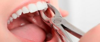
Surgery to remove exostosis
Surgical intervention is indicated for:
- large size of osteochondroma and the presence of persistent pain;
- development of complications (tendinitis, bursitis, vascular, neurological disorders, etc.);
- bone deformations as a result of the growth of exostosis;
- fracture of the base leg;
- malignancy of the tumor.
For children with bone exostosis, surgery is prescribed only in extreme cases. As a rule, it is carried out only if the testimony remains after reaching adulthood.
But if there are contraindications, surgery is not performed. This:
- purulent-inflammatory processes in the area of the upcoming intervention;
- exacerbation of chronic diseases;
- decompensated form of diabetes mellitus;
- acute infectious diseases.
Treatment of exostosis with surgery involves its complete excision with capture of the cartilaginous cap. There are several methods for removing osteochondral growth. A specific one is selected based on the location of the osteochondroma and its size. Usually preference is given to marginal resection of the growth, i.e., its removal within healthy tissue. Only in some cases is it necessary to resort to corrective osteotomy with concomitant resection of osteochondroma, but the cost of removing exostosis in this way is higher, since the operation also corrects bone deformation.
After surgery, complete recovery is observed in 98% of cases. Therefore, almost everyone who has once removed an exostosis forgets about it forever.
Marginal resection is a relatively simple type of surgery. Its essence is to expose the affected bone and remove the entire neoplasm in the front of healthy tissue along with the surrounding capsule using a sharp chisel, drill, oscillating saw or bur. After removal of the pathological formation, curettage of the maternal bone is performed using cutters and bur. In some cases, the extent of resection can be large and create the need for bone grafting using autografts or special implants. It is important to remove the entire cartilaginous cap and neoplasm, otherwise a relapse may develop.
Carrying out a corrective osteotomy involves cutting the mother's bone using an osteotome and removing the osteochondroma. After this, the bone fragments are fixed in the desired position using special plates, screws or knitting needles.
Approaches to surgical treatment of osteochondral exostoses
Key words: osteochondral exostosis, surgical treatment
Introduction. Osteochondral exostoses (osteochondroma) is a disease whose study has a history of almost two centuries, but still leaves researchers with prospects for finding effective approaches and methods of treatment [7–9,11]. Osteochondral exostoses, the most common bone tumors, account for about 50% of all benign bone tumors and about 10% of all skeletal bone tumors [5,6,14]. The actual distribution of these tumors in the general population is unknown because many patients are asymptomatic [14].
According to foreign literature, 90% of these tumors occur in the form of single exostoses, and the remaining 10% - in the form of hereditary multiple osteochondral exostoses [3,4,12].
Osteochondral exostoses are localized predominantly in the metaphyses of long tubular bones, which develop by enchondral ossification and are most often found in the area of the proximal metaphysis of the humerus, distal metaphysis of the femur, proximal metaphysis of the tibia, which correspond to the places of the most rapid bone growth [3,6,7, 10].
Malignant transformation occurs in 1% of cases in patients with single osteochondral exostoses and in 3–5% of cases in patients with multiple osteochondral exostoses [1,2,5,13]. Continued growth, damage, and a cap of hyaline cartilage greater than 1.5 cm in thickness occurring after completion of skeletal growth suggest malignant transformation of the exostosis [2,4,8,13].
Despite the impressive arsenal of surgical methods for treating this pathology, there are still no unified tactics and approaches to the treatment of osteochondral exostoses.
The main goal of our study was to identify the optimal, low-traumatic method and indications for surgical intervention, its effectiveness for early rehabilitation of the patient using the technical capabilities and equipment of the clinic. When determining the indications for surgical intervention, medical indications, aesthetic manifestations, as well as complications caused by exostoses were taken into account. In the selected group of patients, we observed the following complications associated with exostoses: cosmetic defects, vascular and neurological disorders, fracture, impaired movement function, exercise tolerance.
Material and methods. TsTOOR has accumulated quite a lot of experience in the treatment of osteochondral exostoses. Various approaches and treatment methods have been used - from extensive resections, creating a bone defect, to limited interventions - without creating a bone defect.
For the period from 2000 to 2005. About 700 patients with a referral diagnosis of osteochondral exostoses applied to the Center for Emergency Hospital. However, only in 250 patients the diagnosis of osteochondral exostosis was confirmed. Of the 250 patients with osteochondral exostoses, 31 were diagnosed with hereditary multiple osteochondral exostoses; the rest were diagnosed with single osteochondral exostosis.
21 patients underwent surgical treatment using various methods; the rest were not treated due to the lack of indications for surgery or the patients’ refusal to undergo it.
The operated patients were divided into two groups. The principle of the division was the traumatic nature of the operation: the volume of the iatrogenic bone defect created, the type of surgical intervention performed and the timing of postoperative rehabilitation.
The first group included patients in whom surgical intervention led to the formation of a bone defect. In some cases, bone grafting of the defect was performed. The second group included patients who underwent safe surgical interventions without creating a bone tissue defect.
Technique of safe operation: access was made depending on the localization of the osteochondral exostosis, complete exposure of the osteochondral exostosis along with the base and capsule, osteotomy of the formation at the level of its transition into the maternal bone and removal of the osteochondral exostosis in a single block along with the capsule (it is important to remove entire capsule). of the mother was processed using the cutter , without the formation of a bone deficiency or bone defect.
The results of surgical treatment were assessed taking into account the initial severity of the patients' condition, determined by the degree and type of complications caused by exostoses, and the timing of postoperative recovery.
All patients were subject to clinical, radiological, laboratory and diagnostic dynamic examination. Short-term and long-term results were analyzed based on postoperative examinations of patients, taking into account subjective data.
Results and discussion. An analysis of the results of treatment of patients in the first group (14 patients) shows that although there were no relapses of the underlying disease in this group, the patients returned to a full life after an average of 5 weeks, during which they walked with the help of crutches without putting any weight on the limb.
Let's give an example: patient G., 12 years old, clinical case No. 1024/150.
Diagnosis: solitary osteochondral exostosis in/3 of the right fibula, with compression of the peroneal nerve (Fig. 1 A). A segmental resection of the fibula was performed (Fig. 1 B).
Rice. 1. X-ray before (A) and after surgery (B); 1 – solitary osteochondral exostosis; 2– iatrogenic circular bone defect
The patient was discharged 3 weeks after surgery. At the time of discharge, the limb was immobilized in a posterior plaster splint from the third third of the right femur to the toes. No neurological disorders were noted. The patient walks independently with the help of one crutch. During the follow-up examination after one month, he walks with the help of a stick, without plaster immobilization.
Of the 14 patients in this group, one patient with marginal resection without defect replacement experienced a pathological fracture after one month, which was subsequently treated conservatively, and in 2 cases, rehabilitation lasted more than 3 months.
Analysis of the results of treatment of patients in the second group (7 patients) shows that they were mobilized 2–3 days after the operation and were discharged from the center after the wound had healed. The patients returned to a full life on average after 2 weeks, noting that their functional status was significantly improved compared to preoperative; subsequently, during the 1st year, no relapses of the underlying disease were observed.
Another example: patient N., 18 years old, case history No. 2398/494.
Diagnosis: solitary osteochondral exostosis in/3 of the right fibula (Fig. 2 A, B).
Rice. 2. Radiographs before surgery. Broad-based osteochondral exostosis is visible. A – anteroposterior projection; B – lateral projection
A safe was performed using the above technique (Fig. 3 A, B).
Rice. 3. Radiographs after surgery. The absence of a bone defect on the maternal fibula and the impression site on the tibia are visible. A – anteroposterior projection; B – lateral projection
The osteochondral exostosis was removed en bloc (Fig. 4 A, B).
Rice. 4. Macroscopic view of osteochondral exostosis removed en bloc (A). Macroscopic view after sawing of an osteochondral exostosis (B). 1 – fibrous capsule, a cap in the form of a cap made of hyaline cartilage, which functions as perichondrium; 2– wide base; 3– cortical and spongy bone; 4 – hyaline cartilage, which in several areas grows towards the medullary component
The patient was discharged after the wound healed. At the time of discharge, the limb was not immobilized. No neurological disorders were noted. The patient walks independently without additional load.
During follow-up examinations, after 1, 6 and 12 months, he served in the armed forces.
As can be seen from the above examples, with the same treatment result, in the first case, where a wide resection was performed to create a bone defect, the rehabilitation period was extended. Additional fixation was required. In the second case, no additional fixation was used; the patient was activated after three days and discharged once the wound had healed.
Analysis of the above results indicates that in all cases, after surgical treatment, no relapses of the underlying disease were observed. No intraoperative complications or wound infections were noted in both groups.
Thus, as a result of the data presented, we came to the following conclusions:
- With a verified diagnosis of osteochondroma, there is no need for major surgical intervention with marginal bone resection within the healthy bone, which dramatically lengthens the patient’s recovery time, especially when it comes to the lower extremities, which are subject to static loads.
- Extended marginal or parietal bone resection with removal of the root of osteochondral exostosis leads to the unjustified creation of an iatrogenic bone defect, which in some cases has to be filled with a graft. Complete restoration of the bone structure in the area of the bone defect lasts up to 2 years and sometimes pathological fractures occur. This is an aggressive and unjustified approach. This result in the treatment of osteochondroma, taking into account the modern pace of life and the requirements of patients, can hardly be considered satisfactory.
- Indications for surgical intervention for osteochondromas of the upper and lower extremities should be determined by the degree of secondary complications caused by exostoses, and not by the principle that the bone formation should be removed. When deciding on possible surgical intervention, the location of the osteochondroma and the likelihood of its fracture should also be taken into account.
- The results of our study show the effectiveness of safe operations and allow us to achieve similar therapeutic results with an incomparably quick return of the patient to a full life and, no less important, improving the quality of life of patients during the treatment period.
Literature
- Bandiera S., Bacchini P., Bertoni F. Bizarre parosteal osteochondromatous proliferation of bone, Skeletal Radiol., 1998; 27:154-156.
- Bell RS Musculoskeletal images: malignant transformation in familial osteochondromatosis? Can. J. Surg., 1999; 42:8.
- Dahlin DC and Unni KK Bone Tumors. General Aspects and Data on 8,542 Cases. Ed. 4, Springfield, Illinois, 1986.
- D'Ambrosia R. and Ferguson ABJ The formation of osteochondroms by epiphyseal cartilage transplantation, Clin. Orthop., 1968, 61: 103-115.
- Geirnaerdt MJ, Hogendoorn PC, Bloem JL, Taminiau AH, van der Woude HJ Cartial tumors: fast contrast-enhanced MR imaging, Radiology, 2000; 214:539-546.
- Harper GD, Dicks-Mireaux C., Leiper AD Total body irradiation-induced osteochondromata, J. Pediatr. Orthop., 1998; 18:356-358
- James H. Beaty, Canale Terry: Operative Pediatric Orthopedics, 1991, p. 1098-1099.
- Kingsley R. Chin, F. Daniel Kharrazi, Bruce S. Miller, Henry J. Mankin, and Mark C. Gebhardt, Osteochondromas of the distal aspect of the tibia or fibula, J. Bone and Joint Surg., 2000, 82:1269 .
- Lovell WW, Winter RB and Morissy RT Pediatric Orthopedics, Third Edition, 1990, vol. 1, p. 342-343.
- Mark D. Murphey, James J. Choi, Mark J. Kransdorf Imaging of Osteochondroma: Variants and complications with radiologic-pathologic correlation, radiographics, 2000, 20: 1407-1434.
- Milgram JW The origins of osteochondromas and enchondromas. A histopathologic study, Clin. Orthop., 1983,74: 264-284.
- Mirra JM Bone Tumors. Clinical, Radiologic, and Pathologic Correlation. Vol. 2, Philadephia, 1989.
- Ozaki T., Hillmann A., Blasius S., Link T., Winkelmann W. Multicentric malignant transformation of multiple exostoses, Skeletal Radiol., 1998; 27:233-236.
- Resnick D., Kyriakos M., Grennway GD Osteochondroma. In: Resnick D. eds. Diagnosis of bone and joint disorders. 3rd ed., vol. 5, Philadelphia, 1995, p. 3725-3746.
Rehabilitation
After removal of the exostosis, the patient remains in the hospital for 3 days, the sutures are removed on days 7-10. Performing marginal resection does not require any serious restrictions even in the early postoperative period. The patient can completely return to his usual lifestyle after the stitches are removed, and forget about exostosis immediately after the operation.
When performing a corrective osteotomy, complete bone fusion occurs after 6 weeks, but full recovery may require up to 3 months. At this time, it is important to strictly follow all the doctor’s recommendations and not to overload the operated part of the body. All patients who have undergone osteotomy are prescribed drug therapy, exercise therapy, and often physical therapy. The duration of rehabilitation in such cases depends on the type of surgical intervention performed and the individual characteristics of the patient.
Thus, osteochondroma is a very common type of tumor. At the same time, statistics on the frequency of its occurrence are not entirely objective, since exostoses often do not appear throughout a person’s life, and therefore are not diagnosed. However, if the formed formation bothers the patient, causes him pain, or reduces the quality of life, you should not hesitate to contact an orthopedist. A specialist will be able to assess the degree of danger of the tumor and, if necessary, remove it using the most gentle method possible.