Anomalies of teeth - various types of deviations of teeth from the norm (in number, size, shape, structure, etc.), as a rule, congenital or acquired disorders of the functioning of the dental system. According to dental practice, various dental anomalies are observed in 40-50% of children and adolescents and 30-40% of adults. Despite the fact that dental anomalies sometimes require long-term and multi-stage treatment (orthodontic, orthopedic, therapeutic, surgical, etc.), many prefer to turn a blind eye to the presence of problems, some even try to make a feature out of a problem (as in the case of diastema, despite the charm and charm of some public representatives of the fair sex, this is in any case an anomaly).
Examples of dental anomalies from the YuliSTOM dental practice
Anomalies of the dental system can be divided into the following groups:
- anomalies of individual teeth (size, shape, number, position);
- dental anomalies;
- malocclusion;
Below we will look at each group separately.
Anomalies of individual teeth
Anomalies in tooth size
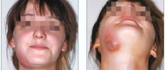
Giant teeth are teeth with disproportionately large crowns. Giant teeth are more common in permanent dentition and less common in primary dentition. Typically, the incisors of the upper or lower jaws are giant, but other teeth can also be giant. The etiology of this anomaly in the shape of the teeth is known; disturbances in the developmental process are assumed, leading to the fusion of the rudiments of the teeth, as well as disruption of the activity of the endocrine system. Giant teeth can cause abnormalities in the position of other teeth, interfere with the eruption of neighboring teeth, and cause crowding of teeth. Sometimes they are located outside the dentition. The main disadvantage of giant teeth is their unusual appearance, which attracts the attention of others.
Small teeth are teeth with disproportionately small crowns that have a regular shape. Small teeth are found in permanent dentition. More often than other teeth, the incisors are small, especially the upper lateral ones. The etiology of this anomaly is unknown; it is assumed that the reasons for this discrepancy in the size of teeth and jaws may be hereditary, i.e. a combination of small teeth of one parent and large jaws of the other. Small teeth are usually spaced at large intervals and disrupt the harmony of the face.
Anomalies of teeth position
Vestibular deviation is the displacement of teeth outward from the dentition. One or more teeth of the upper or lower jaw may be in the vestibular position. Most often the incisors are displaced. The reasons may be: delay in the replacement of milk teeth, lack of space in the dentition, incorrect position of the tooth germ, the presence of supernumerary teeth, bad habits, and impaired nasal breathing.
High or low position of teeth - displacement of teeth in the vertical direction. In the upper jaw, supraocclusion is a high position of the tooth that does not reach the closure plane of the dentition, and infraocclusion is an advanced, low position of the tooth relative to the occlusal plane, and infraocclusion is a low position of the tooth. Sometimes there is supra- and infraocclusion of a group of teeth. The cause may be underdevelopment of the alveolar process or some mechanical obstacle.
Diastema is a gap between the central incisors and is much more common in the upper jaw. The reasons may be: low attachment of the powerful frenulum of the upper lip, the presence of a wide, dense bone septum between the central incisors, adentia, anomalies in the shape and size of the teeth, the presence of supernumerary teeth, incorrect placement of the frontal teeth, early loss of one of them.
Mesiodistal displacement of teeth is the location of teeth in front or behind the normal place in the dental arch. Both frontal and lateral teeth can shift. The cause is early loss of baby teeth, early loss of permanent teeth adjacent to a displaced tooth, incorrect position of the tooth germ, edentulism, and bad habits.
Oral inclination is the displacement of teeth towards the inside of the dentition, towards the tongue or palate. When tilted, the root of the tooth is located in the alveolar process, and only its coronal part is tilted orally; when displaced, the tooth is located body-wise outside the dental arch. One or more teeth may be in this position. The reasons are: delay in the replacement of milk teeth, early removal of milk teeth, narrowing of the dentition, incorrect position of the buds of permanent teeth, the presence of supernumerary teeth, shortened frenulum of the tongue, bad habits.
Rotation of the tooth around the longitudinal axis - most often the incisors of the upper and lower jaw are rotated along the axis. This type of anomaly causes aesthetic and functional disturbances. Sometimes axially rotated teeth injure the teeth of the opposite jaw and loosen them. The reasons may be a lack of space in the dentition due to its narrowing or underdevelopment of the alveolar process, delayed loss of a baby tooth, the presence of supernumerary or impacted teeth.
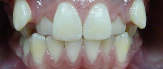
Crowded arrangement of teeth is a close position of the teeth, in which they stand with rotations along the axis and overlap each other due to lack of space in the dentition. The reason is underdevelopment of the alveolar process or the basal part of the jaw or the relatively large size of the teeth, as a result of which the teeth cannot be placed in the correct position.
Transposition of teeth is a positional anomaly in which the teeth change places. The reason is the incorrect formation of the rudiment of teeth.
Tremas are spaces between teeth. There are three types: physiological and pathological. Physiological problems refer to the characteristics of the primary occlusion in its second period; they arise as a consequence of jaw growth. Pathological threes are observed after the replacement of primary teeth with permanent ones in distal and mesial occlusions with protrusion of the upper or lower frontal teeth, with edentia, anomalies in the shape and size of the teeth, anomalies in the location of the teeth, and displacement of the teeth.
Anomalies in tooth shape
Spiny teeth are teeth whose crowns are shaped like a spike. The central and lateral incisors, as well as the lateral teeth of the lower and upper jaws, can have a tenon-like shape. The etiology is not clear; a violation of the development of tooth germs is assumed.
Ugly teeth are teeth of various irregular shapes, which are most often observed on the upper jaw, in its frontal area. The etiology is not clear; a violation of the development of the jaws and tooth buds is suspected.
Anomalies in the number of teeth
Adentia is a congenital absence of teeth and their rudiments. A distinction is made between partial and complete adentia. A violation of the development of the ectodermal germ layer, from which the tooth germ is formed, disturbances of the endocrine system are assumed, and heredity plays a certain role.
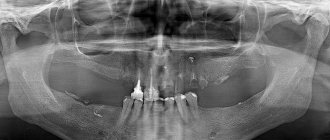
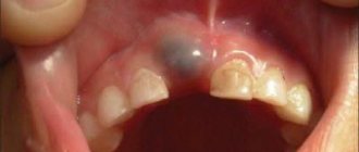
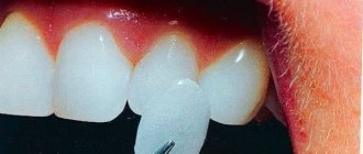
Supernumerary teeth are teeth that are excessive in number. They are most often located in the area of the front teeth. Supernumerary teeth are often tenon-shaped, but may have the shape of adjacent teeth. It is assumed that the cause is disturbances in the development of the epithelial dental lamina.
Examples of supernumerary teeth
Read more about the number of teeth people have in the article “How many teeth does a person have? How many teeth do you have?
Problems that pathology leads to
An awl-shaped tooth in a child or adult is not only an aesthetic and psychological problem. A person with this defect can constantly injure the oral mucosa, which in turn is fraught with stomatitis, gingivitis and periodontitis, the formation of painful and poorly healing ulcers and their degeneration into malignant tumors.
An abnormal form can become a prerequisite for the formation of malocclusion, as well as the cause of poor-quality chewing of food, dysfunction of the maxillofacial apparatus, and problems with the gastrointestinal tract.
On a note! If spike-shaped teeth are supernumerary, then they provoke a phenomenon called crowding. When overcrowding occurs, high-quality hygienic oral care is difficult; small pieces of food are poorly removed from the spaces, which leads to increased accumulation of bacterial plaque, the appearance of halitosis, caries and gingivitis.
The defect is often accompanied by enamel hypoplasia, which means that hard tissues are very vulnerable and sensitive to any external influences and bacteria. The enamel in patients with this problem is quickly destroyed and is susceptible to caries and its complications (pulpitis, periodontitis).
Anomalies of the dentition
Anomalies of the dentition are characterized by a change in the shape of the typical dentition of the upper or lower jaw, which is caused by their narrowing or expansion in various areas and is expressed by crowding of the teeth, their rotation along the axis, vestibular or oral teething, partial edentia, the presence of supernumerary teeth, diastemas and etc. The following irregular forms of dentition are distinguished when they are narrowed:
- acute-angled, when the narrowing is localized in the canine area;
- saddle-shaped, when the narrowing is more pronounced in the premolar area;
- V-shaped, when the dentition is narrowed in the lateral areas, the frontal area acts as an acute angle;
- trapezoidal, when the frontal area is narrowed and flattened;
- common, when all the teeth - frontal and lateral - stand closely together;
- asymmetrical, in which the narrowing is more pronounced on one side of the dentition of the upper or lower jaw, resulting in a crossbite.
The main etiological factors of anomalies in the shape of the dental arches are underdevelopment of the jaws and their deformations caused by diseases of early childhood.
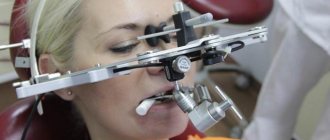
Diagnostics
A wedge-shaped tooth defect is usually identified by a dentist during a routine examination or treatment. He evaluates the location and shape of the pathology, the density of the tooth tissue. It is important to confirm that we are not dealing with dental erosion, superficial, cervical caries or enamel necrosis.
The doctor examines the patient’s dental status (oral hygiene, number of caries-affected, filled and extracted teeth, gum condition) and conducts a thermal test. This is a tooth reaction to temperature stimuli.
Vital staining helps in establishing the diagnosis: the wedge-shaped defect is well stained with iodine solution, but retains its color when treated with methylene blue. It is also important to determine what malocclusion pathologies exist and how they affect the occurrence of the pathology.
To exclude the influence of somatic diseases, the doctor may refer the patient to an endocrinologist and gastroenterologist.
Malocclusions
Malocclusions are deviations from the normal relationship of the dentition of the upper and lower jaws. These deviations can be considered in three directions:
Sagittal
Prognathia (distal bite) - characterized by a mismatch in the relationship of the dentition due to the protrusion of the upper teeth or distal displacement of the lower jaw. Distal occlusion may be partial or total; jaw, skeletal or dental; with or without displacement of the lower jaw. Etiology: congenital structural feature of the facial skeleton, childhood diseases affecting the development of the skeletal system, inflammatory processes in the nasopharynx, etc.
Progenia (mesial bite) - characterized by inconsistency of the dentition due to protrusion of the lower teeth or mesial displacement of the lower jaw. It may be partial or complete; jaw, skeletal or dental; with or without displacement of the lower jaw. Etiology: congenital structural feature of the bones of the facial skeleton, incorrect method of artificial feeding, early loss of primary molars, etc.
Transversal
Transversal narrowed dentition - discrepancy between the width of the upper and lower dentition
Vertical
- deep bite - a closure of the dentition in which the front teeth are largely overlapped by the antagonists. Depending on the vestibular or oral inclination, two types of deep bite are distinguished - vertical and horizontal. Etiology: congenital structural feature of the facial skeleton, childhood diseases affecting the growth and development of bones, early loss of primary molars. The main goals of treatment are to separate the bite, expand the narrowed dentition on the jaw that is lagging behind in development, and, if necessary, move the lower jaw.
- open bite - characterized by the presence of a gap between the teeth with central occlusion. This gap occurs more often in the area of the front teeth. There are two forms of open bite - vertical and horizontal. Etiology: rickets, difficulty in nasal breathing, early loss of frontal teeth, wide diastema.
- crossbite - characterized by the reverse closure of the teeth of the right or left half of the bite. Etiology: delay in the replacement of milk teeth by permanent ones, incorrect position of the tooth buds and subsequent incorrect eruption of these teeth, uneven development of the jaws and dental arches.
Stages of development of a wedge-shaped defect
The wedge-shaped defect develops gradually. The following stages of process development can be distinguished:
- First changes in enamel. A small area at the base of the tooth darkens slightly and loses its shine. Over time, it will develop pigmentation. At this stage, the future defect can only be seen using a special device.
- Superficial lesion. At the base of the tooth there is a noticeable crack, at its widest part not exceeding 3.5 mm. The gums sag, the neck of the tooth is exposed. The patient is uncomfortable with food that is too hot or cold, but the pain quickly goes away.
- Progressive stage. The wedge-shaped defect deepens to 4 mm, becomes yellowish-brown, matte. The shape of the triangle is already clearly visible - the two affected planes of the tooth converge at an angle of 45 degrees. Teeth react painfully to temperature stimuli and acidic foods. It is painful for the patient to brush his teeth. He feels the defect as a kind of step at the base of the tooth, where the remains of soft food are retained. At this stage, the diseased tooth is already noticeable to others when smiling and talking.
- Launched form. The enamel becomes thinner, dentin is affected, and in difficult cases, even the pulp. The wedge deepens to 0.5 cm, the neck of the tooth is exposed. If the destruction reaches the pulp, the neurovascular bundle of the tooth becomes inflamed. Acute paroxysmal pain appears, the tooth reacts to the temperature and taste of food, touch, the chewing process and causes a lot of inconvenience.
Treatment and prevention of dental anomalies
It should be remembered that dental anomalies are a serious disease for patients of all ages and professions and require long-term and thorough therapy to maximize or completely eliminate dental pathologies. Modern dentistry and advanced equipment are capable (if not completely, then maximally) of eliminating the manifestations of dental anomalies. A timely visit to a specialist in order to establish an accurate diagnosis and determine a treatment plan can guarantee a favorable prognosis for recovery. Dentists will be able not only to restore the functional abilities of the jaw, but also to eliminate various aesthetic defects.
Prevention of the formation of dental anomalies begins with taking care of the correct intrauterine and postnatal development of the child. This includes:
- balanced diet;
- timely correction of endocrine disorders;
- regular visits to the dentist to carry out the necessary preventive and therapeutic measures;
It is important to monitor your own health in the first days after the birth of your child.
Early breastfeeding and a balanced diet will help avoid many dental anomalies. We should not forget about a complete preventive examination by the dentist - at least once every six months. Moscow metro station Zvezdnaya, Danube Avenue, 23
The main reasons why pathology occurs
Conical teeth are a problem that can be noticed already in childhood. The pathology may be caused by a genetic factor, a mutation of some DNA genes, burdened by heredity. It can occur due to unfavorable conditions for the development of the child even in the prenatal period, or in the first years of life. For example, if a pregnant woman or the baby himself has had rubella, syphilis or infectious diseases, has metabolic disorders and disorders of the endocrine system.
The anomaly is usually classified as hereditary or congenital1, associated with a violation of the formation and development of tooth buds in the embryonic period. But the problem is often diagnosed in premature babies and those who have low weight or dystrophy. It can develop against the background of an acute lack of vitamin D, because it is this vitamin that the body needs, especially in the first years of life, for the proper formation of bone structures. Another reason that provokes the development of pathology is nephrotic syndrome (kidney disease).