Types of pathology
There are several types of maxillary sinus cysts:
- Odontogenic. The inflammatory process forms in the root system of an untreated dental unit located on the upper jaw. As the tumor increases in size, it destroys the bone and grows into the sinuses. The contents of the cyst are purulent.
- Retention. The reason for the development is dysfunction and obstruction of the glands responsible for the production of mucus.
False cysts are classified into a separate category. In such formations there are no epithelial cells. The appearance of such formations is due to a violation of the structure of the maxillofacial apparatus.
Conservative treatment: when possible
Conservative treatment is only possible for small root cysts. At this time, the canals of the tooth are treated, on the roots of which a cyst has formed. The treatment consists of thorough cleaning and filling of the dental canals and is carried out under a microscope, which ensures the accuracy of all manipulations and allows you to find all the canals in the tooth and fill them. Most often, after endodontic treatment, the cyst disappears, which is recorded on an x-ray taken a few weeks after the operation.
Causes
The key reason for the formation of a maxillary sinus cyst is dental disease. Especially when treatment was not carried out or performed poorly. Pathology develops against the background of advanced caries, periodontitis and pulpitis on chewing units. This is due to anatomical features. Premolars and molars are located closest to the paranasal sinuses and are separated from them by a thin septum.
Until recently, teeth with cysts were subjected to extraction. Today, dentists prefer to perform organ-preserving surgeries. All stages of work are performed under control using a dental microscope.
An endodontist removes the cyst, performs sterilization and filling of the canals.
The formation of a tumor can be caused by a deviated nasal septum, jaw abnormalities, or chronic blockage of the ducts of the nasal glands due to rhinitis or sinusitis.
Diagnosis of the disease
A neoplasm measuring up to 15 mm is diagnosed only with a CT scan. A dentist can suspect the presence of a pathology during a routine examination of the patient’s oral cavity based on existing complaints.
Clinical symptoms
As the cyst grows, the patient develops alarming symptoms similar to those of acute sinusitis. These include:
- acute headache;
- chronic nasal congestion;
- clear or yellow nasal discharge;
- a feeling of heaviness and fullness in the area under the eyes;
- the presence of a viscous mucous lump in the throat after waking up.
Clinical symptoms become more pronounced as the lumen in the nasal cavity closes. If there are concomitant dental problems, pain in the tooth and swelling of the gums occur.
Potential Complications
If left untreated, the infection spreads to adjacent teeth in the upper jaw. There is a risk of bone deformation or jaw fracture, the development of an abscess, phlegmon and other purulent complications.
The cyst is accompanied by sinusitis and chronic sinusitis. The growth of the tumor is dangerous due to visual impairment. The formation of a purulent cyst can lead to the development of meningitis or encephalitis.
Complex treatment
When treating pathology, dentists use medicinal and surgical techniques. The tumor is removed during surgery. Further drug therapy is designed to eliminate the symptoms of the inflammatory process and prevent complications.
Treatment in dentistry
Most patients go to the dentist when the size of the cyst exceeds 1 cm. The dental surgeon removes the bag of pus by cystectomy or cystotomy. The choice of technique is based on the size of the cyst and clinical indications.
The dentist is faced with a number of tasks:
- remove the source of inflammation in the oral cavity and stop the inflammatory process;
- provide drainage for the outflow of pus;
- if possible, save the diseased tooth;
- fill the resulting cavity with bone material;
- close the fistula between the oral cavity and the sinus.
Inpatient treatment in the ENT department
If the appearance of a cyst is caused by ENT pathologies, you should consult an otolaryngologist. Tumors are removed by surgery, laser or gentle endoscopic techniques. During endoscopy, the doctor gains access to the affected area through the nasal passages or an opening in the facial wall of the maxillary sinus. Control of manipulations on the monitor screen is carried out.
conclusions
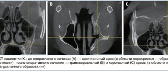
The most objective way to diagnose dense space-occupying formations of the upper jaw is computed tomography (CBCT or MSCT).
If signs of osteoma are detected in the cavity of the upper jaw, the patient must be recommended to undergo surgical excision of the formation. Morphological verification of the removed material is mandatory.
The combination of an endoscopic transnasal approach and navigation equipment in the removal of osteomas of the upper jaw is the most modern and optimal option for treating the pathology.
MestaMidin-nos has proven itself in local therapy as a means to prevent possible postoperative infectious and inflammatory complications. The use of MestaMidin-nos allows you to avoid a course of systemic antibacterial therapy.
The publication was carried out with the support of Solopharm in accordance with internal policy and current legislation of the Russian Federation.
Comments
The other day I visited LORA, because... I'm worried about constant nasal congestion. HE examined and diagnosed “odontogenic sinusitis” and prescribed symptomatic treatment. He said that in order for everything to return to normal, you need to go to the dentist and look for the cause. No cyst was found. Explain what could be the reason and why this is dangerous?
Tanyusha (06/03/2020 at 17:53) Reply to comment
- Dear Tanyusha, the diagnosis of odontogenic sinusitis indicates that the problem should be looked for in the oral cavity. The cause of its development is often periodontitis, perforation of the sinuses during dental treatment, impacted and dystopic teeth. If the exact cause is not identified and eliminated, then the formation of a cyst will not take long to occur.
Editorial staff of the portal UltraSmile.ru (06/08/2020 at 09:16) Reply to comment
Hello. Today I had an MRI about pain on the right side of my face. A cyst was discovered in the maxillary sinus on the right. Tell me where to run and what to do? Thank you.
Victoria (08/17/2021 at 04:57 pm) Reply to comment
Write your comment Cancel reply
Why shouldn’t you treat perforation of the maxillary sinus yourself?
To date, there are no effective treatments for perforation other than surgery. An attempt to cure yourself at home using “traditional medicine” means that time will be lost and the situation will become neglected. You can start the problem before complications arise:
- the sinus cavity becomes inflamed, the infection spreads to the bone tissue, the patient begins to suffer from osteomyelitis of the upper jaw;
- inflammation penetrates into other intracranial sinuses, and there are more foci of infection;
- next to the untreated perforation, the alveolar process is weakened, as a result of which healthy teeth may fall out;
- foci of suppuration develop.
The maxillary sinus is also located in close proximity to the brain. The lack of a timely response and the development of suppuration inside the sinus is fraught with meningitis and meningoencephalitis - these are diagnoses that directly threaten life.
Surgical removal of a cyst in the maxillary sinuses
Other types of cysts and large root cysts that are not suitable for conservative treatment are removed during surgery. Before the procedure, a computed tomography scan is performed, which allows you to accurately determine the location of the lesion and plan the procedure so that it takes place in the least invasive way.
The patient is also prescribed blood tests: morphology with platelets, coagulation, blood group and other additional tests as prescribed by the doctor. If the patient suffers from chronic diseases, consultations are held with relevant specialists (cardiologist, endocrinologist, neurologist). Before the operation, an anesthetic consultation is carried out to determine the dosage and type of general anesthesia.
The operation to remove a cyst in the maxillary sinus is performed using endoscopic or classical methods: this largely depends on the location and size of the cyst being removed. Due to the small post-operative wound, low invasiveness of the procedure and short recovery period, endoscopic sinus treatment is more often used. In case of concomitant infection of the secretion filling the cyst, antibiotic therapy is also indicated.
Description of the procedure
When removing a cyst of the right or left maxillary sinus, the surgeon’s task is to remove the affected mucous membrane of the sinus and other pathologies in its lumen and create a new opening connecting the sinus to the nasal cavity. Several types of surgery are used to remove a cyst:
- cystectomy – excision and curettage of the entire cyst cavity, followed by suturing;
- cystotomy - excision of the anterior wall of the tumor and communication of the posterior wall with the oral cavity.
The procedure is performed under general anesthesia and only in exceptional cases can local anesthesia be used.
Tissue and fluid from the cyst cavity collected during surgery are sent to the laboratory for histopathological examination, which allows us to determine the type of benign tumor and exclude the presence of malignant tumors.
Prevention
Perforation of the maxillary sinus is a problem that is easier to avoid than to fix it later. Since perforation is caused by dental intervention, preventing the problem falls on the shoulders of the dentist. He is obliged:
- responsibly examine the patient before performing procedures;
- clearly understand the anatomical features of the client before major intervention;
- strictly adhere to the intervention technology.
The dentist is also obliged to respond adequately to any signs of perforation that has just occurred due to his fault. If for some reason the doctor has not fulfilled his own duties, then it is up to the patient - he must refuse self-medication and put aside the fear of dentists, in no case try to “endure” the discomfort, but immediately seek help.
Perforation discovered after the fact
If the patient suffered discomfort in the first 2-4 weeks and did not contact the dentist, who, in turn, did not identify the problem at the time of its occurrence, the perforated wound turns into a permanent fistula that is not prone to healing.
Typical signs of chronic sinusitis appear:
- the nose on the side of the fistula is constantly stuffy;
- the parasinus area gives off a dull pain, its waves can roll to the nearest eye and temple;
- pus is discharged from the nostrils and in the mouth (from the fistula);
- possible swelling of the cheek on the side of the fistula with visible deformation of the face.
In most cases, a fistula is felt as air passing between the nose and mouth while talking or sneezing. Pronunciation of a number of sounds becomes more difficult. Liquid food may enter the nose from the mouth. Therapy for an old fistula shows rather weak results, and relapse with such a problem is not uncommon. There is no alternative to surgical intervention - it is necessary to open the maxillary sinus, remove foreign objects and non-viable tissue. The fistula requires excision throughout its entire thickness, the defect is closed with healthy tissues of the patient. After the operation, an antibiotic course lasting one and a half to two weeks is prescribed; in parallel, antihistamines and anti-inflammatory drugs must be used.