16832
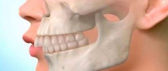
Malocclusion is a fairly common phenomenon that can be confidently interpreted as a diagnosis.
These are not only external defects of the maxillofacial apparatus, but also pathologies that cause certain harm to human health.
At the same time, there are often cases when the harm goes beyond the dental framework - difficulty chewing food leads to dysfunction of the gastric system.
One of these bite deviations is a pronounced defect in the development of the jaw. It manifests itself both in its size and in insufficient width or fragmented crowding of the dentition.
Definition
The concept of “small lower jaw” in orthodontics is not clearly defined.
This diagnosis is classified into several types, differing in the clinical picture of the anomaly.
Micrognathia
Mandibular micrognathia is a defective or too long-term development of an organ that goes beyond the norm.
The defect can affect both the entire jaw and its individual areas, for example, affecting only one lateral part.
Prognathia
It is considered a phenomenon directly opposite to the case described above.
The organ corresponds to the normal parameters, but with the pronounced size of the upper jaw, it seems that it is too small.
Experts often call this disease false prognathia.
Dental formula
In order to improve the convenience of describing each tooth, numbering them, and filling out cards, it is customary to record the order of the teeth using a special formula. There are several varieties of it.
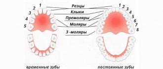
Zsigmondy-Palmer system (quadratic-digital)
Arabic numerals are used, numbering starts from the central incisors in each direction:
- 1 and 2 – incisors.
- 3 – fang.
- 4, 5 – premolars.
- 6-8 – molars.
Milk teeth are designated differently - using Roman numerals:
- I and II – incisors.
- III – fang.
- IV and V – molars.
Two-digit Viola system
Teeth numbering uses 2 digits. The jaws are divided into 4 quadrants. The first digit shows its number.
For adults this is:
- 1 – upper jaw on the right.
- 2 – upper jaw on the left.
- 3 – lower jaw on the left.
- 4 – lower jaw reference.
For a similar description of baby teeth, numbers 5 to 8 are used.
So, there are 8 teeth in each quadrant, its number is shown by the second digit. Thus, the first molar of the lower jaw on the left is designated 35, and the child’s canine from the lower right is designated 43. Therefore, the phrase that “treatment of the 48th tooth is required,” or, for example, the 55th, does not indicate the doctor’s lack of qualifications or what - or pathology in your child, who suddenly acquired so many teeth.
Signs
The main sign indicating the presence of this disease is the visual observability of an anomaly - such a defect is visible to the naked eye, and you do not need to be a specialist in orthodontics to determine such a deviation in a person.
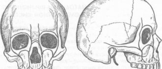
In addition, pathology deforms the natural proportions of the maxillofacial apparatus. The chin becomes sharp and slightly raised upward. Doctors even defined this phenomenon as “bird beard.”
It is important to understand that this is not just a cosmetic defect; micrognathia threatens its “owner” with the development of the pathology of tongue retraction, which is considered a serious disease.
The anomaly causes frequent uncontrollable attacks of suffocation and poses a serious threat to the patient's life.
The disease is often diagnosed based on this characteristic feature - excessive retraction of the chin leads to wrinkling of the skin in its area, and the horizontal fold between the chin and lip is smoothed out.
In addition, underdevelopment of the lower jaw often goes hand in hand with chromosomal mutation processes, which leads to Patau syndrome.
A clear sign indicating the presence of this defect is also the incorrect location of individual fragments of the dentition.
In the case when some organs are missing, neighboring teeth change the direction of growth, trying to spontaneously fill the free space.
Where do teeth come from?
Teeth begin to form and develop when the fetus is still inside the womb (at about 6 weeks). They have their source - the epithelial dental plate. Already by 14 weeks, active formation of dental tissues, which are hard, occurs. Initially, this occurs in the area where the crown will be, and later at the root.
Molars, namely their first rudiments, appear by the 5th month of the embryo. They are located higher than the child’s baby teeth or lower. By the time the child is born, the rudiments are already practically formed in the tissues of the jaw.
Teeth that belong to an additional group (have no predecessors) are formed later. This occurs after about 1 year of life. Why? Because the baby's jaw is still very small and there is not enough space for them.
Causes
This type of anomaly can be triggered by the following factors, some of which are external, and some are internal:
- unbalanced diet of a woman during pregnancy - in this situation, the defect is formed already at the stage of intrauterine development of the fetus;
- chromosomal mutations that cause deviations in the normal growth and development of the baby during pregnancy;
- genetic predisposition;
- Robin's syndrome is a congenital anomaly of the anatomical structure of the facial apparatus;
- too early loss of baby teeth or permanent organs in adulthood , as well as a fairly long change from a temporary bite to a permanent one;
- difficulty breathing through the nose , associated with the structure of the nasal septum or chronic diseases of the nasopharynx;
- mechanical organ injury.
Everything about correct orthognathic occlusion, its signs and characteristics.
Let's talk here about the treatment of dental dystopia.
Follow the link https://orto-info.ru/zubocheliustnye-anomalii/okklyuzii/glubokiy-prikus.html if you are interested in methods of treating deep bites in adults and children.
Milk teeth in children: structure, quantity, timing of eruption
Teeth (deciduous and permanent) are bone formations. They are designed to carry out the process of mechanical processing of food, the so-called mastication, in order to prepare it for subsequent digestion.
As for the anatomical structure of baby teeth, it is in many ways similar to the structure of adult teeth, although there are some important differences.
The part of the tooth located above the gum is called the crown. The surfaces of the crowns can be of different shapes depending on which particular tooth we are talking about, but in any case, in baby teeth they are much smaller in size.
The crown is connected to the root through a neck - a slightly narrowed part, around which connective fibers are located in the horizontal plane, forming the so-called circular ligament.
The root itself is located in a small depression called the alveolus. Vessels that supply nutrition to the tooth and nerves pass through a special hole in the apex of the root. Most people are mistaken in believing that baby teeth do not have roots. In fact, those of them that are intended for chewing food (molars) are also molars, only their roots are independently absorbed by the time they are replaced by permanent ones.
What is inside the crown? A photo of the structure of a baby tooth helps you find out:
- Any milk tooth, like a permanent one, is covered with enamel.
Only in temporary teeth it is much thinner and softer, and not so mineralized, which is why in children caries develops rapidly and can turn into pulpitis or periodontitis in a few weeks.
- Beneath the enamel is dentin, which is also much thinner than in permanent teeth.
This is the highly mineralized underlying tissue surrounding the tooth cavity and root canal. It is slightly inferior in strength to enamel. Dentin in the direction from the center is completely pierced by special tubules, through which impulses are transmitted and all metabolic processes occur.
- Dentin, closer to the root system, covers cement, to which the fibers of the ligamentous apparatus - periodontium - are attached.
- The internal cavity of the crown and root of the tooth is filled with pulp - very soft internal tissue in which nerves and blood vessels are located.
It plays a major role in providing the tooth with nutrients and carrying out metabolic processes. When the pulp is removed, metabolic processes in the tooth become impossible.
In baby teeth, the volume of the pulp is much larger, and the root tubules are wider than in permanent teeth.
In addition to the structural features of baby teeth, parents are concerned about the timing of their eruption and how many teeth should be normal at a given age of the baby. Let's look at these questions in more detail.
Treatment
Methods for eliminating micrognathia depend on the degree of development of the pathology, the age of the patient and can be gentle or radical.
In the first case, treatment is carried out gradually through the use of special bite-leveling structures, and in the second, surgeons are involved in the work.
In children
In childhood, this deviation is easy and in most cases, successfully, treated mainly with therapeutic methods, and with rare exceptions, surgically:
- sanitation of the oral cavity – complete restoration of damaged tooth segments, as well as removal of affected root areas. Restoring the integrity of periodontal tissues using general and targeted spectrum agents;
- pediatric prosthetics – indicated for early loss of mammary organs. The voids are filled by splinting or attaching temporary devices that maintain the correct position of growing teeth and correct the size of the jaw arch;
- correction of language function and normalization of the respiratory system - in the first case, this is surgical cutting of the frenulum, which is done quickly and almost painlessly. In the second - surgical alignment of the partitions, followed by rehabilitation exercises;
- myogymnastics – tonic effect on the muscle tissue of the organ through special gymnastic exercises.
It is advisable to carry out with children 4–6 years of age, when in case of underdeveloped pathology this method can be used as the main method of treating the defect.With positive dynamics, the need for subsequent installation of orthopedic structures, as a rule, no longer arises;
- leveling the unevenness of the chewing surface using the method of grinding (fissures) - an ideal solution for a slight deviation in jaw closure;
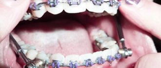
- the use of leveling systems and structures is carried out when the anomaly is too pronounced, when other correction methods are ineffective. Wearing mouth guards and plates, and in infancy special nipples, gradually brings the deviation back to normal.
In adults
Treatment of patients whose bite has already been formed takes longer, and methods for correcting it are most often more radical.
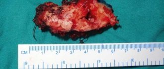
Bone grafting
The essence of the method comes down to artificial extension using a graft. Bone tissue is taken from other areas of the patient’s tissue, which guarantees good survival of the material.
Fixation of the grafted fragments is carried out using screws made of high-quality, durable alloy of titanium components. This is done as follows:
- the lower organ is incised;
- the hard tissue is moved apart and the osteoplast is immersed inside;
- fixed with screws;
- intermediate voids are filled with special plastic chips;
- implantation of the membrane and suturing of the organ.
The main advantages of the method are the rapid survival rate and reliability of the procedure; the jaw augmented in this way retains its functionality for many years.
Disadvantage: individual intolerance to components and osteoplast fixatives.
In the video, watch the process of bone grafting for anomalies of the lower jaw.
Bone grafting plus liposculpture
Plastic surgery of hard bone tissue is carried out similarly to the option described above, with the difference that in parallel, aspiration of the local accumulation of fatty tissue is carried out using a vacuum method.
The technique qualitatively solves the aesthetic problem of underdeveloped chin caused by this pathology, corrects its shape, evens out the oval of the face and, in a sense, resembles the effect obtained from lifting the chin area.
The advantage is a high aesthetic result, the disadvantage is the high cost of the procedure and the presence of a large number of contraindications to its implementation.
Causes of open bite development and its dangerous consequences.
In this article we will discuss treatment methods for microdentia.
At this address https://orto-info.ru/zubocheliustnye-anomalii/ryadov/suzhenie.html we offer details about the treatment of narrowing of the dentition.
Endoprosthetics
It is made with harmless facial implants from non-biological components - silicone, porous polyethylene or cartilage extract. The desired shape is given to the device directly during the operation.
The implant is inserted through deep incisions in the mucous cavity, which leaves no postoperative traces. The product is inserted into the subperiosteum in lateral fragments into the chin area and attached to it with surgical sutures to fix its position.
The disadvantage of this method is the rather long recovery period.
Lipofilling
This option is considered to be a non-surgical correction, which is its main advantage.
The method allows you to give the chin the desired shape through pinpoint needle injection of fat cells into the subcutaneous area, in those areas where an increase in the size of the chin is required.
The entire procedure takes no more than an hour. The rehabilitation period passes quickly and without complications. The only drawback is that 30% of the implanted tissue fragments are rejected by the body, which requires repeated lipofilling 5-6 months after the first procedure.
To learn how chin augmentation is performed using lipofilling, watch the video.
Anatomy of permanent teeth
Each molar consists of certain parts:
- crown. This is the part of the tooth that protrudes from the top;
- the root, it goes deep into the alveoli. At the same time, it is attached thanks to special connective tissue bundles. There can be different numbers of roots (1-5 pieces). This moment affects the number of nerves and channels;
- neck. This part is located between the root and the crown.
Tooth tissues are distinguished by their heterogeneity. The enamel is on top and is known for its durability. Once the tooth has erupted, it is covered with a transparent thin layer. This is the cuticle, which eventually changes to the pellicle. The latter is a film that is created from what saliva produces.
Beneath the enamel is dentin, the tissue of the tooth. Dentin is similar to bone when you study how it is built. However, it is more durable because there is a high level of mineralization. In the area where the root is located, the dental tissue is covered with cement. The latter is rich in mineral compounds and is also associated with periodontium. Collagen fibers are used for this.
As for the part of the tooth that is inside, this is the crown and root canal. They are filled with pulp. This is loose connective tissue; it contains nerve endings and blood vessels.
Prices
The approximate cost of treating a defect of abnormal development of the lower jaw is given in the table:
| Character of the bite | Correction method | Average price (in rubles) |
| Lactic | Sanitation of the oral cavity | From 3 500 |
| Lactic | Orthopedic therapy | From 30 000 |
| Lactic | Fissure grinding | From 11 000 |
| Lactic | Operation | From 15 000 |
| Constant | Bone grafting | From 19 000 |
| Constant | Bone grafting pole liposculpture | From 50 000 |
| Constant | Endoprosthetics | From 40 000 |
| Constant | Lipofilign | From 35 000 |
How to determine that a child will soon have molars?
There are certain signs that indicate that permanent teeth will soon begin to erupt:
- The spaces between the teeth increase. The jaw grows and the free space increases;
- baby teeth become loose as the root gradually dissolves. It cannot be firmly fixed in the jaw tissues;
- in case of loss of a temporary tooth. This confirms that the molar will soon come out as it has pushed out the previous one;
- The gums are slightly swollen and red.
When permanent teeth erupt, the child’s general well-being usually remains the same, the temperature does not rise, and there is no pain.
Prevention
In some cases, the development of such a defect can be completely avoided by following the following recommendations:
- preference should be given to breastfeeding , and if this is not possible, control the hole on the nipple - it should not be large in size;
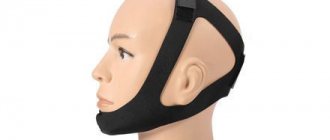
- it is necessary to choose the right pacifier so that its position does not put constant pressure on the weak bone tissue of the gums;
- timely treatment of dental diseases.
Diagnosis and treatment of narrow jaw
Deformed dentition, crowded teeth, malocclusion, unerupted teeth are consequences of an underdeveloped dental system. The earlier these abnormalities are diagnosed, the more effective the treatment will be. To expand the lower and upper jaws, therapeutic methods, mechanical expanders and plates are used. In rare cases, only surgery helps.
In children under 10-11 years old, jaw expansion gives the best results. This is a time of active growth and formation of bone tissue, so the defect can be corrected with the help of orthodontic structures.
Possible problems
Permanent teeth have just appeared, but this does not mean that there will not immediately be any problems associated with them. Parents should be aware of possible dental problems:
- lack of molars;
- pain in the molar area;
- crooked position of molars;
- molars fall out;
- injuries.
For any of these problems, it is important to contact a specialist in time to receive qualified help.