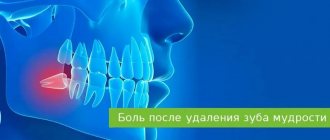
Osteomyelitis of the jaw is an inflammatory and infectious process with purulent contents that covers the structural elements of the jaw bone. Gradually, the disease provokes bone decomposition, that is, osteonecrosis gradually progresses. The pathology requires urgent professional help to avoid the spread of infection to other organs and systems of the human body. Osteomyelitis of the lower jaw is diagnosed twice as often as the same disease, but affecting the upper jaw system.
Causes of osteomyelitis
Penetration of bacteria into bone, which traditionally occurs as follows:
- against the background of tooth damage by caries, that is, by odontogenic means. First, microorganisms penetrate the pulp, after which they enter the hard tissue through the lymph nodes or canals;
- after fur. tooth damage: dislocations, fractures, operations, etc.
- due to the penetration of infection during ENT diseases, skin inflammation, when microorganisms from one source of inflammation pass into the bone.
What types and forms are there?
Osteomyelitis is classified according to several criteria. This is how the disease is distinguished by the location of the infection, the type of microorganisms, routes of penetration and the nature of development.
Depending on the type of pathogenic bacteria, osteomyelitis is classified into two types.
- Specific. The infection enters from another affected organ or system, or from the external environment.
- Non-specific. The infection is opportunistic, as it is constantly present in the human body.
There are three types based on the method of penetration.
- Hematogenous. The infection moves within the body.
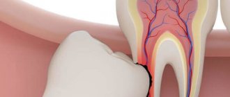
- Odontogenic. Bacteria penetrates through an injured or carious tooth.
- Post-traumatic. Pathogenic microorganisms penetrate the injured jaw bone as a result of contact with objects (during surgery or an open type of injury).
- Based on the location of the lesion, osteomyelitis of the lower and upper jaws is distinguished.
Based on the nature of the infectious process, the disease is classified into chronic and acute forms. Chronic osteomyelitis of the jaw is divided into primary and secondary types.
Symptoms of osteomyelitis of the jaw
Symptoms depend on the form of the disease.
The main symptoms of acute osteomyelitis:
- severe pain in the projection of the tooth;
- swelling of the gums and cheeks;
- facial asymmetry;
- chewing spasms. muscles;
- joint arthritis;
- increased body temperature;
- swelling of the mucous membrane;
- loosening of the pathological tooth and adjacent units;
- general malaise;
- increased sweating;
- pale skin;
- enlargement of regional lymph nodes.
Symptoms of chronic osteomyelitis of the lower jaw:
- the patient’s condition may improve for some time, but after some period the symptoms listed above will manifest themselves, but with greater force;
- purulent fistulas in the oral cavity, which then come to the surface of the skin;
- swelling of the mucous membrane;
- enlarged lymph nodes;
- constant pain;
- it becomes difficult for the patient to point to the exact location of the pain.
Symptoms of osteomyelitis after tooth extraction are very difficult to recognize in the first few days after surgery. The clinical picture is similar to the anatomical recovery of the body after surgery - there may be an increase in body temperature, swelling, and pain. After 2–3 weeks, if the symptoms do not go away, it becomes clear that the operation resulted in an inflammatory process.
Provoking factors
For the development of osteomyelitis, the presence of only reasons is not enough. The infection begins to actively “attack” the bone tissue of the jaw under the influence of provoking factors. The main condition for the occurrence of a pathological process is immunodeficiency. A weakened immune system is unable to resist bacteria, which leads to tissue damage.
Other factors that reduce immunity can also provoke osteomyelitis:
- bad habits (alcoholism, smoking);
- diabetes;
- immunodeficiency diseases;
- poor nutrition, diet, fasting;
- oncological diseases.
In the absence of treatment or incomplete therapy, chronic osteomyelitis of the jaw develops. The chronic form of the disease is difficult to treat and provokes serious complications.
Diagnosis of osteomyelitis
It is carried out exclusively by a dentist, based on clinical and laboratory studies. Differential diagnosis is also involved.
Basic methods for diagnosing pathology:
- general and biochemical blood tests;
- general urine analysis;
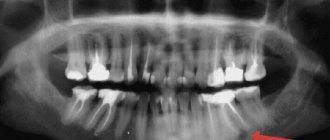
- X-ray of the jaw;
- MRI of the jaw;
- bacteriological examination of purulent contents of fistulas;
- research for diseases that led to bone destruction.
Treatment of periostitis
When treating gumboil, conservative and surgical methods are used. The choice of one method or another depends on the doctor’s experience, the form of the disease and its stage.
A conservative treatment method will be effective at the initial stage of periostitis, as well as in case of inflammation of the periosteum, provoked by weak immunity or the presence of other inflammatory processes in the body. In this case, antibacterial and anti-inflammatory drugs, as well as physiotherapeutic procedures, are used. In this case, the tooth can be saved, and the suppuration is removed from the source of inflammation by cutting the gums and installing drainage.
If purulent inflammation of the periosteum is caused by dental disease (odontogenic periostitis) or if conservative treatment is ineffective, then surgical treatment is performed. To do this, anesthesia is first done: usually, when performing surgical treatment of periostitis, local anesthesia is used. Then the focus of suppuration is opened, for which an incision is made in the gums. After this, all pus is carefully removed and drainage is installed to drain the fluid, which will continue to form for some time at the site of inflammation. To determine the cause of periostitis, an x-ray is taken. Based on the conclusions about the cause that provoked the development of inflammation, the diseased tooth is removed or medicated treatment is carried out.
Self-treatment of flux at home with antibiotics can lead to complications of varying severity. Therefore, at the first signs of periostitis, it is recommended to contact a dental clinic.
Classification of the disease, depending on the location of inflammation. process
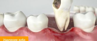
- Jaw osteomyelitis after tooth extraction. The nerve endings of the periodontium and gums remain irritated, pain can be felt immediately or after several days.
- Odontogenic osteomyelitis of the lower jaw is the most common form of the disease at an advanced stage of caries. Quite common in young children.
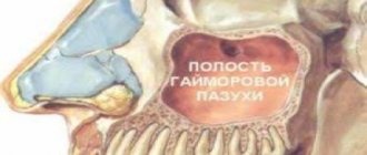
- Odontogenic osteomyelitis of the upper jaw is caused by the anatomical structure of the bone. The infection can provoke inflammation of the maxillary sinuses. If the fang becomes inflamed, it swells, including the infraorbital area.
- Osteomyelitis of the socket is an inflammation of the bed where the extracted tooth was located. A more correct name for the disease is alveolitis, which is accompanied by destructive changes in the bone.
- Hematogenous osteomyelitis of the jaw - affects mainly the upper jaw, after which it affects the cheekbones and nasal bone. The clinical picture is characterized by sepsis, which is accompanied by inflammation in more than half of patients with this diagnosis.
Complications
If you see a doctor in time, make a diagnosis and choose the right treatment, the prognosis will be favorable.
If you ignore all the conditions, complications will arise:
- Meningitis
- Abscess of the brain and lung.
- Orbital phlegmon.
- Sinusitis.
- Vein thrombophlebitis.
- Sepsis.
- Mediastinitis.
Pathological data require immediate assistance to prevent death.
The chronic process, with its long course, affects the soft tissue and bone areas of the maxillofacial area, and is accompanied by:
- traumatization;
- changes in the TMJ;
- the formation of adhesions in the joints and scars on the masticatory muscles;
Such disorders limit chewing movements or lead to immobility.
Effective treatment of jaw osteomyelitis
- it is necessary to eliminate the source of inflammation;
- functional impairments caused by the infectious process should be corrected.
The patient is treated by specialists in the field of maxillofacial surgery. No self-medication or alternative methods will help solve the problem of osteomyelitis - it will only worsen the condition!
Surgical care includes:
- opening and cleaning the purulent focus from pus, draining the area;
- use of antibacterial agents;
- taking painkillers and antibiotics;
- detoxification and anti-inflammatory therapy;
- vitamin therapy;
- gentle nutrition.
As a preventative measure, it is necessary to promptly treat caries, undergo regular dental examinations, and clean teeth from hard and soft deposits.
Symptoms of alveolitis after wisdom teeth removal
The first two days after extraction, the pathological process develops asymptomatically. Two days later, the patient notices an unpleasant throbbing pain, a characteristic putrid odor from the mouth and the appearance of foreign taste sensations that cause noticeable inconvenience. During examination, the specialist notes the absence or small size of a blood clot, the condition of which indicates the natural or pathological nature of the healing of the hole. In the absence of proper treatment, the pain becomes stronger and radiates to the eyes and ears on the corresponding side of the face. The accumulation of colonies of pathogenic microorganisms and food debris in the socket causes unpleasant taste sensations and a pungent odor, which cannot be removed by regular brushing of teeth.
Features of the treatment of acute purulent periostitis
In the case of acute purulent periostitis, the patient is required to be prescribed painkillers, antibiotics and physiotherapy. In addition, the dentist makes an incision in the gum, removes all pus and installs drainage, for which the tip of a rubber strip is inserted into the cavity located at the site of the incision. This prevents the wound from healing very quickly and allows the fluid formed at the site of inflammation to flow out of it for some time without accumulating.
Acute periostitis is usually provoked by a diseased tooth, which must be removed if its crown is severely damaged or there is obstruction of the root canals. In addition, the tooth that is the source of infection is removed if it has become mobile, if its functionality has been lost, or if the conservative treatment method has proven ineffective.
If acute periostitis is caused by inflammation of the tonsils or throat, then it is necessary to eliminate the causes that caused the development of the infection and restore immunity. The most effective approach to treating this form of periostitis is complex, combining surgical treatment, physiotherapy and drug therapy.
Causes of flux after extraction
In dentistry, gumboil refers to an inflammatory process that forms inside soft tissues and manifests itself in the form of swelling, purulent discharge, fever and other unpleasant syndromes. The medical term is periostitis. There is only one mechanism for the development of flux: pathogenic bacteria provoke the development of infection, which penetrates into the gums and periosteum through microtubules.
Dangerous symptoms!
- Deep inflammatory process.
- Invasive tooth extraction, after which the gums are injured. If additional anti-inflammatory therapy is not given, infection may develop.
- Poor-quality temporary prosthesis, which caused gum injury and infection.
- Dry socket syndrome.
Conservative therapy
This includes the following activities:
- general strengthening;
- anti-inflammatory;
- physiotherapy;
- desensitizing.
Anti-inflammatory treatments
Therapy consists of treatment with antibiotics. Antimicrobial drugs should be started as early as possible (for example, gentomycin, ciprofloxacin, lincomycin, etc.). They are absorbed by bone tissue and do not cause allergic reactions.
Patients with severe forms of the disease are simultaneously prescribed several groups of antibiotics, taking into account compatibility and side symptoms.
Due to the fact that osteomyelitis is provoked by aerobic and anaerobic pathogenic microorganisms, it is effective to include antibiotics of the metronidazole group in combination with sulfonamides in the treatment protocol. In some cases, patients are prescribed intravenous administration of enzymes. Competent selection of drugs determines the result of drug treatment, and also helps to minimize the risk of complications and adverse reactions.
Osteomyelitis in mild forms can be cured in 7-10 days. Severe forms require a longer course until the symptoms of inflammation are completely relieved.
