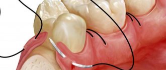
The need for suturing in dentistry
Surgical intervention and, as a consequence, suturing are indicated for the following manipulations:
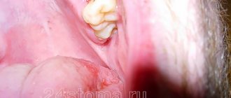
- Removal of wisdom teeth - third molars differ from the rest in the large size of the tooth crown and its root. Late eruption, the position of the “extreme” in the dentition very often leads to a violation of the physiological location of the tooth or even its impaction (partial or complete failure to erupt). In this regard, the procedure for removing a wisdom tooth is often accompanied by complications - extensive damage to the soft tissues of the oral cavity and alveolar process, ligaments, and blood vessels. To avoid this kind of consequences, dentists apply stitches to reduce the wound.
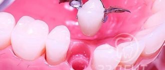
- Implantation - its implementation is always accompanied by the need for surgical intervention. A deep incision in the gum requires suturing.
- Maxillofacial operations - elimination of the consequences of traumatic injuries, congenital defects of the dentofacial apparatus.
- Removal of tumors of various etiologies - the integrity of the soft tissues of the oral cavity is damaged, often requiring layer-by-layer sutures.
Atheroma in a child
When examined by a specialist, it is extremely important to distinguish atheroma from lipoma. Atheroma in a child is a smooth, round neoplasm located deep in the skin. When palpated, it is soft and motionless. In most cases, the cyst does not cause any discomfort to the baby. There is no threat to health. When an inflammatory process occurs, the situation is completely different. The main symptoms include: redness of the tumor, pain when pressed, discharge of blood or pus. With such manifestations, urgent medical attention is required.
On our website Dobrobut.com you can make an appointment with a doctor and find out how to treat atheroma of the eyelid in a child. Consultations are conducted by specialists with many years of experience. If necessary, a diagnosis and course of treatment will be prescribed.
Types of sutures and materials used in dentistry
To facilitate and accelerate the healing process of postoperative wounds in the oral cavity, the following types of sutures are used:
- nodal - used when it is important to ensure equal tension on both edges of the wound (for example, between teeth);
- continuous - when a violation of the integrity of soft tissue occurs along several dental units;
- single - each stitch is knitted separately from each other.
Threads used for suturing in dentistry can be made of absorbable and non-absorbable material.
Absorbable threads:
- from catgut - which contains a foreign protein, which is perceived by the body as a foreign body, and therefore often causes inflammation of the damaged soft tissue of the patient’s oral cavity;
- “Vicryl” is a synthetic material made from polyclatin and polyglycomic acid, which does not cause the risk of developing inflammatory processes even during complex operations.
Threads made of absorbable material do not require the removal of sutures; they disappear within 2-5 weeks.
Material for non-absorbable threads:
- silk is an easy-to-use material, highly durable, but has a significant drawback - it causes inflammatory processes in soft tissues if not removed 7 to 10 days after surgery;
- monofilament is a thread made of polytetrafluoroethylene, which is durable and does not cause inflammation, but has a rigid structure that can damage the mucous membrane. To exclude this possibility, a bandage is applied to the wound surface;
- polyester is a woven thread often coated with silicone for elasticity and smoothness. A very durable material that does not lead to inflammatory processes, but requires careful fixation in the form of knots.
The main types of treatment for atheroma without surgery
Atheroma is a benign tumor formed as a result of blockage of the sebaceous glands.
Atheroma forms mainly on the neck, face, groin area and back. The neoplasm is considered relatively safe, since it has no tendency to degenerate into cancer. Below we will tell you whether it is possible to remove atheroma of the scalp with a laser? and “is it possible for the tumor to re-form, as well as what earlobe atheroma and other forms of the disease look like. Causes of the disease:
- improper skin care;
- injuries, skin cuts;
- excessive use of deodorants;
- hormonal disorders;
- hypersweating (hyperhidrosis);
- pimples, blackheads;
- inflammation of the epidermis;
- metabolic disease.
The main method of treating pathology is surgery. Below we will talk about the types of treatment for atheroma without surgery.
When and how is the procedure for removing sutures in dentistry performed?
The time for suture removal is determined by the dentist, guided by the process of regeneration (healing) of the soft tissues of the mucous membrane, which depends on:
- on the age of the patient;
- complications of surgical intervention;
- presence of complications.
Usually, sutures are removed 5-10 days after they are applied.
The dentist removes sutures as follows:
- the surface of the wound is disinfected with an antiseptic;
- if the patient has a low pain threshold, local anesthetics are used (but usually this is not necessary);
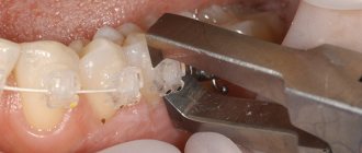
- Using surgical scissors and anatomical tweezers, the dentist carefully cuts and removes the threads from the healed wound;
- a visual examination is carried out to check the integrity of the wound;
- Disinfection of the suture prevents the risk of wound inflammation.
Sometimes sutures are removed due to the risk of suppuration in the area of the operation. In this case, the sutures are removed, sanitizing procedures are performed and appropriate antimicrobial therapy is prescribed.
Suppurating atheroma on the face
Ordinary atheroma in itself is not dangerous. The exceptions are cases of localization on the face and suppuration. Causes of festering atheroma on the face: infection, complications after a poorly performed operation, advanced form of the disease.
Symptoms of the disease:
- general weakness, enlarged regional lymph nodes;
- temperature increase;
- redness in the area of the tumor;
- swelling;
- cyst enlargement.
Treatment of suppurating atheroma is only surgical. The surgical intervention consists of two stages. Your doctor will tell you at a personal appointment about the day on which the sutures are removed after surgery to remove atheroma.
Hygienic oral care after suture removal
To quickly restore the integrity of the soft tissue of the oral cavity, the patient must:
- Use a soft toothbrush to brush your teeth daily;
- use anti-inflammatory rinses;
- rinse the mouth with warm water or a decoction of medicinal herbs after each meal;
- Avoid eating hard, sour, salty, spicy foods that can irritate the mucous membrane.
Timely removal of sutures, maintaining oral hygiene, and performing procedures prescribed by the dentist will ensure good wound healing and speed up recovery.
Diagnosis and treatment
After examining the patient, the doctor will prescribe a histological examination that will help differentiate atheroma from limoma, hygroma and fibroma.
The main treatment method is surgery, during which the tumor will be removed. The intervention takes place under local anesthesia in a hospital setting. The surgeon makes an incision through which the atheroma is removed. The wound is sutured and a sterile dressing is applied. The stitches are removed on the 10th day.
Laser removal of a tumor can be carried out in three ways: photocoagulation, laser evaporation of the capsule, laser excision with the shell.
Radio wave removal is indicated for small atheroma sizes. Radio waves affect tumor cells, causing them to die. A crust forms at the site of the cyst, which disappears over time. Your doctor will tell you more about how radio wave removal of atheroma on the back is carried out during your consultation.
After removal:
- The area after surgery should not be wetted for three days;
- the wound is treated daily with an antiseptic;
- You must wear a bandage for a week;
- It is mandatory to take antibiotics (as prescribed by a doctor);
- During the recovery period, a course of physical therapy is recommended.
The wound healing time depends on the location and size of the cyst.
Postoperative complications
The most common complications after dental implantation are:
- Pain syndrome. The norm is mild soreness of the gums in the area of implantation for 1-2 days. In cases where severe pain does not go away even after taking the generally recommended painkiller, you should consult a doctor, because this is most often a symptom of a complication.
- Inflammation.
- Seams coming apart.
- Hemorrhages. After surgery, ichor often separates from the gums, the amount of which quickly decreases and disappears. It is enough to remove it with cotton swabs. But if the bleeding becomes more intense, you should consult a doctor immediately.
There are also complications such as inflammation of the sinuses, peri-implantitis (inflammation of the tissue around the implant, leading to destruction of the supporting bone tissue), mucosal hyperplasia (pathological growth of the mucous membrane in the area of the implant), damage to the implant and prosthesis.
Swelling after dental implantation
In the first three days after implantation, swelling may occur in the gum tissue. This is the body's natural reaction to surgery. Most often, a tumor is an indicator that blood is actively flowing to the operation site.
But if you want to avoid the discomfort caused by swelling, you can reduce the risk of its occurrence. To do this, in the first two days, apply cold to the operation site: for 5-10 minutes with breaks of 20 minutes. Do not apply ice directly to the skin; it is best to wrap it in a towel. In rare cases, the tumor will require the use of antihistamines, antibiotics (to prevent infection) or treatment with antiseptic solutions.
It will also help to avoid swelling by eliminating sudden stress loads on the body: physical overload, hypothermia, overheating.
What happens after surgery
After the surgeon has finished working with the ears, suture threads will first be applied to these places, holding the edges of the wound together. Then the patient is put on a special bandage that has a fixing effect. Its main task is to hold the ears in place and protect them from displacement, loss of shape, or infection.
The bandage can be either a regular compression bandage or a mask bandage. The first product looks like a regular decorative headband, is comfortable to wear, has good throughput, i.e. "breathes". The mask bandage covers the entire neck of the patient and passes under the chin, along the ears and through the back of the head. This option is no less reliable and functional, but wearing a mask can be a little difficult in the summer, when the air temperature rises high.
Wearing a bandage after otoplasty is mandatory.
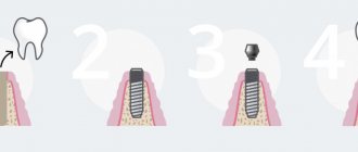
Simultaneous (immediate) implantation
This prosthetic method is a one-stage implantation. In this case, the dentist removes the patient’s damaged tooth and immediately installs an artificial root.
Implantation immediately after tooth extraction is only possible if the bone tissue has sufficient volume to accommodate the implant - for example, if the tooth was damaged or lost in an accident. But tooth loss much more often occurs due to diseases when bone tissue is lost. Therefore, this method is not always available.
Dental implantation is an achievement of modern dentistry that allows you to restore lost teeth and use them for the rest of your life. And if you carefully follow your doctor’s recommendations, your smile will always bring joy to your loved ones!
How long does it take to heal?
Bone tissue and gums heal after dental implantation on average from three to six months, and the period is different for the upper and lower jaws. The lower jaw will heal in three to four months, but the upper jaw will take longer: four to six months. This is due to the greater bone density of the lower jaw.
Sometimes the healing rate deviates from the average time frame, because it depends on the individual characteristics of the patient’s body: the speed of regeneration, the strength of the immune system, the pain threshold. Even such factors as gender, age, lifestyle, diet and daily routine of the patient are important.
In any case, recovery after surgery takes some time. Following medical recommendations will help protect yourself from complications during healing.
How to remove stitches correctly
In most cases, the use of disinfectants is not necessary; they can irritate the wound and cause discomfort to the dog.
The owner will need:
- Surgical scissors. In the absence of such, you can use a similar model with rounded edges.
- Surgical tweezers.
- Cotton swabs.
- Iodine, chlorhexidine.
- Clean towel (clean rags, gauze).
On the day the sutures are removed, the animal should be calmed so that it does not become nervous. It is enough to hold a small dog by the head with one hand or swaddle it, but with a large one you will have to tinker, quite possibly, ask someone at home for help so that the dog cannot move. Nervous and aggressive dogs should wear a muzzle.
Next, the animal should be placed in a comfortable position so that it lies on its side in a relaxed, natural position.
How to remove post-operative stitches for a dog:
- Clean the healed wound from dried blood and other contaminants using a towel moistened with warm, soapy water.
- Find the longest ends of the threads, pinch them with your left hand and pull them towards you.
- Cut the threads with scissors between the skin and the knot.
- Press the skin next to the seam with the fingers of your right hand, pull the ends of the threads with the fingers of your left hand, and quickly and carefully pull them out.
- If there are threads left at the site of the healed wound, remove them using tweezers.
- Treat the postoperative area with iodine.
This concludes the suture removal procedure, and if the owner did everything correctly, then there should be no complications.
Withdrawal algorithm
Stitches are removed from a sutured wound in medical institutions as follows:
- Remove the bandage.
- The edges of the injured area are assessed, the stitched area is examined, and the stitches are counted.
- Iodopirone or 70% alcohol treats the skin near the scar.
- A secondary narrow treatment of the tissue contraction site itself is performed.
- Each stitch is cut one by one, and the suture is removed from the wound by pulling with tweezers.
- Depending on the doctor's decision, not all stitches can be removed.
- Having finished removing the threads, the wound area is treated with an antiseptic.
- A sterile napkin is applied on top, secured with a bandage, or a bandage is applied.
- After removing the sutures, the surgeon gives recommendations on how to care for damaged skin surfaces.
You can remove suture material from a small wound at home; the algorithm of actions will differ. Initially, you need to prepare everything you need to remove the threads.
- Organize a workspace for tools that will be useful when removing sutures. Remove all foreign objects from it, wipe off the dust, and treat the surface with alcohol. On a sterile napkin (tray), place a bandage, cotton wool, plaster, antiseptic (iodine, medical alcohol). The operation site should be well lit.
- Boil a pan of water. Dip metal tools (scissors, tweezers) into water. Boil for 3-5 minutes. Place on a clean, dry towel over the area where surgery will be performed. After the instruments have dried, they must be wiped with alcohol.
- Wash your hands with soap and dry. Wear sterile rubber gloves. Carefully remove the bandage (plaster) from the surface of the wound.
- Before removal, inspect the scar surface for redness and inflammation. Treat the damaged area and the skin around the periphery of the seam.
- Using tweezers, carefully pick up the stitch knot and lift it 2–3 mm. Place the jaw of the scissors under the thread and cut the suture material on the side of the knot.
- Gently pull the thread at the knot so that the cut part opposite the knot is pulled through the skin. Carry out such manipulations with each stitch. Sharp pain during suture removal may indicate incorrect action. Unpleasant sensations and discomfort when pulling the threads are considered acceptable.
- Treat the wound with an antiseptic. Apply a sterile bandage over it. If there are special doctor's recommendations, they must be followed.
After removing the surgical suture, you must follow the rules for caring for the postoperative wound:
- Perform dressings twice a day (morning/evening).
- Protect from injuries and unnecessary tension - at first it can lead to divergence of the edges.
Features of caring for internal sutures (in the vagina and cervix)
Sutures on the internal genital organs made with absorbable threads (such suture material is called catgut, in modern institutions, vicryl or safil) do not require complex manipulations. They do not need to be removed or washed with any solutions; it is enough for the young mother to ensure complete rest and strictly adhere to the rules of personal intimate hygiene so that the infection does not enter the organs of the reproductive system, since discharge in the postpartum period (especially the first twenty days) is a favorable environment for reproduction pathogenic microbes.
Perform any intimate procedures after washing your hands first - this applies to washing your genitals, visiting the toilet, changing sanitary pads, etc. After each visit to the toilet, remove the used pad and wash yourself, so that the direction of movement goes from the vulva to the rectum with warm water and soap.
Remember - the intestinal microflora should under no circumstances get on the genitals, which is why it is so important to follow this sequence of actions. After treating the perineum with water, gently pat it dry with a clean or disposable towel, collecting all the moisture. If you use linen products, change them at least once a day, and more often as they become dirty. Empty your bladder every three to four hours, even if you don't feel the urge to urinate. You will have to avoid taking a bath in the first month after giving birth.
Antibiotics after dental implantation
For preventative purposes, the doctor prescribes a course of antibiotics after surgery. In most cases, this is inevitable, since surgical trauma, even with the modern level of development of medicine, carries a high risk of infection. Therefore, it is better not to ignore antibacterial drugs prescribed by a doctor if you do not want to additionally place unnecessary burden on your immune system to fight infection.
Most often, the course of antibacterial therapy does not exceed 7 days, and broad-spectrum drugs are prescribed - amoxicillin, amoxiclav, cephalosporins. They are characterized by low toxicity and have long proven themselves to be well tolerated by the human body.
Numbness after dental implantation
For most patients, anesthesia wears off within four to five hours after implant placement. Then oral sensitivity should return. If numbness persists after this period, then you have cause for concern because it may indicate nerve damage. This is only possible when the operation was performed on the lower jaw, because there are no large nerves in the upper jaw.
Other symptoms of mandibular nerve injury are lack of facial skin sensitivity, excessive salivation, and difficulty speaking and eating. Timely diagnosis will help to establish the nature and extent of the damage and manage the treatment with medications rather than surgical methods.