Warts are classified as benign neoplasms.
They can appear on various parts of the skin and mucous membranes.
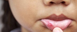
If a wart appears on the lip, it represents a cosmetic defect and can also cause discomfort to its owner, worsening the quality of life.
Forming on the lip, the tumor affects speech and interferes with eating.
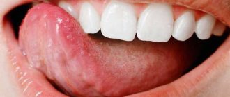
A wart can cause pain, especially if it is localized on the mucous membrane inside the lip.
If measures are not taken to eliminate it, the growth may become injured, which will lead to a secondary infection.
Types of warts
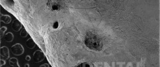
A wart is a growth that rises above the surface of the skin or mucous membrane.
Its dimensions, as a rule, are no more than 1 centimeter in diameter.
The tumor can appear on any part of the body, including the face.
On the face, most often a wart forms on the lip, above or below the lip, on the buccal surface of the oral mucosa.
A warty growth can appear on both the upper and lower lips.
The following types of neoplasms are distinguished:
- Common (vulgar) warts. These are growths that have a dense structure, soft to the touch, flesh-colored or yellowish in color. New growths form on the red border of the lips. They are not painful, but tend to itch. Vulgar warts appear on the lips not only in adults, but also in children.
- Flat- shaped growths may appear on the outside of the lips or on the oral mucosa. Their surface is smooth, oval or round in shape. The color of the growth resembles the color of the skin and almost does not rise above it. The location is also the red border of the lips. Flat warts do not hurt, but they are very itchy.
- Hanging neoplasms are located on a thin stalk. At the beginning of development they resemble a bump. They have a brown, pinkish tint or close to the color of the skin.
- Filiform (acrochords). These are small growths that appear after 35 years, but can occur earlier.
- Genital warts . The localization of such growths is the mucous membrane. The new growths are elongated and arranged in groups, resembling a cockscomb or cauliflower inflorescences. The growths are prone to injury, which causes inflammation and bleeding.
The wart forms gradually and almost imperceptibly.
- At the beginning of development, a small compaction forms, which gradually increases.
- A white dot forms on the surface of the mucous membrane of the lip or in the mouth, which gradually turns into genital warts.
To date, approximately one hundred types of papillomavirus have been discovered.
Some types of growths have a benign course, while others are capable of malignancy.
They can provoke the development of cancer.
In addition to the types of growths listed above, there are also senile warts (keratoma, keratosis).
The difference between keratosis is that its appearance is not associated with papillomavirus, and keratoma does not provoke the formation of cancer.
In their development, senile warts go through several stages:
- ✓ brown spots appear, but the wart has not yet formed
- ✓ formation of papules and nodules
- ✓ appearance of keratoma - when you try to scrape off the scales, bleeding appears
- ✓ transition to the cutaneous horn, which is characterized by excessive growth and keratinization of the neoplasm
According to some experts, keratosis and senile warts belong to different diseases.
Symptoms
The tumor grows and develops quite slowly, so for a long time the patient may not even be aware of its presence in the mouth. Fibroma of the oral mucosa looks like a hemispherical growth rising above the plane, covered with pinkish tissue. If you press it, pain or other discomfort does not appear. The surface is smooth, there are no irregularities or roughness on it.
The appearance of ulcers with such a diagnosis is very rare. In such cases, an infection is usually associated with the subsequent development of the inflammatory process. Swelling, redness, erosion occur, and pain is felt. The pain persists even if you do not touch the pathological area.
If you do not injure the formation, it may not change its size for quite a long time and remain in a stable state. If it is exposed to constant traumatic effects, there is a high risk of malignant degeneration, which is dangerous to the life and health of the patient.
The difference between a wart and keratoses and cancer
Keratosis may be similar in appearance to melanoma (a type of skin cancer).
Melanoma can begin as a warty growth or as a seborrheic keratosis.
A dermatologist can distinguish them from cancer.
To exclude an oncological process, a biopsy is performed.
The peculiarity of seborrheic keratosis is that the formation is waxy, flat, and there is no pain when touched or at rest.
With cancer, a growth similar to a keratosis may change shape or color.
In this case, you will need to consult a specialist.
Classification
The disease is divided into several types depending on different characteristics.
Considering the structure of the tumor, which is classified as squamous cell carcinoma, the following types are distinguished:
- Keratinizing cancer is observed in 95% of cases of malignant lesions of this organ. The course in this case is more favorable: slow exophytic growth and moderate germination are noted. Metastases rarely form, and ulcers appear late in the development of the disease.
- Non-keratinizing cancer - with this form the prognosis is less favorable. Tissues located nearby are affected quickly, ulcers form early, and metastases often develop. According to statistics, 5-8% of patients are diagnosed with lymphogenous metastases, when a malignant process develops in the mental and submandibular lymph nodes, in the area of the jugular vein. In 2%, hematogenous metastasis is noted. In this case, metastases develop in the lungs.
Considering the symptoms that appear during the development of the disease, the following forms are distinguished:
- warty - manifests itself against the background of diffuse productive dyskeratosis;
- papillary;
- ulcerative and ulcerative-infiltrative - these forms are the most malignant.
There are also several stages of lip cancer:
- The first (initial stage) - at this stage the size of the formation is no more than 2 cm, the lymph nodes are not affected. The formations are dense and covered with a crust.
- Second , the tumor reaches no more than 4 cm, the lymph nodes are not affected.
- Third - the formation reaches no more than 4 cm, while the nearby lymph node may already be affected by metastases .
- 4A – the size of a dense formation is no more than 4 cm. In this case, metastases in the lymph nodes can reach 5 cm. They spread to other organs and tissues.
- 4B – the formation spreads to the pharynx, base of the skull, large metastases appear. The disease spreads through both the blood and lymphatic routes.
- 4C - the tumor not only grows into the pharynx and base of the skull, but metastases develop and are transferred through the bloodstream.
Causes of warts
The appearance of warts is caused by infection with papillomavirus or HPV.
Since the skin on the lips is quite thin, it is therefore easily permeable to viruses.
It should be noted that signs of infection with the human papillomavirus do not always occur.
The better the immune system, the less likely it is that symptoms of the disease will appear.
And yet, most of the world's population is infected with HPV.
The incubation period for the development of the disease can be several months or years.
As soon as the immune system fails, the first signs of infection appear.
Activation of the virus is accompanied by the formation of warts.
As a rule, growths appear on the area of the skin or mucous membrane where the pathogen entered the body.
Infection can occur both through sexual contact and through household contact.
Can the virus be transmitted by kissing on the lips?
The most common cases of infection are:
- intrauterine infection of the fetus if a pregnant woman is infected with papillomavirus
- shaking hands or kissing an infected person (if there are wounds or abrasions, the risk of infection increases)
- the use of personal items of an infected person (toothbrush, dishes, lipstick) is unlikely, since the pathogen is not stable in the environment
- sex accompanied by oral sex
- visiting common areas (swimming pool, sauna, shower)
People at risk are those who:
- patients with diabetes mellitus or other pathology of the endocrine system
- suffer from obesity and metabolic disorders
- taking antibiotics for a long time
- have chronic diseases
- suffer from increased sweating, especially on the feet, as this is where plantar warts often form
A malfunction of the immune system can occur for the following reasons:
- If there is a deficiency of essential substances in the body
- After suffering stress or physical exhaustion
- In case of hormone imbalance: during puberty, during pregnancy, with the onset of menopause, during treatment with hormonal drugs
- In case of chronic lack of sleep and overwork
- Due to a recent infectious disease
- If a person abuses smoking and alcoholic beverages
Flat warts: features and treatment options
In their clinical practice, doctors of various specialties often encounter human papillomavirus (HPV) - associated diseases of the skin and mucous membranes. The sharp increase in the infection rate of the population, the diversity of clinical pictures, and the characteristics of the course of these conditions arouse interest and active discussion about the tactics of managing such patients among a large number of clinicians. The relevance of the problem of HPV infection, along with the negative trend of spread, is associated with a significantly pronounced negative effect of the virus on the immune system, which leads to its long-term persistence, reluctance to therapy and frequent recurrence of the disease.
Papillomaviruses were allocated to a separate family Papovaviridae, which, according to modern concepts, consists of 16 genera, representatives of five of which are pathogenic for humans [1, 2]. Virions do not have an envelope; their diameter is 50–55 mm. The capsid has the shape of an icosahedron and consists of 72 capsomeres. The genome is represented by double-stranded circularly twisted DNA and includes about 8000 base pairs [3]. During the replication cycle, the viral genome expresses 8 to 10 protein products. Early proteins that control viral replication, transcription and cellular transformation are represented by oncoproteins E6 and E7. The E1 and E2 proteins regulate viral DNA replication and gene expression. Late proteins L1 and L2 are structural proteins of the virion. Protein L1 forms capsomeres [4]. Invasion of the virus occurs through microdamage to the skin and mucous membranes with infection of predominantly immature, dividing cells of the basal layer, followed by replication of the virus and assembly of viral particles in the differentiated cells of the surface layer of the epidermis/epithelium. The entire development cycle of the infectious process is closely associated with the division of cells of the integumentary epithelium of the skin and mucous membranes and is not accompanied by signs of inflammation. In this case, HPV can have a productive or transformative effect on the epithelium. With productive exposure, benign neoplasms arise - papillomas, warts and condylomas of the skin and mucous membranes. The result of the transformative effect is dysplasia of varying severity, the progressive development of which leads to cancer [5].
Currently, more than 100 types of HPV have been identified that can infect the skin and mucous membranes and provoke the development of changes characterized by papillomatous growths. The human papillomavirus has tissue specificity—the ability of certain types of HPV to infect tissue specific to their localization. In this case, the type of virus determines the clinical features of the infectious process.
One of the most common pathologies resulting from infection of the skin and mucous membranes with papillomaviruses are warts, which are benign epithelial tumors.
Among warts, there are 8 clinical varieties, each of them is associated with certain HPVs: vulgar warts (1–4, 27th, 29th, 57th HPV genotypes); deep palmoplantar warts (1st, 3rd, 27th, 29th, 57th); mosaic plantar warts (2nd, 4th); cystic warts (60th); flat warts (3rd, 10th, 28th); “butcher’s” warts (7th); focal epithelial hyperplasia (13th, 32nd); verruciform epidermodysplasia (5th, 8–10, 12th, 15th, 19th, 36th) [1].
The group spread of warts, as a result of direct and indirect contact with patients, is characterized by a high incidence rate, amounting to 7–12% in adults, and up to 10–20% in school-age children [1]. The incubation period varies from 1 to 6 months, but can be over three years. Within two years, up to 40–65% of warts regress on their own. In other cases, they continue to increase in size and over time may become more resistant to therapeutic effects [1, 6].
The most common types of warts encountered in clinical practice are vulgar and flat warts. Vulgar warts, which are predominantly caused by HPV types 2 and 4, clinically present as multiple painless dense round gray papules with a diameter of 0.2–0.5 cm with an uneven, keratinized surface of flesh-colored or yellow-brown color, most often located on dorsum of the hands. However, rashes can also be located on other areas of the skin [7].
Flat warts, most often localized on the back of the hands, forearms, face and mucous membranes, are clinically presented as small multiple papules the color of normal skin. They occur in any age category, but are especially common in children and adolescents.
Currently, there are quite a lot of methods for treating warts; they are divided into destructive, chemical, and immunotropic. Depending on the specific clinical situation, preference is given to a certain method of therapy.
The most common methods for removing warts are the use of salicylic acid and cryotherapy with liquid nitrogen. The manual by J. Sterling et al. salicylic acid has been named the drug of choice for the treatment of flat warts on the face, as well as flat and common warts on the hands. Over-the-counter medications contain less than 20% salicylic acid, while prescription medications may contain up to 70% salicylic acid. However, 15–20% salicylic acid is usually sufficient to cure a wart. The use of salicylic acid preparations is considered first-line therapy in the treatment of common non-genital warts [1]. The effectiveness of this effect is quite high; cure is observed in 70–80% of patients [1, 8].
Given the ability of retinoids to influence keratinization processes, accelerating wart removal, some authors recommend their use orally and topically as a second line in the treatment of flat warts [6, 9].
Of the destructive methods in practice, cryotherapy using liquid nitrogen applications is the most widely used. The method is based on rapid freezing of intra- and extracellular fluid, which is subsequently accompanied by cell death and lysis during thawing. Most researchers estimate its effectiveness at 70–75% and recommend its use in the treatment of flat and simple warts as first-line therapy. The method does not require anesthesia or special equipment, large material costs, and is quite simple to implement, which greatly facilitates its use.
Laser therapy, which leads to necrotization of the tissue area with the wart as a result of coagulation of blood vessels, according to some authors, leads to a positive result in up to 50–80% of cases, but the recurrence rate is quite high and amounts to 4–22% [10]. At the same time, it should be remembered that long-term non-healing wounds are fraught with the addition of a secondary infection and the formation of scars at the site of removal. Therefore, it is recommended to use laser for plantar warts as a second-line therapy, and for common and flat warts as a third-line therapy [9].
The ability of HPV to persist in the human body and the formation of secondary immunodeficiency makes it advisable to include in the complex of treatment of patients drugs that disrupt viral replication and improve regeneration. Therefore, in world practice, local agents with antiviral and immunomodulatory effects are widely used to treat warts, the effectiveness of which increases significantly when used sequentially or in parallel in combination with methods of destruction.
At the same time, despite the seemingly wide variety of methods for treating warts, none of them provides a one hundred percent guarantee of a complete cure for the patient. Until now, despite the fundamental discoveries and achievements of modern medicine, for papillomavirus infection, unlike other viral lesions of the skin and mucous membranes, there are no specific treatment methods with an almost complete absence of systemic therapy. Every year, the statistics of HPV-related diseases are steadily worsening, the frequency of recurrence of the pathological process remains high, which dictates the need to search for drugs and new techniques that increase the effectiveness of therapy. This problem becomes especially relevant when it comes to young patients, where it is necessary to eliminate the risk of unwanted drug reactions and complications as much as possible. According to many studies, the most effective method of treating HPV-induced skin tumors is a combination one, combining the simultaneous or phased use of local destructive effects and the systemic use of antiviral and immunoprotective agents. Since only destruction, according to a number of authors, gives a high percentage of relapses within six months, and warts in most cases appear on the same areas of the skin as before. This is probably explained both by the stages of the HPV life cycle, the DNA of which can be detected at a distance of up to 1 cm from the visible boundaries of the tumor, and by a violation of general and local immunity [4].
The purpose of this study was to evaluate the effectiveness and safety of combination therapy for flat warts in children, including the interferon drug Genferon Light and cryodestruction with liquid nitrogen.
Material and research methods
We observed 73 children aged 2 to 9 years with a diagnosis of human papillomavirus infection of the skin, with clinical manifestations in the form of HPV-induced flat warts. The duration of the disease varied from two months to 2.5 years. Almost 90% of children (65 patients) had previously been treated for this disease with local antiviral drugs for 2–3 months, without effect. Warts were localized mainly on the face and the back of the hands. The number of elements on the skin ranged from 2 to 9. All patients were divided into two groups, 35 and 38 children in the first and second, respectively.
In the first group, patients were treated with a destructive method using liquid nitrogen in the form of cryomassage. The applicator with a cotton swab was placed parallel to the surface of the skin and moved with rapid rotational movements with slight pressure over the treated area until the skin became slightly pale. The procedure was repeated 3–5 times depending on the patient’s skin reaction at short intervals (1–2 minutes). More pronounced, raised warts were frozen additionally, with the applicator positioned perpendicular to the lesion, without pressure, for 10–15 seconds, without affecting the surrounding skin. The number of procedures varied from 4 to 5–6. A repeat session was carried out after the reaction (hyperemia) caused by the procedure disappeared. The interval between procedures was 3–4 days.
In the second group, in addition to cryomassage (cryodestruction), an immunomodulatory drug with an antiviral effect, Genferon Light, was simultaneously prescribed according to the following regimen: 1 suppository rectally 2 times a day with a 12-hour break for 10 days before destruction and for 10 days after destruction, in appropriate cases age doses (a single dose for children under 7 years old was 125,000 IU, for children over 7 years old - 250,000 IU). Genferon light, specially created for a special category of patients (pregnant women and children), contains a reduced dose of active ingredients (interferon α-2b (IFNα-2b) at a dose of 250,000 IU and taurine at a dose of 0.005 g). IFNα-2 has pronounced antiviral, immunomodulatory and anti-inflammatory activity. The amino acid taurine has antioxidant and membrane-stabilizing properties, which significantly increases the biological activity of IFNα. All patients were recommended to wipe their skin with a 2% solution of salicylic alcohol as skin care during the treatment period. To prevent the occurrence of hyperpigmentation at the site of removed warts, it was recommended to avoid active insolation for 5–6 months and use sunscreen. The effectiveness of the treatment was assessed by the absence of clinical manifestations of the disease during the year.
results
At the end of therapy, a complete absence of skin rashes (as a result of exfoliation of the stratum corneum along with warts) in the first group was observed in 23 children, which amounted to 65.7%. In 2 (5.7%) patients in this group, flat warts were partially preserved, in 6 (17.1%) children there was a relapse within 1–2 months, in 4 (11.5%) there was a relapse within six months, which required in the future, additional therapy will be prescribed in the form of a combination of cryodestruction with the immunomodulatory drug Genferon light.
In patients of the second group, who received Genferon Light in addition to cryomassage, the effectiveness of therapy was 92.1% (35 children), relapse was recorded in 3 (7.9%) children by the end of the year of clinical observation, and reinfection could not be excluded.
Tolerability of Genferon Light was good in all children. No side effects from the therapy were recorded.
Thus, the inclusion of the immunomodulatory drug with antiviral effect Genferon Light in the complex treatment of flat warts in children can significantly increase the effectiveness of therapy, avoid complications and unwanted drug reactions, which is extremely important in this category of patients.
Literature
- Khlebnikov A.N., Selezneva E.V., Dorokhina O.V. // Bulletin of Dermatology and Venereology. 2015. No. 1. pp. 122–128.
- Manykin AA Papillomavirusa // Medical virology under editions Lvova DK 2008. P. 269–276.
- Kungurov N.V., Kuznetsova Yu.N., Gorbunov A.P., Tolstaya A.I. Combined method of treatment of palmoplantar warts // Pharmacotherapy in dermatovenerology. 2011. No. 2. P. 62–69.
- Molochkov A.V., Khlebnikova A.N., Lavrov D.V., Gureeva M.A. Genital papillomavirus infection. Tutorial. 2010. 10 p.
- Rogovskaya S.I. Human papillomavirus infection in women and cervical pathology. M.: GEOTAR-Media, 2005. pp. 15–17.
- Belyaev V.V., Myasnikov L.L. Plantar, flat, vulgar warts: modern approaches to treatment // Clinical dermatology and venereology. 2012. No. 6. P. 55–58.
- Stirschneider Yu. Yu., Volnukhin V. A. // Vest. dermatology and venereology. 2013. pp. 65–70.
- Cockayne S., Hewitt C., Hicks K. et al. EVerT Team. Cryotherapy versus salicylic acid for the treatment of plantar warts (verrucae): a randomized controlled trial // Br. Med. J. 2011: 342: d3271.
- Bacelieri R., Johnson S. Cutaneous Warts: An Evidence-Based Approach to Therapy // Am Fam Physician. 2005, Aug 15; 72(4):647–652.
- Mavrov G.I., Shcherbakova Yu.V., Chinov G.P., Nagorny A.E. Methods for diagnosing and treating skin lesions caused by the human papillomavirus // Dermatology and Venereology. 2010. No. 2. P. 49–60.
E. I. Yunusova1, Candidate of Medical Sciences L. A. Yusupova, Doctor of Medical Sciences, Professor G. I. Mavlyutova, Candidate of Medical Sciences Z. Sh. Garayeva, Candidate of Medical Sciences
GBOU DPO KSMA Ministry of Health of the Russian Federation, Kazan
1 Contact information
Diagnosis of warts
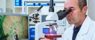
The main diagnostic method is to examine the patient's face for the presence of warts on the lips and oral mucosa.
To confirm or refute the presence of HPV, it is enough to take tests:
- ✓ Blood tests for antibodies
- ✓ To conduct a histological examination (biopsy), a small part of the tumor is taken for analysis
- ✓ The PCR technique allows you to detect the genetic material of a pathogen with one hundred percent probability
Before getting rid of the growth, you should visit an oncologist.
A specialist will use dermatoscopy to determine the condition of the wart to rule out signs of malignancy.
What is the best way to treat?
Warts can be removed in almost any way. The surgical method, electrocoagulation, laser and cryodestruction, as well as radio wave surgery are used.
Radio wave surgery
, in my opinion, is a more effective method that allows you to remove wart tissue under visual control. When using this method, the tissue is not burned, but is excised along with a small piece of healthy skin. Thus, the likelihood of warts reappearing becomes minimal.
See all photos before and after removal
Histological examination during removal is mandatory if there is the slightest doubt.
Clinical manifestations of warts on the lip
A wart on the lip does not appear the next day after the papilloma virus enters the body.
The incubation period can last from six months to several years.
This often occurs when the immune system malfunctions.
Having penetrated the skin or mucous membrane, the virus integrates its genetic material into human cells and thus begins to multiply.
As a result, characteristic neoplasms appear at the site of virus introduction.
The pathogen provokes warty growths, disrupting the processes of keratinization of the skin.
Oncogenic strains of papillomavirus change the genetic apparatus of cells in such a way that a malignant formation is formed.
What does it look like?
Warts are small, dry, flesh-colored lumps. They have a rough surface and are sometimes painful. On the hands, this can lead to a pronounced cosmetic defect.
When warts appear on the feet, severe pain appears, making it difficult to walk. Another type of wart, genital warts, develops on the genitals. This can make intimate relationships difficult. It is important to note that in women, condylomas of this localization can be caused by strains of the virus that increase the risk of cervical cancer.
Such situations require immediate treatment.
Is it necessary to remove a wart on the lip?
Removal of the tumor is carried out if there are indications for this.
The growths sometimes disappear spontaneously.
Therefore, if the wart is small and does not cause much concern, then there is no need for removal.
The decision to excise the growth must be made by a specialist.
If a wart causes discomfort in the form of itching, burning, pain, grows, becomes inflamed, or changes in appearance.
If it has increased in size, changed shape and color, or is bleeding, then it must be removed by resorting to excision by one of the methods.
Removing a tumor at home is not safe.
As a result of incorrect actions:
- It may not be possible to remove the wart completely, which will cause its reappearance
- the risk of injury to healthy tissues that surround the growth increases
- scars and cicatrices may form
Wart treatment
Treatment of any growth on the lips begins with a medical examination and determining whether the growth is malignant.
Next, the doctor selects a method of therapy.
In case of a malignant form, the patient is referred to an oncologist, who will decide how to treat the patient.
Treatment of the disease is complex.
Treatment consists of the main stages: removal of the growth and drug therapy.
Removal of the tumor can be done in one of the following ways:
- ✓ Using pharmaceutical products
- ✓ In a medical institution where modern excision techniques will be used
- ✓ Folk remedies. The use of alternative medicine methods must be agreed with a doctor
Routes of infection
Infection is possible not only through sexual intercourse, although this is the most common cause. HPV infection is also possible through household means:
- when using common hygiene and tableware;
- through contact with an infected person in the acute stage in the presence of microtraumas;
- the possibility of HPV transmission through household items is still being debated.
The transplacental route of infection is rare - from mother to child, even at the stage of intrauterine development of the fetus.
Pharmacy products for removing warts
You can buy medications at the pharmacy that will help you deal with warts at home.
Among them:
- Super clean . The product is an alkaline solution that has a chemical effect on the growth, causing cauterization.
- Cryopharma, Wartner . These agents cause cryodestruction of the wart; as a result of cold exposure, the neoplasm is frozen.
- Solcoderm . A drug containing acids that have a cauterizing and mummifying effect on pathological tissues.
- Immunomodulators – Viferon, Oxolinic ointment will help increase immunity and the body’s fight against the virus.
- Antiviral agents – Isoprinosine, Interferon.
The use of cauterizing agents on the lips and face is extremely undesirable; application of these agents to the oral mucosa is especially dangerous.
Wart removal in the clinic
The clinic uses modern hardware techniques to excise growths:
- Laser therapy . The method allows you to quickly and without blood remove the growth. There is no discomfort during the procedure, as pain relief is performed. During one session, you can get rid of several tumors in a very short time. The method is suitable not only for adults, but also for children. The likelihood of relapse is minimized. Skin defects in the form of scars after laser use are very rare.
- Radio wave therapy . The technique is considered the safest. When excising a wart using a radio wave, there is no contact with the skin. It is not always possible to completely remove the growth. The procedure leaves no traces of impact.
- Electrocoagulation . The procedure requires anesthesia. Using an electrode to which a high-frequency electric current is applied, the wart is burned out, followed by cutting off the growth with a special metal loop. Scarring may appear after removal.
- Cryodestruction . Liquid nitrogen is used and the wart is treated until it turns white. As a result of exposure to low temperatures, the growth tissues die. There is no need for anesthesia for the manipulation. The disadvantage of the technique is the lack of control over the depth of nitrogen exposure. For this reason, frequent relapses occur.
- Surgical. Removal is done using a scalpel. The disadvantage of the procedure is the formation of a scar, so this technique is used only for certain indications (in case of malignancy of the process and large size of the wart).
How to recognize warts: symptoms and signs
An inexperienced person may confuse warts with other skin growths, for example, moles, calluses, melanomas.
The main differences between warts and moles:
- moles have a dark or black tint, while warts have a light color;
- warts grow tightly together with the skin, moles are separate structures, as if glued to the body;
- moles are soft and smooth to the touch, warts are hard, hard and rough.
It is also easy to distinguish a wart from a callus. When pressing on the growth, painful sensations will occur, and if it peels off, traces of hemorrhages will be visible underneath it. Under the callus is new, tender skin.
You can distinguish a wart from a melanoma by color and shape. This dangerous disease is characterized by heterogeneous red and black shades, proliferation and an uneven contour.
It is not difficult for a dermatologist to make the correct diagnosis using a visual examination. But a good specialist will not be content with just a simple inspection. He will definitely use a special magnifying device - a dermatoscope. If there is a suspicion of a pathogenic process, scraping of the surface layer will be required.
In the case of anogenital warts (located around the anus and on the genitals), consultation with a gynecologist or proctologist is necessary.