Even the most experienced doctor is not able to make an accurate diagnosis by visual examination and from the patient’s words, so additional research methods come to the rescue. In our clinic, patients have the opportunity to undergo a number of examinations aimed at diagnosing pathologies, one of which is radiography.
The essence of this method is that X-rays pass through the tissue. Mineralized tissue blocks x-rays, so the tissue appears light in the image. Soft fabric allows rays to pass through and, accordingly, the fabric in the photo is dark.
When is a jaw x-ray taken?
Tell the patient exactly whether he needs and can have x-rays of his jaws
, should his attending physician. General indications for such a diagnostic procedure are:
- Suspicion of jaw development abnormalities (in childhood or adulthood). These may be congenital clefts of the hard/soft palate, impaired growth and proper development, or malocclusion.
- Suspicion of infectious-inflammatory and specific pathologies. These include osteomyelitis, periostitis, syphilis, actinomycosis, tuberculosis or necrosis of the jaw bone.
- To identify various acquired defects resulting from injury or removal of oncological formations.
- Suspicion of the presence of a false joint.
- Suspicion of a benign or malignant neoplasm.
What can you see in the photographs?
The following can be seen on x-rays of the upper and lower jaws:
- defects - cracks, fractures, presence or absence of bone fragments;
- pathological changes in bone tissue - areas of tissue thinning, compaction, thickening or thinning of the cortical plate;
- areas of necrosis - sequestra;
- sclerotic process;
- various periosteal layers, osteophytes (bone growths);
- tumor-like neoplasms.
What types of dental x-rays are there?
Depending on the recording method, it can be digital or analog. Analog devices use cassettes with X-ray film, while digital devices use matrices. The digital method is more accurate and informative, so it is gradually replacing the film method.
According to the coverage of the dentition, x-rays are:
- Biteful. Used to study the crown part of the tooth. You can diagnose caries and tartar.
- Sighting. The image covers from one to four teeth. A targeted x-ray visualizes the inside of the tooth and its roots, which is why it is prescribed for intracanal treatment.
- Panoramic. Covers all teeth, bone tissue and maxillary sinuses. In the resulting image you can see the condition of the entire dental system. Necessary for implantations, surgical procedures and prosthetics.
- 3D pictures. This is a three-dimensional image of the entire jaw, in which you can study the dentition in different projections.
There are several methods of carrying out:
- Standard. A film is applied to the problem tooth, a picture is taken with a radiation tube, after which it is developed.
- Radiovisiography. Prescribed for targeted dental x-rays. Instead of film, a matrix is used, from which the information is read into a computer, then the image is displayed on the monitor.
- Orthopantomography. Allows you to take panoramic photos. Can be film or digital. A curved object (jaw) is displayed unfolded in a plane. To take pictures, you need a special device - an orthopantomograph.
- CT scan. The tomograph takes a series of images in different projections. The program processes them and creates a three-dimensional model of the jaw. This is the most accurate and informative x-ray technique.
How is the procedure performed?
X-ray of the jaw of an adult or child is a simple procedure. It can be done in different projections in order to better examine the structure of the bone tissue and make an accurate diagnosis.
X-ray of the lower jaw
To obtain general information about the condition of the lower jaw, an X-ray is taken in a direct projection. It allows for primary diagnosis of inflammatory, oncological and traumatic diseases. The patient is positioned as follows: lying on his stomach, face down, resting the tip of his nose and forehead on the cassette. The X-ray machine sensor is installed at the occipital protuberance.
A lateral view of the lower jaw is taken to assess the condition of the body, ramus and teeth of the desired side. The patient lies on his side, with his cheek on the cassette located at a slight angle.
To diagnose diseases of the lower jaw, images are also taken in the axial projection. The patient takes a position lying on his stomach, stretching his head forward with his chin as much as possible. At the same time, it is pressed against the cassette by the front surface of the neck and lower jaw.
X-ray of the upper jaw
To assess the condition of the bone tissue of the upper jaw and the chin area of the lower jaw, a nasomental placement is performed. To do this, the patient lies on his stomach, face down, resting the tip of his nose and chin on the cassette. The sensor is installed perpendicular to the cassette, 2 pictures are taken - with the mouth open and closed.
Types of X-ray examinations
- Sight shot. The image captures one or more teeth and shows the condition of the periodontal tissues. Used to diagnose periodontal tissues and check the quality of root canal fillings.
- Panoramic photo (orthopantomogram). This image shows the condition of soft and hard tissues, the temporal and mandibular regions, and the maxillary sinus. Used to plan future treatment.
- Computed tomography of the maxillofacial region. This study is highly accurate. Makes it possible to identify hidden pathologies and tissue defects. A huge advantage is that this diagnosis shows the location of the mandibular nerve, which facilitates surgical intervention.
X-ray of the jaw is an easy-to-perform procedure that does not require preparation. In order to better examine the image, x-rays are taken in different projections. For an accurate x-ray of the upper jaw, a nasal-chin position is used. X-rays of the lower jaw are taken in lateral, direct and axial projections.
Panoramic shot of the jaw
A panoramic X-ray, or orthopantomogram, displays the entire upper and lower jaw in a direct projection. In such a picture you can see almost all the anatomical features of the patient’s dental system, all kinds of neoplasms, defects, fractures of tooth roots, and so on. An orthopantomogram allows you to identify the following pathologies:
- damage to tooth roots by caries;
- hidden carious cavities;
- cysts, granulomas, tumors;
- pathological changes in the roots and peri-root space.
X-ray of the lower and upper jaw of an adult: what does it show?
X-ray examination is a method for diagnosing the upper and lower jaws, which makes it possible to detect pathological foci, their shapes, sizes and depth of tissue damage.
An X-ray is taken in the following cases:
- Differentiation of diseases;
- For preventive purposes, which allows timely detection of the disease;
- Treatment quality checks;
- Determining the position of teeth in the dentition;
- Before orthopedic, orthodontic, therapeutic treatment;
- Tissue structure assessments;
- Before implantation;
- Exclusion of contraindications before treatment;
- If pathology in the sinuses is suspected.
X-rays in dentistry have virtually no contraindications. The only contraindication is the presence of bleeding.
Decoding the results
An x-ray of an adult's jaw is usually interpreted by a radiologist.
Other specialists may also be involved in this work: dentist, facial surgeon, otolaryngologist. Specific pathologies of the upper and lower jaws have certain radiological features:
- Chronic osteomyelitis. Depending on the type of disease, the images may show foci of resorption of various shapes with a shadow of necrotic masses inside. In more severe cases, communication of the sequestra with the oral cavity is visualized.
- Acute osteomyelitis. When examining the image, you can see areas of bone tissue resorption that do not have clear boundaries.
- Fractures or cracks in the jaw bones. This pathology reveals itself by the fact that thin, elongated shadows are quite clearly visible in the photographs. In such cases, it is important not only to identify the fracture, but also to see the bone fragments, if any, and to understand how much they are displaced.
- Chronic periostitis. In the image with such a pathology, periosteal thickenings will be visible. If the disease has become severe, areas of ossification of the periosteum and new bone tissue along the edge of the jaw are visualized.
What is a dental x-ray?
The concept of “dental x-ray” can be considered in two aspects. In a narrow sense, it is a planar image of one tooth or the entire jaw, displayed on film or paper. This definition was appropriate a century ago, when X-ray diagnostics were just beginning to be used in dentistry. Today, the concept of “dental x-ray” is more than just an image on film. In a broad sense, this is a set of diagnostic methods that use X-ray radiation. This includes both film and digital radiographs.
X-ray diagnostic devices consist of an emitter and a receiver. In the very first devices, the radiation source was a ray tube, and the receiver was a film onto which the image was projected. Now, along with film, sensitive matrices are used. They allow you to take digital photographs. Their sensitivity results in higher quality images and requires less intense radiation.
The principle of the method is based on the fact that rays passing through our body are weakened. The denser the tissue or organ, the more the X-ray beam will be scattered. Teeth and bones have the highest density. They absorb radiation better, which appears as white spots in the image. By deciphering them, your dentist will be able to make a diagnosis and prescribe treatment.
Is it possible to take dental x-rays during pregnancy?
The only contraindication for X-ray diagnostics is pregnancy. X-rays do not harm the mother's body, but can harm the fetus. Radiation is dangerous for cells that are in the process of dividing. In the body of an adult, these processes are not so pronounced, while in the fetus the formation of tissues and organs occurs. X-rays entering a dividing cell can damage parts of the DNA chain, as a result the new cell will be different from the old one, which can cause pathology. Therefore, women during pregnancy should avoid any X-ray diagnostics.
Advantages and disadvantages of x-rays
Dental X-ray has become a leading diagnostic method due to its advantages:
- Information content. X-rays allow you to study the internal structure of the entire dentition. The accuracy of the data obtained can reach tenths of a millimeter.
- Easy to carry out. X-ray does not require preparation from the patient. The procedure is prescribed after the initial examination, it is immediately carried out, a diagnosis is made and treatment is prescribed.
- Availability. There are x-ray rooms in almost every dental clinic.
- Efficiency of the procedure. The picture is taken instantly; CT and orthopantomography will take 20-30 seconds.
- It has virtually no contraindications. Dental X-rays are performed for both children and adults. Contraindicated only for pregnant women.
- Relatively low cost. The price of the service depends on the region, clinic and equipment it uses. Overall, this is a cheap diagnostic method.
The only disadvantage of dental x-rays is the certain dose of radiation that the patient receives. However, it is not that big. If you do not abuse x-rays, it will not have any effect on your body.
How many times can a dental x-ray be taken?
According to legal regulations, the annual radiation dose from X-ray diagnostics should not exceed 1000 μSv (microsievert). If you divide this figure by the dose received after each procedure, it turns out that in a year you can do:
- up to 500 targeted images with a radiovisiograph,
- up to 100 targeted shots per film,
- up to 80 panoramic digital images,
- up to 40 film orthopantomograms,
- up to 20 computer images.
How dangerous is a dental x-ray?
When X-ray diagnostics are performed, the patient receives a certain dose of radiation. It depends on the type of x-ray and the equipment used to take the picture. For a targeted film image it is up to 15 μSv, radiovisiography - up to 3 μSv; digital orthopantomography - up to 17 µSv, film - up to 30 µSv; computed tomography - up to 60 μSv.
We receive about 4000 μSv per year from natural background radiation, that is, every day we receive the same radiation dose as during visiography. When making a flight, we rise to a certain altitude, where the background radiation is higher. In one plane trip you can get up to 20 µSv, it’s like doing an orthopantomogram. If you are reading this text on an old cathode ray monitor, in a few hours it will emit the same amount of radiation as a radiovisiograph in an x-ray room during a targeted shot.
X-rays are safe for the patient if they are not exceeded. To prevent this from happening, always undergo x-ray diagnostics in one place. This will make it easier to calculate how much radiation you received in a year.
Indications
- Diagnosis of jaw injuries (fracture or dislocation)
- Diagnosis of congenital malformations or malocclusions
- Diagnosis of infectious bone lesions such as tuberculosis, syphilis, osteomyelitis or necrosis
- Detection of pseudarthrosis or arthritis of the joint
- Diagnosis of jaw bone tumors (benign and malignant).
- Finding out the causes of swelling and pain in the jaw area
- Periodontitis
- Cystic formations
- Dental pathology, including wisdom teeth
- Sinusitis
How are dental x-rays done?
The procedure does not require preparation, it is done quickly and painlessly. All manipulations take place in the X-ray room. The shooting process will vary slightly for different types of x-rays.
Bitewing x-rays and spotting x-rays are done in a similar way. A matrix or x-ray film wrapped in light-proof paper is applied to the problem tooth. A radiation tube is brought to the tooth and an image is taken. Information is transferred from the matrix to the computer. The photo can be printed or saved digitally. If film was used for x-rays, it is first developed in a dark room. This takes a little longer.
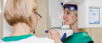
For panoramic and 3D images, special devices are used - orthopantomograph and tomograph. The procedure is done standing or sitting, it depends on the design of the device. You will have to remove metal jewelry, dentures, and piercings before the x-ray. You will be wearing a lead apron to protect you from x-rays.
Next, the patient's head is fixed in the desired position, and then filming begins. It lasts 20-30 seconds. The emitter and receiver will move around you, they will make noise. Don't pay attention to this, your task is to sit still. Any movement can affect the accuracy of the photo. At the end of the procedure, the film is developed, and the digital data is processed by a computer.