Today, only the laziest have not listened to magnetic resonance imaging. This diagnostic technique is considered the most informative and accurate for identifying pathologies and drawing up a correct medical report. Doctors of different specialties can refer you for examination if the patient has characteristic symptoms. What if you need to undergo an MRI, but you have metal implants in your body or crowns on your teeth?
Even today, there is a widespread belief that patients with such devices in the body should not undergo tomography. However, in reality this is not entirely true. A similar situation existed decades ago, when at that time patients were fitted with prostheses made of steel, cobalt or nickel. At that time, conducting electromagnetic research could harm the patient's health.
In 2022, this situation has changed dramatically. Today, individuals with endoprostheses, screws, fixation plates, pins, breast and dental implants are allowed to undergo MRI. It all depends on the composition of these items. In the article, the team will look into the intricacies of diagnostics for patients with implants. Go!
Operating principle of tomography equipment
First of all, you need to understand how exactly an MRI device works. The equipment is designed for targeted screening of soft tissues, bones and blood vessels. Advanced devices in diagnostic centers today have enormous technical capabilities. Thus, the power of the equipment is usually 1.5–3 Tesla, thanks to which doctors can examine anomalies whose dimensions are less than 0.2 mm.
MRI technology is based on the generation of radio waves and the ability to magnetize hydrogen elements present in different tissues of the body. The nuclei are able to respond to the influence of a magnetic field, that is, enter into resonance, and a computer program records all the data and converts it into detailed images. The research results are generated from many virtual sections of the selected department, for example, the brain, the vascular system, the abdominal cavity, and so on. Based on them, the radiologist draws up a medical report and a preliminary diagnosis.
How to prepare for an MRI procedure
- when planning a visit to the MRI room in advance, do not wear metal jewelry (hairpins, bobby pins, rings, earrings);
- For the procedure, if possible, wear clothes without metal decorations or buttons (otherwise you will have to take them off before the procedure);
- It is better for women not to wear a bra with metal underwires (in most studies it will get in the way and you will need to remove it before the test);
- if you are wearing a corset, then immediately before the MRI you will have to take it off in the locker room;
- You will not be allowed into the MRI room on a wheelchair or with crutches (with the exception of a wooden or special non-magnetic medical gurney for transporting bedridden patients);
- empty your pockets of change, key rings, lighters, keys and magnetic plastic cards (this can jump out of your pocket near the magnet and cause injury);
- You cannot take your phone with you into the MRI machine (unless, of course, you plan to buy a new one).
With which implants are MRIs allowed?
Tomographic examination can be performed for people with hip and knee joint replacement. An important rule for passing screening is the material from which the structure is made. It can be metal or ceramic, but their magnetic susceptibility should be minimal. Such material will avoid displacement of the structure or its overheating during diagnostics.
MRI is performed on patients with hernia meshes, dental prostheses, implants built into the chest and joints. All these elements are created from a material that does not in any way affect or interact with magnetic vibrations. All this allows us to make the tomography process safe for humans. It is important to consult with your doctor and diagnostician before scanning. Doctors will assess the likely risks and recommend precautions.
How to find out if you can have an MRI
Remember that an MRI can be done with the permission of a specialist. Only he will determine whether you need this research and whether it will harm you. Perhaps the doctor will make a diagnosis without magnetic resonance imaging. Spinal spondylosis and deforming osteoarthritis of stages II-IV can be detected using conventional radiography.
Comparison of visual diagnostic methods. MRI is on the right.
What are endoprostheses made of in the 21st century?
All plates with pins used in traumatology and orthopedic departments consist of various alloys. Different implants contain different amounts of paramagnetic and ferromagnetic materials. Its properties depend on the composition of the product. Not all prosthetics are created only from metal. Most are made of ceramic or polyethylene. The latter does not interact in any way with magnetic vibrations, which means it is 100% safe for MRI. However, there is one point. Ceramics usually contain aluminum oxide, but it does have some magnetic susceptibility. Therefore, it is necessary to notify the doctor about the presence of any prostheses in the body.
There are several possible combinations of materials in built-in structures:
- ceramics;
- metal;
- ceramics with polyethylene;
- metal together with polyethylene;
- ceramics with metal alloy.
Artificial compositions of joint additions include:
- titanium;
- zirconium;
- cobalt;
- chromium;
- tantalum.
Having studied the component parts of the implant, it will become clear exactly how it will respond to electromagnetic resonance.
The magnetic abilities of endoprostheses are determined not only by the material used to make the product, but also by its shape and dimensions. Pins and steels, the length of which reaches more than 20 cm, can heat up above the permissible values.
Examination with contrast
Contrast is used to improve visualization; in approximately 20% of all diagnostic cases it is necessary. Gadolinium-based drugs are used, which are safer than iodine-containing substances.
An intravenous injection is performed after routine imaging. For this, the patient is “rolled out” of the device and given a regular injection, after which the test is repeated.
Discuss with your doctor whether contrast is necessary, since in this case the cost of the examination doubles.
The procedure is indicated for suspected multiple sclerosis or for vascular examination. To obtain accurate information about myocardial viability or congenital pathologies, it is also advisable to perform an MRI of the heart with contrast.
Contraindications for MRI
When prostheses with plates are securely attached to the bones and do not move, then implants of a different location can easily move under the influence of a magnetic field. Therefore, under such circumstances, MRI is not performed - it is strictly prohibited. The main constructions for which screening cannot be done are:
- artificial heart valves;
- clips on vessels of different locations;
- ear implants;
- electronic pacemakers;
- artificial eye lens;
- Ilizarov device;
- insulin pump;
- impressive sized metal structures.
For consultation and clarification of all questions regarding diagnostics, call the hotline. A representative of the support center will help you choose a clinic for tomography, tell you about the restrictions, and will be able to book a place for an appointment with a doctor with a bonus of up to 1,000 rubles.
Possible complications and precautions
MRI in the presence of electronic implants can seriously harm a person or even lead to his death. Performing the study on persons with coronary walls and clips on cerebral vessels can provoke massive bleeding, which will lead to death. Endoprostheses made from some alloys may move out of place or heat up during an MRI, causing burns.
MRI installation before the procedure.
People with certain types of implants are strictly prohibited from undergoing magnetic resonance imaging. But for patients with implants made of “dangerous” alloys, you can still try to perform the study. As a precaution, a button is placed in the person's hand. If he feels a strong burning sensation, he presses it and the study is stopped.
Fact! Metal prostheses tend to “fade”, making the image of nearby tissues unclear. Therefore, it is pointless to try to obtain an MRI image of the replaced joint or bone held together by fonts or plates.
How does MRI affect implants?
Since the study is based on the use of magnetic vibrations, most patients have a question: “Is it possible to undergo tomography with dentures?” Innovations regularly occur in the dental field, prosthetic technology develops and new materials appear to create a beautiful smile. Previously, dental crowns were made only from copper or gold, but today patients have access to a large range of materials, for example, metal-ceramics or titanium. What's the difference?
Past methods that used copper and gold did not allow MRI patients to obtain detailed and clear images. The results were simply distorted, but there was no negative impact on health. Now dentists are massively using titanium alloys with admixtures of other particles, which has made dentures more durable and lighter. By nature, this substance is inert, so it does not:
- undergoes oxidation;
- generates toxic elements;
- responds to the influence of a magnetic field.
Because of this, people with titanium crowns can receive clear, detailed images after a CT scan. All this applies not only to dentures, but also to structures installed in bones and joints. Metal-ceramics during electromagnetic examination also does not affect the patient’s health or the screening results.
However, in addition to the built-in structure itself, the body contains pins, screws and plates with which it is secured. It is in them that a problem can arise. Implant fastener parts are made from various ferromagnets - iron alloys that have a certain magnetic susceptibility. The influence of resonance on them during an MRI scan can distort information and affect the patient’s well-being during diagnostic manipulation. All side effects can be avoided if you notify your doctor in advance about the presence of a prosthesis or other object in the body.
How is MRI performed?
During the procedure, you must lie on a special mobile table. The head is fixed, the limbs are secured with belts to better maintain immobility. The table rolls into the annular part of the tomograph, where the examination will take place. Monitoring sensors may be attached to the body, and sometimes a device is given to the patient's hand to squeeze to let doctors know that you feel unwell.
Open type tomographs are made in such a way that the magnet does not surround the entire body, and there is enough space around the device.
During the procedure, crackling and clicking sounds are heard - this indicates normal operation of the device. Patients are given earplugs or headphones to improve comfort and reduce noise. It is important to lie still. During some examinations, specialists may ask you to hold your breath.
During the diagnosis, the patient is alone in the office. Doctors monitor the condition through a special window.
In rare cases, you may feel a tingling sensation in your mouth if there are metal fillings, and patients also report a feeling of warmth during the procedure.
Myths about MRI
Our team has collected for you the top 3 common false theories about magnetic resonance scanning:
- During an MRI, the implants heat up and move. The specific gravity of the metal fastening elements of the structures is minimal, which means there are no defects in the examination results and no discomfort for the patient. But it is imperative to notify doctors about the material from which the product is made. Safe prostheses are considered to be those made of zirconium dioxide, ceramics or other expensive alloys. If you have a metal-ceramic structure, you need to clarify which metals and impurities were used in the creation.
- MRI cannot be performed during pregnancy. This is only half a lie. Due to the fact that in the first trimester the formation of tissues and organs in the unborn child begins, any effect on the fetus (chemical, physical or biological) must be excluded. In late pregnancy, tomography allows you to determine the position of the baby and determine the size of the mother’s pelvis. The results of the examination allow the obstetrician to prepare for childbirth without consequences for the child and the pregnant woman.
- MRI with prostheses is painful. Complete lie. Tomographic examination is one of the most painless and safe diagnostic techniques. MRI is allowed even with titanium inserts. Scanning can only be denied if the structure is implanted with steel, since it can heat up and move under the influence of magnetic vibrations. Only such a process can lead to injury to the subject.
If you have implants or not, but want to undergo an MRI, use. Our platform is designed to quickly search and select a clinic for diagnostics. The service presents dozens of medical centers, collects current prices for tomography and real reviews from visitors. To make an appointment, just call the hotline. When you reserve a place with a doctor using our resource, you will receive up to 1,000 rubles as a gift!
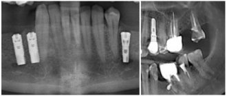
Is it possible to do an MRI with an endoprosthesis?
Endoprostheses used in orthopedics and traumatology are made from alloys that have different percentages of ferromagnetic materials, so different endoprostheses have different magnetic properties.
Since endoprostheses are very securely fixed in the bone, even the strongest magnetic field will not be able to move them or disrupt the integrity of the implants, such as vascular clips, which can be torn out of their place during an MRI. If the prosthesis is made of a ferromagnetic alloy, depending on its size and shape, it can only warm up in a magnetic field, so the doctor evaluates the possibility of scanning in this case individually.
MRI with a titanium prosthesis or one made of another paramagnetic is not prohibited, because titanium implants are not susceptible to the magnetic field of the device.
You can find out what alloy the prosthesis is made of in the product data sheet.
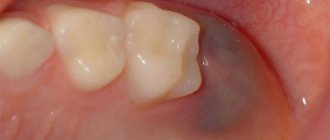
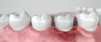
MRI and dental implants
Is it possible to do MRI with iron crowns? If the patient has an iron alloy crown installed, it is not recommended to perform an MRI of the head structures, because Under the influence of a powerful magnetic field, such a crown can heat up, causing the patient a burning sensation and pain.
If crowns or prostheses are made of metal-ceramics, an MRI of the head can be done, but in this case various artifacts may appear on the images, which reduces the reliability of the study.
The alloy material from which the crowns are made does not affect the possibility of performing MRI and the effectiveness of screening other parts of the body - the spine, internal organs, soft tissues and joints.
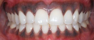
The answer to patients' questions about whether MRI can be done with braces similarly depends on the composition of the alloy and the size of the braces. Since such structures are firmly installed in the oral cavity, they cannot move under the influence of the equipment field, however, braces of about 15-20 cm in size can heat up and cause discomfort to the patient.
Most often, bracket systems do not affect the scanning result, even if the bracket is swallowed, an MRI can be done (we are talking about a small object).
In any case, if the patient has dental implants made of polymer alloys, iron and other materials, the possibility and feasibility of conducting an MRI of the brain, pituitary gland, orbits, its vessels, and soft tissues of the face is assessed by a radiologist.
Is it possible to do an MRI after stenting?
An MRI after stenting may be ordered by your attending physician. However, the radiologist must be informed that the patient has stents installed in the body and what material they are made of. Currently, alloys without ferromagnetic properties that do not respond to the influence of magnetic fields are most often used.
If the stent is bioabsorbable or made of stainless steel, cobalt and other inert metal alloys, MRI is not prohibited. However, you must adhere to the instructions for the stent, which may indicate that MRI cannot be performed during the first time after the stenting procedure. Moreover, this may apply not only to the area where the stent is installed, but also to MRI of other areas, since the magnetic field is equally strong throughout the room where the tomograph operates.