To the reference book Palpation of the larynx and neck is one of the diagnostic methods that are used in otolaryngology in the treatment of diseases of the ENT organs.
Author:
- Oganesyan Tigran Sergeevich
ENT pathology expert
3.75 (Votes: 4)
Palpation of the larynx and neck is a visual diagnosis of diseases of the pharynx and larynx, which allows you to assess the condition of the skin, the severity of the local vascular network, the shape and position of the organ.
Much becomes clear already during a conversation with the patient, for example, by the sound of his voice (the presence of nasality, rattling, shortness of breath and other characteristic symptoms), the complaints presented (their dynamics, concomitant diseases in the anamnesis). After examining and questioning the patient, the doctor proceeds to palpation of the larynx and neck. Depending on the indications, it can be superficial or deep.
Where does red throat come from?
A red throat is hyperemia of the pharyngeal mucosa - a symptom that accompanies various diseases of the pharynx in adults and children. Hyperemia can only be in the area of the arches, tonsils or posterior wall of the pharynx; all areas can also be involved at once, including the soft palate. The process can also be one-sided or two-sided.
But it is worth noting that the assessment of this symptom is sometimes not objective, since it is influenced by the color perception of both the patient and the doctor, and external lighting. Therefore, the patient should consult a doctor in order to carefully collect the entire history of the disease and identify the cause of the red throat.
Features of the structure of smooth muscles
The muscles of this group are found in almost all important internal organs, such as the intestines, stomach, uterus, and they are also present in the walls of blood vessels, skin and eyes. Smooth muscles perform involuntary movements, obeying only automatic signals from the nervous system.
Muscle cells have a spindle-shaped shape; they shorten due to the sliding of their filaments. The speed of this process is much slower than that of skeletal muscles, due to which they are able to remain in a tense state for a long time without expending a lot of energy.
An important feature of smooth muscles is their ability to maintain their shape, changed by stretching or deformation, as well as high plasticity, which is important for the functioning of internal organs. The muscles of this group are characterized by the slowest contraction and relaxation, which can last up to several tens of seconds. They can also remain in a state of tone for a long time, practically without getting tired. The main functions of smooth muscles: •
- maintaining pressure in hollow internal organs (ureter, intestines, uterus);
- contracting, they ensure natural peristalsis of organs and their emptying;
- regulate pressure in blood vessels;
- in the organs of vision they provide dilation and constriction of the pupil;
- located on the skin, they promote the secretion of subcutaneous fat.
Prolonged stretching of smooth muscles leads to their tension, which, in turn, plays an important role in promoting the contents of the gastrointestinal tract and ureters. The smooth muscles of the walls of blood vessels, contracting, affect the body's blood supply and blood pressure.
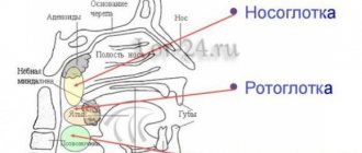
Anatomy of the pharynx
The pharynx is located behind the nasal and oral cavities; begins at the base of the skull and reaches the lower edge of the 6th cervical vertebra, where it narrows in a funnel-shaped manner and passes into the esophagus.
There are three sections of the pharynx:
- upper – nasopharynx
- middle – oropharynx
- lower – laryngopharynx
Nasopharynx
In the nasopharynx there is an accumulation of lymphoid tissue, which forms the pharyngeal tonsil - adenoids. Also in the nasopharynx are the tubal tonsils, which are located around the pharyngeal openings of the auditory tube.
This part of the pharynx is visualized only with the help of instruments or video endoscopic examination.
Oropharynx
The oropharynx is precisely that part of the pharynx that attracts attention when examined by an otolaryngologist and is accessible for examination by the patient himself.
It is located behind the oral cavity and is delimited by the pharynx. The pharynx is an opening formed above by the soft palate and uvula, on the sides by the anterior and posterior arches, and below by the root of the tongue. Between the anterior and posterior palatine arches there are another lymphoid accumulations - the palatine tonsils.
The palatine tonsils, arches, and uvula are subject to friction at the time of swallowing, due to which their mucous membrane may appear redder (hyperemic), in contrast to the entire oral cavity, gums, etc. But this is just a physiological factor.
The mucous membrane of the posterior pharyngeal wall contains elements of lymphoid tissue that visually resemble granules. The mucous membrane of the oropharynx has a dense network of vascular and lymphatic capillaries with rich sensory innervation, and therefore pathological processes are accompanied by symptoms - pain and discomfort in the throat, sensations of dryness, etc.1
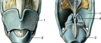
hypopharynx
The laryngopharynx begins at the level of the upper edge of the epiglottis and passes into the initial part of the esophagus.
This part is also visualized only using instrumental and video endoscopic methods. This area is visually inaccessible to the patient.
Diseases of the oral mucosa are accompanied by various pathomorphological changes. Inflammation is one of the most common pathological processes of the mucous membrane of the mouth and pharynx and is a manifestation of the body’s protective reaction to the influence of a pathogenic factor. The course and outcome of the inflammatory process depends on the reactivity of the body, localization, activity and duration of action of the pathogenic factor.2
Why surgery is needed
Benign formations of the laryngopharynx do not manifest themselves in any way at the initial stage and are more often discovered when the patient seeks treatment for other diseases of the ENT organs. However, as they grow, various discomforts arise in the throat, nasal sound, changes in voice timbre and even complete aphonia appear. Volumetric tumors provoke breathing difficulties and sometimes cause asphyxia.
Despite their benign nature, certain types of formations, for example, chondromas and extramedullary plasmacytomas, can transform into a malignant process. The treatment of such pathologies is surgical. The method of surgical intervention is selected depending on the clinical characteristics.
Causes of a red throat
| CAUSES | A COMMENT | ||
| causes: | Infectious causes | a comment: |
|
| causes: | Non-infectious causes | a comment: |
|
In 70% of cases, inflammation of the pharyngeal mucosa is caused by ARVI viruses (rhinoviruses, adenoviruses, coronaviruses, influenza and parainfluenza viruses), as well as other viruses (enterovirus, herpes virus, Epstein-Barr virus for infectious mononucleosis, and others).
The cause of inflammation can be bacteria:
- group A beta-hemolytic streptococcus (GABHS)
- Staphylococcus aureus
- Streptococcus pneumoniae
- Haemophilus influenza
It should be remembered that a red throat can be a manifestation of infectious diseases such as measles, scarlet fever, and rubella. In some cases, differential diagnosis with other infectious diseases is required.
Benefits of treatment at GMS Hospital
Removal of benign tumors of the pharynx and larynx at the surgery center of the GMS clinic is carried out on an outpatient basis, using endoscopic technologies. Endoscopically-assisted operations have the following advantages:
- minimal surgical trauma - healthy tissues are not injured;
- targeted accuracy - all modified tissues are removed along with the growth zone;
- minimal blood loss - simultaneous coagulation of blood vessels ensures the absence of bleeding both during and after the intervention;
- short recovery - the patient returns to normal life after 4-7 days;
- the ability to remove all tumors in one operation;
- high clinical result.
Since the operations are performed through the oropharyngeal approach, pain is minimal or completely absent. You can make an appointment with an otorhinolaryngologist at GMS Hospital by leaving a request on the website or by phone.
Diagnosis of red throat
To identify the cause of a red throat, the patient must consult an otorhinolaryngologist (ENT) or therapist.
If a child has a red throat, a pediatrician’s consultation is necessary.
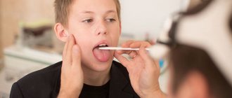
To identify lesions of the oropharynx, certain rules must be followed. First of all, the inspection requires sufficient lighting (fluorescent lamp, frontal reflector, etc.). The doctor carefully collects anamnesis to make the correct diagnosis.
An otorhinolaryngologist may prescribe a consultation with other specialists: a gastroenterologist, a dentist, and others.
Various laboratory tests are performed: bacteriological examination (culture from the back of the throat and tonsils and PCR), blood tests (complete blood count, ESR, C-reactive protein).
Cost of removal of benign tumors of the pharynx and larynx
The prices indicated in the price list may differ from the actual prices. Please check the current cost by calling +7 495 104 8605 (24 hours a day) or at the GMS Hospital clinic at the address: Moscow, st. Kalanchevskaya, 45.
| Name | Price |
| Biopsy of the oral cavity and oropharynx | RUB 10,767 |
| Opening of the epiglottis abscess | RUB 54,999 |
| Opening abscesses, phlegmon of the laryngopharynx | RUB 64,995 |
| Removal of a foreign body from the ear, nose, throat | RUB 8,075 |
| Removal of fibromas, papillomas, pharyngeal polyp (for 1 piece) | RUB 14,494 |
Dear Clients! Each case is individual and the final cost of your treatment can only be found out after an in-person visit to a GMS Hospital doctor. Prices for the most popular services are indicated with a 30% discount, which is valid when paying in cash or by credit card. You can be served under a VHI policy, pay separately for each visit, sign an agreement for an annual medical program, or make a deposit and receive services at a discount. On weekends and holidays, the clinic reserves the right to charge additional payments according to the current price list. Services are provided on the basis of a concluded contract.
Plastic cards MasterCard, VISA, Maestro, MIR are accepted for payment. Contactless payment with Apple Pay, Google Pay and Android Pay cards is also available.
Western standards of treatment (evidence-based medicine)
Continuous staff development
Regular interaction with leading Russian and foreign medical institutions
Modern medical equipment and advanced diagnostic and treatment methods
Unified standard of service
We work around the clock 24/7/365
Make an appointment We will be happy to answer any questions Coordinator Oksana
How is the operation performed?
All interventions are carried out via intrapharyngeal access using endoscopic equipment. Local anesthesia is predominantly used. The otolaryngologist carefully inserts an endoscope into the laryngopharynx and carefully examines all parts, assessing the size and location of the tumor. Then, the doctor identifies the tumor and removes it using micro-instruments, while coagulating the vessels.
- cysts, fibromas, neurogenic formations are removed by peeling them together with the capsule;
- for teratomas, the doctor first ligates the base of the tumor, stopping its blood supply, and then excises it, cauterizing the bed;
- local hemangioma, expanding into the lumen of the organ, is excised, followed by diathermocoagulation, laser or cryotherapy. For large hemangiomas growing deep into the tissue, sclerosis or occlusion of the vessels feeding it is used.
Simultaneously with the removal of altered tissues, coagulation of blood vessels is performed to prevent bleeding. Modern endoscopic stands used at the GMS Hospital surgery center allow us to examine in detail the relief of the laryngopharynx mucosa and perform targeted removal of the tumor along with the growth zone. The removed tissues are sent for histological analysis.
You have questions? We will be happy to answer any questions Coordinator Tatyana
Features of the rehabilitation period
Endoscopic removal of tumors of the pharynx and larynx is a minimally invasive operation with a short rehabilitation period. After the intervention, the patient is transferred to a day hospital ward, where he will remain under medical supervision for several hours. For two weeks, you need to limit physical activity and exclude thermal procedures (bath, hot bath, sauna).
You also need to adhere to a certain diet:
- exclude coarse, hard, spicy, sour and any other foods that irritate the mucous membrane;
- Avoid hot and cold drinks and food.
Most often, medications are not required after endoscopic surgery. But under some circumstances, an otolaryngologist may prescribe a course of antibiotic therapy, immunomodulatory and antiviral therapy. You can get more detailed information about the removal of benign tumors of the hypopharynx by making an appointment with an otolaryngologist, by phone or online.
Indications for surgery
A direct indication for surgical intervention is the presence of a benign formation in the pharynx or larynx. You definitely need to consult an otolaryngologist if the following symptoms occur:
- sensation of a foreign body in the throat;
- change in voice timbre (hoarseness, nasality, hoarseness);
- swallowing disorder;
- systematic sore throat;
- coughing not associated with inflammation of the ENT organs;
- admixtures of blood in saliva and sputum (with hemangiomas).
The decision on the operation and surgical technique is made by the otolaryngologist based on the examination and the results of a comprehensive examination.