- Questions and answers
- Stock
- Reviews
- Causes
- Types of cysts
- Symptoms
- Treatment
| Prices | Causes | Types of cysts | Symptoms | Treatment |
A dental cyst is a pathological neoplasm that occurs as a result of the body’s defensive reaction to infection or injury. A cyst occurs when pathogenic microorganisms that cause inflammation enter the jaw bone tissue through the root canal. Gradually, the cells involved in the disease process die, forming a cavity in the bone tissue. To prevent the spread of the process, our body forms a capsule with dense walls around the necrosis zone.
The resulting neoplasm contains inflammatory fluid, dead cells, decay products and bacteria. Sizes range from a few millimeters to several centimeters. Small ones (less than 5 mm in diameter) are called granulomas. Granuloma, cystogranuloma and cyst are stages of the inflammatory process in the bone tissue of the jaw. As long as it is small, there will be no manifestations. The number of microbes in it continues to grow, but since this happens under the control of the immune system, the process develops unnoticed. Immune cells are sent to the cyst through the blood vessels that feed the bone tissue. This is how the body tries to cope with the problem. But, if the immune system weakens, the disease enters the acute phase.
The development of the disease can lead to serious consequences. An increase in size threatens a fracture (especially of the lower) jaw, loss of teeth involved in the inflammatory process, and degeneration into a malignant tumor. Exacerbation of the disease leads to the development of periostitis, purulent abscess or even osteomyelitis. If at this stage the patient does not receive qualified medical care, sepsis may begin. This is an extremely serious condition, characterized by infection entering the blood and subsequently spreading throughout the body.
What is a cyst? Jaw cyst
A cyst is a cavity that is lined with epithelium and filled with fluid or soft material.
The formation of teeth (odontogenesis) is a complex process in which connective tissues and epithelium (enamel organ, dental follicle and dental papilla) participate.
The enamel organ refers to an epithelial structure derived from the oral ectoderm. The dental follicle and dental papilla are ectomesenchymal structures, because they are partly derived from neural crest cells.
For each tooth, odontogenesis begins with the apical (affecting the tip of the tooth root) proliferation of the epithelium of the oral mucosa, known as the dental lamina. The dental lamina gives rise to the enamel organ, a cap-shaped structure that subsequently takes on the shape of a bell. After the formation of the enamel organ, the dental lamina cord usually fragments and degenerates. However, small islands of dental lamina may remain after tooth formation. They are believed to be responsible for the development of some odontogenic cysts and tumors.
The enamel organ has four types of epithelium. The inner lining of the enamel organ is called the inner enamel epithelium and becomes the ameloblastic layer that forms tooth enamel. The second layer of cells adjacent to the inner enamel epithelium is the intermediate layer. Adjacent to this layer is the stellate reticulum, followed by the outer enamel epithelium. The enamel organ is surrounded by loose connective tissue known as the dental papilla. Contact with the epithelium of the enamel organ causes the dental papilla to produce odontoblasts, which form dentin. As odontoblasts lay down dentin, they induce ameloblasts to form enamel.
After initial crown formation, a thin layer of enamel organ epithelium, known as Hertwig's root sheath, grows at the apex of the tooth root. This epithelial expansion later becomes fragmented but leaves behind small nests of epithelial cells known as Malassez remnants in the space of the periodontal ligament. They are the source of epithelium for most periapical (radicular) cysts, but do not cause any odontogenic neoplasms, with the exception of squamous odontogenic tumor.
Causes of pathology
The appearance of a cyst at the root of a tooth means that infectious processes are occurring in the body (dental canals). Through the upper part of the root, the infection enters the periodontal tissue structures.
There may be several reasons for the development of such a disorder:
- active course of caries or pulpitis, which transforms into periodontitis;
- chronic form of periodontitis;
- painful and difficult eruption of the figure eight;
- a previous infectious disease, the causative agent of which reached the periodontal tissue through the bloodstream;
- a chronic form of inflammation occurring under an artificial tooth element;
- suffered injuries to the jaw bones (teeth), blows, falls;
- the course of a chronic form of sinusitis with purulent complications;
- poor-quality (or absent) root canal treatment.
Why do jaw cysts form?
Odontogenic cysts (developing or inflammatory) get their name from the nature of their origin. Most jaw cysts are lined by epithelium, which is derived from odontogenic epithelium.
The occurrence of such cysts is usually associated with unerupted teeth (third molars of the lower or upper jaw, second premolars of the lower jaw, and canines of the upper jaw). They can also form near additional teeth and in combination with odontomas. The occurrence of odontogenic cysts is rarely associated with baby teeth.
Most often, jaw cysts are discovered between the ages of 10 and 30 years. Men, especially white-skinned men, suffer from them more often.
In most cases, dental cysts do not manifest themselves in any way and are an accidental finding obtained during an X-ray examination. But, in some cases, they can reach significant sizes, which lead to the expansion of bone tissue, but even they may not give pronounced symptoms until a secondary infection occurs.
Types of cystic formations on the tooth
Dental cysts are divided into several classifications: in terms of the cause of their appearance, by the place of formation, by size.
Due to education
- radicular pathology (usually formed on the upper half of the tooth root, or, in more rare cases, on its side) - is a consequence of periodontitis;
- retromolar (paradental) cyst – is a consequence of severe eruption of the “figure eight” (with the inflammatory process accompanying its formation);
- residual neoplasm – formed after incomplete or incorrect tooth extraction, due to which parts of the root remained in the gum;
- keratocyst (primary) – formed due to improper growth and formation of the dental element, it consists of the remains of cells of tooth-forming tissues;
- follicular formation (tooth-containing) - is a consequence of the course of an infectious process that has affected immature rudiments or supernumerary dental elements.
By place of appearance
- in the area of wisdom teeth;
- on the front dental elements (purulent processes that occur at the roots of the “fours” can spread to the maxillary sinuses);
- at the base of artificial dental elements.
By size
- granuloma (cystogranuloma) – reaches 0.5 mm in diameter;
- dental cyst – exceeds a diameter of 0.5 mm.
How do jaw cysts appear?
In most cases, the cyst does not cause significant symptoms. Its development can be provoked by incorrect treatment of dental diseases or caries.
Odontogenic cysts are usually distinguished by type of origin:
· dentofacial cysts – their occurrence is associated with the crown of a tooth that could not erupt;
· keratocysts – are a consequence of Nevoid basal cell carcinoma syndrome;
radicular or radicular cysts – are of inflammatory origin and most often result from a reaction to necrosis of the dental pulp;
· bifurcation buccal cyst – typical for children 5-10 years old, it is formed in the area of buccal bifurcation of the first molars of the lower jaw;
· primary cyst – in most cases it is a keratocystic odontogenic tumor;
· orthokeratinized cyst – also refers to a subtype of keratocystic odontogenic tumor;
· eruption cyst – usually formed from a degenerating dental follicle and forms in the gum when the tooth erupts;
· newborn gum cyst – formed from the remains of the dental plate on the gum of a newborn;
· adult gingival cyst – is a variant of the lateral periodontal cyst;
· lateral periodontal – a non-inflammatory cyst on the side of the tooth, formed from the remains of the dental plate;
· calcifying cyst is a rather rare pathology, which is characterized by cystic and neoplastic signs;
· glandular cyst is a formation with a respiratory epithelial lining and potential relapse; in its manifestations it is similar to the central variant of poorly differentiated mucoepidermoid carcinoma.
Odontogenic cysts are difficult to detect at an early stage. It gives virtually no symptoms. The patient may be alarmed by tooth displacement or a change in the color of the diseased tooth. If the cyst has reached a significant size, the patient may notice protrusion of the bone structures.
A long asymptomatic course can lead to the formation of inflammatory processes, which are dangerous due to the development of suppuration and can provoke pathological fractures of the jaw bones.
If a cyst has formed in the upper jaw, it can cause nosebleeds, headaches and impaired nasal breathing.
The main symptoms associated with the presence of an odontogenic cyst (pain, fever, inflammatory changes in the oral cavity) usually appear in the later stages of the disease.
Content:
- Classification of pathology
- Causes of the disease
- Symptoms of the disease
- Diagnostic measures
- How to treat
- What will happen if left untreated?
- What can you do to avoid getting sick?
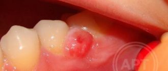
A jaw cyst is a hollow benign formation localized in the jaw bone.
There is liquid content inside it. Very often, the structure develops unnoticed by a person for a long time and is accidentally discovered during an X-ray diagnosis for another reason. If a cyst in the jaw becomes inflamed, the patient immediately feels it. Suppuration can provoke the occurrence of periostitis, sinusitis, osteomyelitis, and fistula.
Unfortunately, most often the formation has to be removed surgically. The doctor performs a resection of the apex of the tooth root and at the same time fills the cavity cleared of exudate with a special biological composite composition.
How are odontogenic cysts diagnosed?
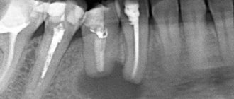
The leading method for identifying odontogenic cysts is radiography, which is capable of visualizing jaw cysts at an early stage of their development. On an x-ray image, the cyst is distinguished by the presence of clear boundaries, and the formation itself gives a characteristic shadow of a round or oval shape, immersed in the sinus of the tooth root.
Ultrasound examination also helps to recognize odontogenic cysts.
As already mentioned, pronounced symptoms are characteristic of the late stage of development of a pathological formation, therefore it is difficult to diagnose a cyst at the initial stage, relying only on symptoms.
The final diagnosis is made on the basis of histological examination. It is important to differentiate an odontogenic cyst from other pathologies (adenomatoid odontogenic tumor, ameloblastic fibroodontoma and calcifying epithelial odontogenic tumor).
The CT scan method is widely used in the diagnosis of jaw cysts to confirm the presence of calcifications along the wall of the cyst, as well as tiny spots that are usually not found on x-rays. In addition, computed tomography is necessary during the surgical planning stage.
Danger and consequences
Incorrect or absent treatment of a cystic tumor on a tooth can lead to serious complications, including:
- deformation or destruction of root tissue and tooth loss;
- inflammatory processes occurring in the area of the periosteum (otherwise, periostitis);
- the formation of osteomyelitis of the jaw bone tissue (developing cystic pathology provokes the occurrence of purulent-necrotic processes, which lead to the destruction of bone tissue);
- development of a chronic form of sinusitis;
- spontaneous deformation or fracture of the jaw (with a significant increase in the dental cyst);
- formation of phlegmon (acute purulent infection).
In the most advanced cases, death is possible, since the purulent contents of the cavity can enter the bloodstream and cause blood poisoning (sepsis).
What is the prognosis for the disease?
How successfully the situation in a patient with an odontogenic cyst will be resolved depends on at what stage the cyst was discovered, how severe the symptoms were and how it was treated.
As a rule, the use of surgical treatment gives a positive prognosis. Therapeutic treatment provides a positive prognosis only if it is started at the initial stage of the disease.
A negative prognosis may be associated with detection of the disease at a late stage: odontogenic cysts can provoke the development of serious pathologies that cause deformation of the maxillofacial tissues.
LifeTime Warranty
The Microscopy Department provides treatment under the LifeTime Warranty format.
We have eliminated possible errors at each stage:
- Endodontic treatment of dental cysts is carried out only using high-precision computer diagnostic
- All work is performed under multiple magnification of a microscope - diagnostics, removal of old fillings, direct treatment and filling of canals, placement of new restorations
- The Center uses of super-tight filling materials from the world “gold standard” line - both for filling canals and for restoring the shape of teeth
When a treatment case is closed, guarantee documents are drawn up confirming the quality of the work performed without complications or relapses. The patient is invited to free annual preventive examinations.
What treatment methods for jaw cysts exist?
The choice of treatment method for an odontogenic cyst directly depends on the symptoms it causes, as well as the results obtained during instrumental and laboratory diagnostics.
If surgical treatment is chosen (cystotomy or cystectomy), the maxillofacial surgeon performs complete removal of the cyst. In some cases, it is necessary to remove the cyst along with the affected parts of the tooth root. Treatment is carried out in a hospital setting.
If the choice falls on therapeutic treatment, the doctor will carry out procedures aimed at reducing inflammation. This is a long process, taking at least six months.
The first step is to drain the contents of the cyst using a special drainage tube, which is inserted into a small incision in the tumor. As the contents drain out and the tumor shrinks, the size of the tube is adjusted downward.
After removing the contents of the cyst, the dentist cleans the root canals and administers medications that destroy the tumor tissue. At the end of all procedures, the doctor uses a special solution aimed at accelerating healing.
Treatment is monitored radiographically.
Both after surgical and therapeutic treatment, the patient requires preventive measures that will help avoid the re-formation of an odontogenic cyst.
Price
The cost of treatment or removal of a dental cyst consists, like many other surgical interventions, of a number of factors. The complex of diagnostic procedures performed, types and methods of treatment, conservative preparation of canals, preoperative preparation, the operation itself, the osteoplastic materials used, postoperative observation - all this affects the final cost of treatment in these clinical situations.
A dental cyst is a serious pathology. In all cases, even in cases of complete absence of any symptoms, it poses a health hazard. With regular visits to the dentist, you can be sure: a dental cyst, if there is one, will be detected in time, and subsequent treatment will allow it to be eliminated without leaving a trace, preserving the health of the tooth.
According to antiplagiat.ru, the uniqueness of the text as of October 16, 2018 is 99.9%.
Key words, tags: X-rays, tooth extraction, orthopantomogram, periodontitis