Dental hyperplasia is an excessive increase in the structural components of dental tissues due to their excessive formation. The exact causes of such defects, including the most pronounced one, enamel hyperplasia , have not yet been established, although it is assumed that they lie in the area of impaired cell differentiation during the development of tooth germs and the growth of dental tissues. Provoking factors can be various effects on the reproduction and differentiation of dental tissues: metabolic disorders, violations of correlations in the process of growth and reproduction of tissue components, etc. Based on this, it is clear that for the identification and treatment of dental tissue hyperplasia, it is very important timely preventive examinations and sanitation of the oral cavity.
Dental hyperplasia can be hereditary (for example, focal enamel hyperplasia) or acquired (for example, hypercementosis - excessive formation of cement in the roots, observed against the background of certain pathologies). It is estimated that this defect of varying severity is observed on average in 1.5% of the population.
Dental hyperplasia in children: causes and treatment
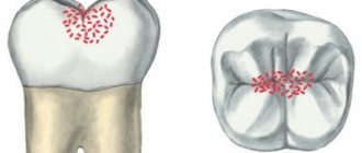
Dental hyperplasia externally looks like small growths on the enamel, formed due to excessive tissue formation.
In children, the defect is detected during examination by a dentist or by radiographic methods. Tubercles measuring 3-5 mm are located in the neck of the tooth, near the roots and are often covered by the gums. There is also localization on the crown. The causes of hyperplasia are not fully understood. Among the possible options for the formation of an enamel defect:
- improper development of the rudiments of mammary and permanent units;
- pathologies of dental tissue growth;
- metabolic disorders;
- diseases of the endocrine system;
- heredity.
The incidence of hyperplasia in children is less than 1%.
The defect does not bother the patient and therefore does not require special treatment. If there are “enamel drops” on the surface of the chewing teeth, the doctor will recommend fissure sealing to reduce the risk of caries.
If the localization of the anomaly is unsuccessful and the gums are injured, the pediatric dentist will carefully remove the tubercles with special burs, treat the enamel with remineralizing compounds, and return the dental crown to its anatomically correct shape.
Increased tooth sensitivity
In dental language - Hyperesthesia. If you feel a sharp pain lasting 5-20 seconds, then be sure that this diagnosis has reached your teeth. The disease manifests itself in the form of sensitivity of one or more teeth, or is generalized, affecting almost all teeth.) There are several types of hypersensitivity
- reaction to hot and cold.
- reaction to sugar, salt and acids.
- reaction to everything, including tactile stimulation.
Hyperesthesia almost always leads to many other dental diseases. Sensitivity between teeth is a symptom of the development of dental caries. There are many reasons for sensitivity, but the main ones are:
- Carious process
- Tooth wear
- Enamel chipping
- Wedge-shaped defect
- Hypoplasia
- Erosion of tooth enamel
Note that this disease is a direct consequence of loss of tooth integrity. Treatment of the disease depends on the causes of its occurrence (caries - filling; chipping of enamel - tooth restoration, etc.)
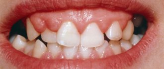
Hypoplasia of primary teeth: what is it and its causes
Hypoplasia is characterized by thinning of the enamel and a pronounced change in relief. On the crown of a baby or permanent tooth there are depressions, dots, pits, and grooves. Affected teeth differ from healthy ones already at the moment of eruption - the enamel is covered with white, yellowish or brown spots.
Hypoplasia of primary and permanent teeth is observed in 40% of patients. And the number of children with this pathology is growing steadily. Scientists associate the development of the anomaly with poor ecology, lack of a balanced diet, and hereditary factors. And all because the development of the defect occurs already at the stage of formation of the tooth germ.
A tooth—deciduous or permanent—is made up of pulp, dentin, enamel, and other tissues. Special cells, ameloblasts, are responsible for the proper development of enamel. In the absence of pathologies, one ameloblast forms one enamel prism. And it is these “prisms” that make up the outer tissue of the erupted tooth. As soon as the ameloblasts complete the task assigned to them, the destruction of the building cells occurs. After this, the tooth enamel of the erupted unit loses its ability to recover.
Ameloblasts are only the first stage in creating strong enamel of a child’s tooth. After eruption, it takes about a year for another stage of formation - final mineralization - to be completed. If all stages of development are successful, the child pleases his parents with strong, snow-white, healthy teeth.
Failure at any stage means potential enamel hypoplasia.
Causes of tooth enamel hypoplasia in children
There are three risk factors for the development of hypoplasia:
- features of fetal development during pregnancy;
- illnesses of the child himself;
- external circumstances.
The health of the expectant mother is the basis for the child’s excellent well-being. The risk of hypoplasia can be provoked by:
- Rh conflict - negative Rh factor of the mother and positive Rh factor of the child;
- severe pregnancy, toxicosis;
- acute viral infections or toxoplasmosis during pregnancy;
- taking tetracycline drugs during pregnancy;
- smoking, drinking alcohol.
Among the diseases of the child himself, the risk of defects in the formation of teeth increases:
- CNS lesions, Down syndrome;
- rickets, vitamin deficiency;
- digestive disorders;
- diseases of the endocrine system.
External factors include poor nutrition of the child and injuries to the jaws, which prevent the normal development of tooth germs.
Intrauterine factors provoke hypoplasia of primary teeth. The rest have an equally negative impact on both temporary and indigenous units.
Plaksina Margarita
My patients really like dental remineralization. Pastes rich in minerals have a pleasant taste and smell, and the procedure itself is painless and even pleasant.
Can you prevent tooth sensitivity on your own?
Of course, yes, if you keep your mouth clean, observing all oral hygiene standards, which will help stop gum loss and, as a result, periodontal disease. Thorough flossing and flossing as recommended by our dentist can help reduce the chance of avoiding this disease. A diet without consuming acidic foods also helps stop the development of this symptom. Ignoring the disease can lead to other health problems, leaving you vulnerable to tooth decay and gum disease.
Hypoplasia of primary teeth: treatment
Since one of the reasons for the abnormal development of enamel is low tissue mineralization, doctors have found an opportunity to help young patients. During treatment, two goals are set - to stop pathological changes, reduce or eliminate the severity of existing defects.
The following methods are used in pediatric dentistry in Moscow:
Enamel remineralization
The procedure allows you to replenish the deficiency of missing minerals and strengthen the child’s teeth. At the appointment, the doctor sequentially treats healthy and damaged teeth with a special gel. Treatment is continued at subsequent appointments or even at home. For home procedures, the doctor prescribes the child the optimal paste composition.
Fluoridation of teeth
Treatment of hypoplasia differs in the choice of drug. When fluoridating, the dentist uses a gel with a high fluoride content - up to 65-70%.
The method of restoring baby teeth with hypoplasia is chosen by the dentist after assessing the condition of the enamel and the indications.
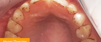
Dental fluorosis
Dental disease that occurs when drinking water and food with a high fluoride content, and in large cities and during the usual inhalation of combustion products (exhaust gases from cars, factories, etc.). It is one of the most common non-carious diseases affecting tooth enamel. One of the reasons for the appearance is the entry of fluoride elements into the blood and manifests itself in the form of stains on the tooth, of different sizes, shapes and colors. The disease usually destroys the incisors. When treating the disease, professional whitening is used, or restoration of the damaged part of the tooth with a composite material or an onlay (orthopedic design).
Hypoplasia of permanent teeth in children: treatment
Sometimes the process of hypoplasia cannot be stopped by applying mineral and fluorine-containing compounds. In such cases, pediatric dentists offer options for reconstructing tooth enamel or even the crowns of permanent teeth themselves.
Restoration is carried out using one of the following methods:
Filling children's teeth
Using grinding, dentists clean the affected area and then restore the surface using high-quality, reflective filling compounds. Installing fillings makes teeth straight, white and neat, and also reduces the rate of spread of the lesion and reduces the risk of complications.
Prosthetics
Installation of full crowns on affected permanent teeth or production of veneers. For children, prosthetic options are selected by the doctor according to the indications and the current clinical situation.
Plaksina Margarita
Unfortunately, children with hypoplasia are sometimes brought to the appointment too late. It is not always possible to save a tooth; it has to be removed. In the case of a primary malocclusion, the child is given an orthodontic ring to maintain space for the permanent unit. This is necessary so that neighboring teeth do not take up free space and spoil the bite.
Why does tooth sensitivity (dentin hypersensitivity) occur?
When the root of a tooth becomes exposed, it does not have a layer of enamel like the crowns of your teeth. The roots have a very vulnerable coating - cementum, which, after the loss of the top layer, leaves the dentin exposed. Excessive brushing with highly abrasive dental paste causes abrasion of the surface of the tooth enamel and exposes dentin. A very acidic diet with lots of acidic fruits and pickles can also cause tooth decay and dissolve the surface of the tooth, exposing dentin. It is important to visit your Doctor periodically if you have any symptoms of sensitivity so that he can examine your mouth and identify any symptoms associated with dentin sensitivity and suggest you the best treatment plan. When teeth react painfully, brushing them can be painful, and if you don't brush your teeth because of pain, it can lead to the destruction of enamel and gum disease. Reactions to heat, cold, sugar, sour and salty foods and drinks are also a clear sign of tooth decay, or a sign of a broken tooth, in which case your dentist will treat you comprehensively. Some of the most common causes of sensitivity are:
- Gum recession due to age or improper brushing of teeth
- Acidic drinks (such as soda), which cause enamel erosion and dentin attack
- Brushing with very low quality toothpaste, improper brushing, and brushing more than three times a day will result in loss of enamel.
- Gum disease that can lead to gum recession
- A broken or fractured tooth can expose dentin
Additionally, some dental procedures may cause sensitivity. Procedures such as teeth whitening, professional teeth cleaning, braces or fillings are known to cause sensitivity during or after the procedure.
Hypoplasia of dental enamel in children without treatment
Treatment of enamel hypoplasia in primary teeth is required, since in the absence of timely intervention, the root units will suffer. Among the likely consequences:
- development of caries, pulpitis;
- violation of permanent occlusion due to tooth displacement;
- increased abrasion of tooth enamel;
- painful reaction to cold, heat, sour, sweet;
- tendency to form fistulas;
- absence of rudiments of permanent units;
- loosening, destruction and loss of teeth.
Pathological abrasion of teeth
Characterized by severe loss of hard tissue on all teeth. Traditionally, three types of abrasion are distinguished: horizontal, vertical (extremely rare) and mixed. In this case, tissue loss occurs either in the horizontal plane, then the cutting edges of the teeth, cusps and chewing ones are erased. There are several subtypes of increased abrasion and they depend on the depth of the tissues involved in the process and the diminishing tissues. Often this disease is promoted by: traumatic occlusion, bruxism and frequent chewing of very hard foods. Minor abrasion of two or more teeth is a consequence of increased, systematic load on the teeth. If you have teeth worn down by time? If yes, then you definitely need two dentists, an orthopedist and an orthodontist, since it is impossible to get rid of tooth wear without a dentist. I would like to note that solving this disease is not the cheapest pleasure, which means installing braces at a young age will allow you to maintain healthy teeth and a very significant budget.
Types of gum hyperplasia
Periodontal tissues can grow in different ways, involving different areas of the gums. There are several types of hyperplasia:
- localized: the growth of new cells occurs in a limited area, next to one tooth or group of teeth (only on one or both sides);
- generalized: the gums grow over the entire surface on one or both jaws;
- papillary: new cells are formed only in the area of the gingival papilla, the pathology does not affect other gingival tissues;
- marginal: the gingival papilla and the area of the gingival margin grow;
- diffuse: growth of both marginal and attached gums occurs (an increase in periodontal tissue to the entire height from the gingival margin to the mucogingival border).
Hyperplasia may be limited. The increase in tissue in this case is similar to a tumor, strictly isolated, and has a wide base or pedicle. It looks like a growth or bump.
How do you know if you have gum disease?
Gum disease is most common in adults. If detected in the early stages, the problem can be corrected with minimal consequences, please contact your dentist if you notice any of the following symptoms:
- Gums that are red, swollen, or swollen or tender
- Gums that bleed when brushing or flossing
- Teeth that look longer because your gums have receded
- Gums that have separated or pulled away from your teeth, creating a pocket
- Changes in the way your teeth fit together when you cleave them together
- Pus between teeth and gums
- Constant bad breath
- How is gum disease treated?
When you detect the first symptoms of gum disease and proper oral hygiene, you will rid your teeth of plaque and avoid the development of the disease. Professional cleaning with a Doctor is the only way to remove plaque that has accumulated and turned into tartar. Your Doctor will clean your teeth and remove tartar from above and below the gum line.
How to diagnose and how to treat gum hyperplasia?
The diagnostic algorithm is focused on not confusing the disease with other pathologies. Therefore, the examination should be trusted only to a qualified dentist. The Nurimed Clinic offers a modern algorithm for confirming hypertrophy:
- examination of the oral cavity (allows the doctor to suspect a problem);
- X-ray examination to exclude similar pathologies;
- histological analysis of tissues taken for biopsy (a conclusion on the benign quality of the process and cellular composition is required).
After confirmation of the diagnosis, which is impossible without histology, treatment of gingival hyperplasia is carried out. Modern dentistry offers safe, painless surgical removal of overgrown gum tissue. The dentist peels off and removes excess tissue, after which the mucous membrane is sutured. This procedure is considered minimally invasive and does not require special recovery. In some cases, the dentist prescribes a number of medications to prevent complications.
An additional stage of treatment is sanitation of the oral cavity. Manipulation allows you to eliminate infectious processes that could cause hyperplasia. Also, professional cleansing can reduce the risk of inflammation and relapse of hypertrophy.
In the case of drug-induced fibromatosis, it is recommended to consult with your doctor and, if possible, change medications. To prevent re-growth, the cause of hypertrophy must be eliminated: the bite is corrected, tartar is removed, inflammation is treated, the balance of hormones is controlled, and so on.
Sensitivity due to gum disease, what is it?
Gum disease
is an inflammation of the gums that can progress to affect the bone that surrounds and supports your teeth, which also causes sensitivity, which we often confuse with tooth sensitivity. It is caused by bacteria in plaque, which always forms on tooth enamel. If plaque is not removed by daily brushing and flossing, thick plaque will form and bacteria will then infect the entire mouth. This can lead to weakening of the gums and, as a result, teeth falling out or being removed by the dentist.
Stages of gum disease:
- Gingivitis: This is the earliest stage of gum disease and is caused by the formation of plaque on the gums. If daily brushing is not carried out promptly or does not meet all hygiene standards, plaque produces toxins (poisons) that can irritate the gum tissue, causing gingivitis. You may notice some bleeding during dental hygiene. If gum problems are detected early, the damage may be localized because the bone and connective tissue that holds the teeth in place are not yet affected.
- Periodontitis: At this stage, the bone and fibers are irreversibly damaged. Your gums may begin to form a pocket under the gum that traps food and plaque. Proper dental treatment and improved home care usually help prevent the disease from developing. Advanced periodontitis: In this final stage of gum disease, the fibers and bone that support the teeth are destroyed, which can cause the teeth to shift or become loose. This can affect your bite, and if aggressive treatment cannot save them, your teeth may need to be removed.
What to do if you have sensitive teeth?
See your dentist first. The doctor will offer you all treatment options. It is also important to tell your Doctor if the cause is not dentin (root) hypersensitivity, the tooth may be reacting due to a more serious problem. To treat increased reactivity to stimuli, your Doctor will definitely recommend using a low abrasion dental paste designed for such clinical cases - desensitizing toothpaste. These toothpastes make your teeth less sensitive if you brush them twice a day, and also contain fluoride to protect your teeth from decay. In addition, your dentist may prescribe a fluoride gel, fluoride rinse, or a high-consistency fluoride toothpaste that is specially formulated to make your teeth less sensitive and provides additional protection against tooth decay. These procedures are carried out at home and are inexpensive. Other procedures for sensitive teeth that your dentist or hygienist may suggest in your dental office.
Manifestations of the disease
The following clinical symptoms allow you to recognize gum fibromatosis:
- In the initial stages, swelling or enlargement of tissue of a tumor nature.
- On the mucous membrane you can see thickenings that gradually increase.
- Increased gum volume without signs of swelling.
- Filling interdental spaces with soft tissue.
- Overlapping of the teeth with the edges of the gums.
- Injury to overgrown areas during cleaning (this can cause bleeding and inflammation).
- Increasing intensity of tissue color (the gums become bright pink).
If you notice any signs of fibromatosis, you should immediately contact your dentist. If you ignore the problem, unpleasant complications may develop:
- decreased quality of daily hygiene with an increased risk of developing infections;
- pain when eating;
- redistribution of chewing load;
- development of periodontal diseases.
Structure of tooth enamel
Tooth enamel is the main protector of teeth and completely covers their crown part. The average thickness of the tissue is about two millimeters: in the chewing area the layer is slightly thinner, and on the sides of the teeth, on the contrary, it is thicker. The color of tooth enamel depends on its density and the quality of dentin, as well as on some individual characteristics of the body, but this type of tissue is essentially transparent.
The composition of tooth enamel includes, for the most part, inorganic substances (more than 95%). The percentage of water content is very small (approximately 3%): this is partly why such high strength is achieved. Inorganic materials primarily refer to hydroxyapatite crystals (a mineral containing fluorine, magnesium, carbon and other elements). The presence of hydroxyapatite crystals also explains the fact that the enamel is vulnerable to the effects of an acidic environment, i.e. The consumption of foods that increase the acidity of saliva is contraindicated. This is especially true for calcium ions, on which the integrity and density of the enamel largely depends.
Types of disease
Doctors call two types of gum hyperplasia:
- Focal, which is practically not registered. It looks like single gingival growths observed in the area of the lingual gingival zone or in the area of the tubercle of the upper dentition. Moreover, single growths can cover all gums, or just one;
- Generalized gingival hyperplasia. It is registered quite often. It is visualized as granular growths, which eventually grow together into one, subsequently covering a significant area of the tooth surface.
Necrosis of hard dental tissues
Necrosis or death of hard tissue is a lesion of enamel and dentin, manifested by the formation of multiple defects on the surface of the teeth. The disease is non-carious in nature, develops after teething and is equally common in men and women. Necrosis causes a change in cell structure, which manifests itself in the destruction of hard tissues and pulp vessels. For successful treatment, it is important for the dentist to establish the cause that caused the necrosis, so the patient must undergo a comprehensive examination, including an endocrinologist and gastroenterologist.
Causes of necrosis of enamel and dentin:
- hormonal imbalance;
- digestive disorders;
- work in hazardous industries and contact with toxic substances;
- exposure, including radiological treatment
Symptoms of necrosis of hard dental tissues
At the early stage of this disease, the enamel loses its shine and becomes covered with chalky spots, which gradually acquire a dark brown color. The matter is not limited to a change in color - the hard tissues under the spots soften, destruction of enamel and dentin occurs, which ends in the destruction of the supragingival part. The process of necrosis involves several teeth at once, which become sensitive to external irritants. The foci of the disease are located, as a rule, in the cervical part.
Treatment of hard tissue necrosis
The pathology is non-carious in nature and its symptoms resemble erosion and a wedge-shaped defect. In the early stages, calcium-containing preparations are applied to strengthen hard tissues and reduce sensitivity. In case of severe damage, orthopedic treatment cannot be avoided, during which the affected tissue is removed and the tooth is covered with a crown. At the same time, disease therapy and the elimination of negative factors that led to the development of necrosis must be carried out.
Wedge-shaped (abfraction) defect
Non-carious lesion of teeth, in which a defect is formed on the neck of the tooth that looks like a wedge. In the early stages, a wedge-shaped defect is difficult to diagnose, since the tooth retains its structure and color, so dentists, as a rule, detect deep lesions accompanied by darkening of the tissue and pain felt by the patient while eating. Often, a wedge-shaped defect develops against the background of periodontal tissue diseases (periodontitis and gingivitis) and is usually diagnosed in older and middle-aged people, mainly on canines and premolars (small molars).
Causes of wedge-shaped defect:
- deterioration of tooth nutrition due to age-related problems with blood supply;
- metabolic disorders, including due to thyroid diseases;
- improper distribution of the load on the teeth, for example, chewing on one side;
- bruxism or teeth grinding;
- improper brushing technique;
- brushing your teeth immediately after eating acidic foods;
- increased stomach acidity;
- gum diseases, including gingivitis and periodontitis.
Symptoms of a wedge-shaped defect
The disease is non-carious in nature, that is, it is not associated with pathogenic microflora, and therefore develops rather slowly. The enamel in the gingival zone is mainly affected: in the early stages, the strongest tissue of the human body becomes thinner with the formation of a pit, but remains smooth and retains color. Gradually, the enamel is completely destroyed, dentin is exposed, and the teeth react sharply to sour and sweet, hot and cold foods, “aching” pain also appears when brushing your teeth.
Treatment of wedge-shaped defect
In case of significant damage, they resort to filling the defect cavity with a material that has a certain degree of elasticity. If the tooth is not strengthened in this way, its supragingival part may break off. Another effective way to protect a tooth with a wedge-shaped defect is to perform microprosthetics and install a veneer on it. In case of a small depth of the defect, remineralization (saturation with calcium and fluoride) of the enamel is carried out in a dental clinic. It is recommended to maintain the mineral composition of teeth yourself using toothpaste and mouth rinse selected by a specialist. In addition, it is important to master the correct technique for brushing your teeth and avoid using hard-bristled brushes, as well as toothpastes with abrasives, that injure enamel and gums.