Classification of fluorosis
The disease is divided into degrees of severity:
- The first is doubtful fluorosis. These are barely noticeable white spots on the enamel that become better visible when the surface dries.
- The second is very weak. Opaque white spots occupy up to 25% of the enamel surface.
- The third is weak. The lesion covers from 25 to 50%.
- The fourth is moderate. The surface of the tooth is completely damaged, abrasion and brown spots are noticeable on the enamel, and the relief is modified.
- Fifth – heavy defeat. There are significant brown areas and serious enamel destruction.
Manifestations of the disease
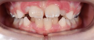
Symptoms depend on the form and severity of the disease. At the very beginning (at the stage of spotty or streaked form), areas with a changed color appear on the surface of the tooth. Stripes and lines are also visible. The enamel itself becomes rough, and small bulges appear.
Subsequently, small spots lead to the formation of larger pathological areas. Inhomogeneous mineralization of tooth enamel occurs. It breaks down and takes on a dull, matte color. At the same time, the smoothness and shine disappears.
A person with fluorosis complains:
- to increase sensitivity to temperature stimuli;
- cosmetic defect, as the color of the enamel becomes yellowish or light brown;
- fragility and rapid wear of teeth;
- frequent chipping;
- multiple caries.
X-rays reveal areas with reduced density, which indicates the process of demineralization, that is, a decrease in mineral substances in the enamel structure. It is for this reason that coloring substances are able to penetrate the porous structure of the tooth surface, which leads to the appearance of pigmented areas.
Forms
The streak form is characterized by barely noticeable stripes in the subsurface enamel layer. They are hardly noticeable and form on the vestibular surface of the incisors.
In the spotted form, chalky spots alternate with areas of healthy enamel. The incisors are most often affected. Sometimes the formation takes on a yellowish-brown tint. But the enamel always remains smooth and dense, with an intact structure.
The chalky mottled form has various symptoms. Distinct pigmented spots are visible on the enamel. The enamel may be yellow, with spots and dots, and other minor defects. It quickly wears away, exposing pigmented dark brown dentin.
The erosive form is accompanied by unaesthetic erosions on which there is no enamel. Destructive is characterized by a violation of the shape of the crowns of the teeth due to abrasion of hard tissues. They become brittle and break off easily, but due to the formation of replacement dentin, the tooth cavity is not opened.
Clinical researches
Clinical studies have proven that regular use of professional toothpaste ASEPTA REMINERALIZATION improved the condition of the enamel by 64% and reduced tooth sensitivity by 66% after just four weeks.
In addition, it has been proven that in order to eliminate increased sensitivity of teeth in people suffering from type 2 diabetes mellitus, long-term use of the domestic toothpaste “Asepta plus remineralization” is advisable, which is important to consider in the complex treatment of the dental pathology in question to ensure psychological balance, quality of life, restoration of chewing function and satisfaction with treatment of such patients.
Sources:
- Features of personal response in the prevention and treatment of hypersensitivity of teeth against the background of type 2 diabetes mellitus A.K. YORDANISHVILI, Doctor of Medical Sciences, Professor, North-Western State Medical University named after. I.I. Mechnikov, Military Medical Academy named after. CM. Kirova, International Academy of Sciences of Ecology, Human Safety and Nature, N.A. UDALTSOVA, Candidate of Medical Sciences, Associate Professor, St. Petersburg State University of the Government of the Russian Federation; dental clinic No. 29, Frunzensky district of St. Petersburg, O.V. PRYSYAZHNYUK, head. surgical department No. 2, dental clinic No. 29, Frunzensky district of St. Petersburg
- Report on the determination/confirmation of the preventive properties of personal oral hygiene products “ASEPTA PLUS” Remineralization doctor-researcher A.A. Leontyev, head Department of Preventive Dentistry, Doctor of Medical Sciences, Professor S.B. Ulitovsky First St. Petersburg State Medical University named after. acad. I.P. Pavlova, Department of Preventive Dentistry
- Report on determining/confirming the preventive properties of toothpaste “ASEPTA PLUS” GENTLE WHITENING” Author: doctor-researcher A.A. Leontyev, head Department of Preventive Dentistry, Doctor of Medical Sciences, Professor S.B. Ulitovsky First St. Petersburg State Medical University named after. acad. I.P. Pavlova, Department of Preventive Dentistry
Subtleties of differential diagnosis of fluorosis
It is very important to distinguish fluorosis from enamel hypoplasia, especially from its patchy form. Chalky spots in both diseases are located symmetrically, in areas of the crown that are not typical for caries. As a rule, these are the labial and lingual surfaces, cusps and cutting edge. With fluorosis, they have a pearly white tint, are shiny, do not cause pain during probing, and gradually turn into healthy enamel.
With hypoplasia, the spots are white and dense, also shiny, but with clear boundaries. Under the influence of UV rays, with fluorosis, chalky spots give a light blue glow (pigmented ones - red-brown), with hypoplasia - light yellow. Fluorosis is not prone to changes in spots, while formations with hypoplasia often require treatment for caries in children, since they change and progress.
Signs and symptoms of dental fluorosis
The disease can be of two types - endemic or occupational. Symptoms of endemic dental fluorosis are represented by damage to molars. Depending on the complexity of the disease, brown or white spots and erosions appear on the teeth, with increased abrasion and fragility of the enamel.
Signs of professional dental fluorosis are usually represented by decreased joint mobility, with pain in the joints and bones. Often, in severe forms of the disease, disturbances in the functioning of the autonomic nervous system, muscle weakness, and problems with the liver occur.
Treatment of fluorosis
The treatment regimen depends on the severity of the disease, general health and the influence of endemic factors. Therapy solves the following problems:
- reduce excessive intake of fluoride into children’s bodies from drinking water and food products;
- weaken the toxic effect of increased concentrations of fluoride on the body (the child is prescribed calcium preparations that bind fluoride and remove it).
Topical treatment for fluorosis depends on the severity. For any clinical picture, it is recommended to brush your teeth twice with toothpastes that contain calcium glycerophosphate, for example “Pearls” or “Calcium Complex”. At the first stage, no further action is required.
In the presence of pigmented spots, whitening is sometimes carried out followed by remineralization - only after 16 years under the supervision of a dentist. There are other methods of combating age spots, for example, removing pigmented enamel using microabrasion.
In erosive and destructive forms, when there is loss of hard tissue, the anatomical shape of the tooth can be restored with composite materials. Orthopedic treatment is sometimes indicated.
Diagnostic methods
Diagnosing dental fluorosis is not difficult. The doctor relies on the clinical picture characteristic of each degree of damage. Differential diagnosis includes:
- Mild opacity of the enamel is detected by drying the surface to assess the condition of the protective layer in the area with altered pigmentation.
- Vital staining method - in case of fluorosis, the affected areas of the enamel are not subject to staining with methylene blue, unlike a carious defect.
- Luminescence study - mild types of the disease glow weakly in ultraviolet light; in moderate and severe forms, partial quenching of fluorescence occurs.
- Microradiography is aimed at determining the depth of damage to dentin tissue.
Drinking water must be sent to the laboratory for analysis to determine the percentage of fluoride compounds.
Prevention of fluorosis
Prevention of fluorosis should be carried out in geographic areas where the fluoride content in drinking water exceeds 2 mg/l. Ideally, it is desirable to solve this problem collectively - to eliminate the etiological factor. If this is not possible, drinking water is defluoridated using a reagent or filtration method. Such water is also mixed with water from artesian wells or mountain rivers to reduce the concentration of fluoride. You can also remove fluoride from water at home by freezing, boiling and settling, and filtering through a layer of magnesium oxide.
Individual prevention involves the use of imported clean water for cooking. It is very important to limit the consumption of foods high in fluoride - strong tea, sea fish, seafood. In the winter-spring period, children from 2-3 years of age at risk are prescribed calcium supplements for a month, as well as ascorbic acid.
What causes dental fluorosis?
Dental fluorosis is caused by consuming too much fluoride during the formation of permanent teeth in children. This occurs before the age of 8 years. To avoid it, supervise tooth brushing so that children do not use too much toothpaste, mouthwash, and learn to spit out excess toothpaste rather than swallow it.
How do you know if your child has dental fluorosis?
Since there are many possible reasons for changes in the appearance of teeth, it is better to seek professional help. Your dentist can check your teeth for fluorosis or other problems. Regular visits to the dentist ensure complete control over the occurrence of this disorder.
How common is fluorosis?
Fluorosis first attracted the attention of scientists and doctors in the early twentieth century. Researchers were surprised by the high prevalence of what was called "Colorado Brown Stain" on the teeth of Colorado Springs natives. The stains were caused by high levels of fluoride in the local water supply. People with these spots also had an unusually high resistance to tooth decay. Fluoride is an important mineral for the normal development of children. Our mouths contain bacteria that combine with sugars in foods to produce acid that harms tooth enamel and damages the teeth themselves. Fluoride protects teeth, but increases the likelihood of fluorosis, which is why this disease is quite common these days.
How much fluoride should a child take to protect their teeth without risking fluorosis?
Children who use fluoridated dental products (toothpastes, rinses, etc.) will receive the fluoride they need for healthy teeth. There is no need to monitor your intake of water or food containing fluoride, since the body receives low doses of fluoride from these sources.
Causes and mechanisms of development
Fluorine is a microelement that enters the body with water and food and is involved in many physiological processes. It is found in high concentrations in human bone tissue and teeth. The incoming fluoride is not completely absorbed; the main share is fluoride dissolved in water. If a lack of a microelement increases the risk of dental caries, then an excess becomes the cause of the disease.
Fluorosis is most often observed in children during the eruption of permanent teeth. This applies to children, including those who previously lived in regions with high concentrations of fluoride in water. It is enough to live in such a region for up to 3-4 years. Excess fluoride also affects the buds of permanent teeth. In this case, milk teeth are not affected by the disease, since their rudiments are formed in utero, and excess fluoride is retained by the placenta and is not passed on to the baby.
An element concentration of 1.5 mg per liter of water will not lead to the development of fluorosis in adults. Drinking water containing 6 mg of fluoride per liter leads to illness.
Ask a Question


