Simple surgery
As noted above, the operation will not require an impressive amount of time. Dentiki dentists act in stages:
- First of all, the doctor examines the patient and inquires about the presence of allergic reactions to drugs, acute and chronic diseases. The specialist also clarifies whether the patient is taking medications during this period.
- Next, the doctor sends the client for an x-ray. Depending on the situation, a targeted or panoramic shot may be needed.
- The dentist begins active work with pain relief. After all, painless extraction is impossible without the use of anesthesia.
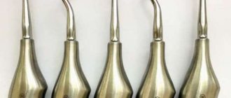
- Now the dentist begins to loosen the problem tooth. Carefully pushes back the gum with a special tool, grabs the crown with forceps or pries it up and gently rocks it. These actions allow you to widen the hole and break the periodontal ligaments.
- At the most important stage, the doctor removes the element of the dentition along with the root from the alveoli.
- At the end of the procedure, he will have to stop the bleeding. The damaged area is covered with a cotton swab moistened with an antibacterial compound. After 20-30 minutes it can be removed. This time is usually enough for the bleeding to stop and a blood clot to form.
When deciding which teeth can or cannot be removed (pulled out), you should know that if a pathological formation is observed at the root tip, another x-ray should be taken immediately after surgery. In cases where an x-ray confirms that part of the cyst or granuloma is still inside, it will be planned to remove it.
Dentics also practices non-traumatic methods using an ultrasonic scaler.
Complex extraction
Such operations include the removal of figure eights, elements with curved roots, a destroyed crown, or samples (incisors, canines, molars) previously subjected to resorcinol-formalin therapy. This method of treatment leads to the coloring of individual units in a pinkish color and the fragility of their walls.
The duration of the process can be 1 hour or more. Often the root and coronal zones are sawed to remove them from the bone tissue. In case of retention, the gums are first peeled off. Completely destroyed or broken fragments are initially “turned out” with a special elevator, since it is impossible to get them out with ordinary forceps without preparation, and excessively sharp actions can lead to additional damage to the oral cavity. At the end of the operation, sutures are required.
If a purulent process is detected, the neoplasm is opened and the outflow of the accumulation of pus is organized. Next, the drainage system is removed and secured to prevent suppuration.
Case 2. Perforation of the tooth canal.
Also often referred to are patients whose tooth canal was unfilled and treated by the so-called. “blindly”, without sufficiently enlarging the surgical area. In this case, the dentist’s instrument could come off the hard material of the canal and cause damage to the root canal - perforate it. In this case, two foci of inflammation occur:
- the first focus of inflammation occurs at the root apex due to an untreated root canal,
- the second focus of inflammation occurs in the area of canal perforation.
The doctor recommends tooth extraction, but in such a situation it is also possible to avoid extraction and save the tooth!
How to do it right:
Contact a clinic that uses a powerful dental microscope in medical practice. Is the tooth removed in this case? - No.
Using a dental microscope, the doctor can look into the root canal, see all the filling material, carefully remove it, unfill the root canal, treat the perforation area, reach both sites of inflammation, eliminate the inflammation and fill both the canal and the previously caused perforation.
Thus, we save the patient’s tooth without resorting to unnecessary removal.
Sick teeth: save or remove
First of all, I want to say that every doctor should understand that preserving the tooth itself is, for many reasons, better than removing it. Implantation is not a panacea. You can always get an implant – in any case and at any age, when there is no other option. But trying to save your tooth is an art!
Today, many doctors say that conservative treatment of large cysts is impossible. They are removed and a resection of the root tips is performed - that is, the cyst is removed along with part of the root. Then artificial bone material is placed there. To do this, you need to cut the gum. That is, the treatment of such pathologies in the “standard order” is surgical. The life expectancy of such a tooth is 5 years. After this, a relapse very often occurs, that is, the inflammatory process begins again.
I am a supporter of endodontics, in other words, I try to treat teeth where a cyst or granuloma has formed conservatively through a canal, without cutting the gum. And I, of course, have my own technique in these cases: in fact, it is enough to rinse the canal well and remove the infection with therapeutic drugs. In this case, of course, it is necessary to know anatomy, chemistry, and also microbiology - that is, to understand what microflora and what to kill with, because the success of treatment depends on this. For the doctor, in theory, it is no secret that in the case of cysts and granulomas, the causative agent of infection is gram-positive and gram-negative cocci. And believe me, if the doctor understands these things, then there will be nothing magical in endodontic treatment. True, such treatment takes from 1 month to six months, depending on the size of the cyst. Today there is an opinion that such teeth can be treated in one visit, without prior calcium treatment. Based on my own experience, I believe that it is difficult to achieve positive dynamics in this case, we’ve been through it - we know! After all, it also happens: the tooth seems to be improving, and then suddenly there is a sharp relapse. Therefore, once a month the patient visits the clinic, I rinse the tooth, apply medicine, perform an X-ray control and... put a temporary filling. And so on until a positive result is confirmed by the results of a computed tomography scan. After six months, a crown can be placed on the tooth. In difficult cases, we install a temporary crown for six months, after which we perform another computed tomography scan and after that we cover the tooth with a permanent crown. After this treatment, I have no relapses in 100% of cases.
Or here’s another example: a fracture of the crown of a tooth below the level of the gum (gingival attachment). You can, of course, remove the tooth, as is usually the case. But I prefer to clinically lengthen the neck of the tooth due to additional tissue located in this area. How? I'll try to explain. The gum has a movable layer that covers the neck of the tooth, and there is also a layer partially attached to the bone tissue. Using a laser (that is, surgically), I remove the gum around the tooth, thus lengthening its neck and providing space for the installation of an orthopedic structure designed to protect the tooth. I know that for many this sounds like something unreal. Fortunately, I can afford to save teeth with a crown fracture even below the gum attachment, but only if this fracture does not reach the level of the interradicular septum. If the interroot septum is affected, then infection will constantly get there. As a result, the tooth will still have to be removed.
Yes, such treatment is not for one day. After all, it takes 2-3 weeks just to form a new gum. In general, the entire process can take from 1 to 2 months - in complex cases (more often when the painters break, that is, large molars in the area of the palatal or lingual wall), in simple cases - 2-3 weeks. But during all this time the patient will need to come to me 2 times. First time I lengthen my neck. The second is when I restore the wall of the tooth, so that the gums grow correctly: this will provide enough space where you can then put a stump inlay, and put a crown on top. But this is the work of an orthopedist...
With root fractures, a lot depends on how badly the root is damaged. Root fractures in single-rooted teeth are not dangerous, especially if the root is broken, for example, 4 millimeters vertically. In these cases, we combine surgical and orthodontic treatment approaches. And then we restore the tooth using orthopedics. But if the crack is deep, then there is no point in saving such a tooth, because complications may arise later.
Perforations of teeth can be closed in any part of the roots, provided that the tooth is sterile (not periodontitis): with a competent approach, the prognosis of such a tooth will be 100% successful. But if the perforation is located in the area of the interradicular septum, a large cyst has formed there, then this perforation can, of course, be closed, but the success of treating such a tooth will be 50 to 50. It is important to seal the periodontium hermetically so that infection does not get there. I close the perforations with a special cement that was created for this purpose and is also used to close wide apical foramina. It seals the perforations hermetically, hardens quickly, and behaves well in a humid environment. However, I would like to make a reservation right away: working with perforations is only possible under a microscope - after all, I need to see how I close the hole! This is the job of an endodontist. Perforations are treated in one visit, meaning you don’t have to wait several weeks or even months for results. But if the bottom of the cavity is severely broken by a bur, when the tooth is not only perforated, but also the area of the interradicular septum is open and infection constantly gets there, then closing such perforations may be impractical.
When removing an instrument from a tooth root, it is also necessary to use a microscope. Although often, for example, when an instrument is broken in a bend in a canal, even a microscope is powerless. What can I say here? We use... intuition and think through every step well. Sometimes it is necessary to use instruments that were not originally designed for work in the canals, or even several instruments, creating “in-house” designs that ultimately allow the removal of the recalcitrant fragment. In general, everything “that comes to hand” is used - in the good sense of this expression. Why not if it can help? When doing this work, it is very important not to overexpand the channel. In principle, I work without expanding the tooth canal - I work only with the tool! The channel remains intact on all sides. There is also the so-called by-pass technique - its essence is that we “bypass” the instrument broken in the canal, leaving it in the tooth canal system. If there is full access to the canal and the instrument can be bypassed by properly rinsing the canal with aggressive solutions, cleaning it and passing it with “cement,” then the foreign body no longer causes any harm to the tissues, since it ceases to be a source of additional infection. And this is also considered a positive result of treatment! To be honest, many doctors look and do not understand how I manage to remove instruments from the apical foramina - for them it is a mystery, shrouded in darkness =).
Case 3. An instrument broke in the canal
We often come across cases when patients come to us who, during treatment in other clinics, have had a broken instrument in the root canal.
But the patient has no idea about this, since the attending physician himself did not notice it. When treating inflammation, in the absence of a microscope at hand, this remaining instrument interferes with the full treatment of the root canal. And the doctor, without seeing this instrument, cannot remove it, making full root canal treatment possible. Therefore, the patient is recommended to have the tooth removed. But is it necessary to remove the tooth in this case? - No.
How to do it right:
Contact a clinic that has a dental microscope in its arsenal.
Under high magnification, the doctor will be able to see the problematic root canal and see the remaining fragment of the instrument. Then carefully remove it, treat the root canal and eliminate inflammation. This allows the patient to save the tooth without having to remove it. Effective treatment of the above situations has become possible thanks to the use of dental microscopes in dental practice. Don’t skimp on your dental health; go to clinics that are equipped with microscopic treatment technologies.
Caring for your oral cavity after surgery
The speed of wound healing depends on how you follow the rules of hygiene. Also, proper care allows you to avoid infection of the hole and aggravation of problems.
- Do not eat food for the next 4 hours after the procedure. The first time, try to eat something soft: cream soup or puree. Under no circumstances should you eat hot food. The same goes for drinks.
- Don't touch the hole. Neither with language, nor with any objects. For effective healing, it is very important to maintain the integrity of the blood crust that has formed on the wound.
- You won't be able to brush your teeth for a few days. To keep your mouth clean, rinse your mouth with a baking soda solution (half a teaspoon per glass of water). A decoction of chamomile, calendula or oak bark also disinfects and promotes healing.
- During the first weeks, brush your teeth only with a soft or ultra-soft toothbrush so as not to irritate your gums. Use the toothpaste recommended by your doctor - it will promote healing.
- For two to three days after surgery, try to refrain from smoking, drinking alcohol, and using the sauna. All this can negatively affect the condition of the crust while it has not yet hardened.
Full recovery will occur in 2-3 weeks, then you can return to your normal rhythm of life. In complex surgeries where you have to wait for bone tissue to grow, recovery may take up to 14 weeks . But still, after the first three, you will be able to eat normally, and your gums will not be sensitive to temperature.
Case 4. The source of inflammation at the root of the tooth - the so-called. cyst
Imagine the situation.
A tooth that previously did not have a filling or was previously treated endodontically begins to hurt. An examination is carried out, X-ray control is done, but often a more informative cone-beam computed tomography CBCT or, as many say, a 3D x-ray is performed. A dental cyst is detected. The doctor recommends removing the tooth because there is a large area of inflammation at the top of the tooth root, caused by an infection in the root canal. The infection gets beyond the root tip and causes inflammation. This inflammation can be treated by removing the infection from the root canal. It is easier for a doctor who does not have the knowledge and treatment methods to remove a tooth.
How to do it right:
There is no need to remove the tooth. In the case of a dental cyst, it is necessary to carry out diagnostics, examination and high-quality treatment under the control of an operating microscope with isolation of the working field, followed by hermetically sealed restoration of the tooth at the stage of endodontic treatment. The recommendation is simple:
before removing a tooth, consult a professional. You may not need tooth extraction! This means that sinus lifting, implantation and prosthetics procedures following the removal will not be necessary.
Pulling out milk elements
The first incisors and canines fall out on their own with age. This happens during the period of change in the temporary bite. However, there are often cases when it is necessary to resort to extraction before the onset of natural loss. The need for it is determined by a dental specialist. Parents just have to listen to his recommendations and not put off solving the problem until “later.”
It is a big mistake to consider that various pathologies of milk samples are insignificant and that it is not necessary to take action to eliminate them. Yes, teeth will certainly fall out on their own over time, but pathological deviations may well be transferred to the developing permanent elements. As a result, they will erupt already sick.
Premature tearing is resorted to when inflammation occurs, which is provoked by an advanced form of caries, pulpitis or periodontitis. In all these cases, it is no longer possible to restore the crown. If there is more than 1 year left before the bite changes, the installation of dentures is necessary. Otherwise, the series will inevitably become distorted, which is fraught with consequences.
The most difficult thing in working with children is dealing with fragile dental walls. The child should not be in pain during the procedure. It is very easy to crumble a thin coronal surface with large forceps, so instead they use a special smaller and lighter version.
Contraindications to wisdom tooth removal
Despite all of the above, there are situations when the mouth cannot do without the “eight” or the operation cannot be performed for other reasons:
- you need to install a bridge supported by the third molar;
- adjacent molars become loose, and a wisdom tooth is used as a base for applying a splint;
- due to the absence of chewing molars, the chewing function falls on it;
- period of pregnancy and breastfeeding;
- cardiovascular pathologies;
- impaired blood circulation;
- allergy to anesthetic;
- hypertensive crisis.
Delete or Treat?
There are two approaches to the treatment of periodontitis:
- Remove the problematic tooth and place a crown on an implant in its place. Such treatment, according to doctors, is faster and easier, and the result is more predictable.
- Doctors who recommend fighting for a tooth insist that your own tooth is always better, and it’s never too late to get an implant. They suggest using “every option” to save the tooth.
However, dentists do not always clearly explain that treating a “problem tooth” will require significant time and financial costs, but may not lead to the desired results. If efforts to save the tooth turn out to be in vain, then the cost of “prosthetics on an implant” will have to be added to the cost of “treatment”, since the absence of even one tooth in the jaw affects the position of neighboring teeth.
The choice of “treat” or “remove” is not very simple, since it depends on many factors. Let's figure out what guides a dentist when offering his treatment plan.
Is it possible to restore a damaged tooth?
Yes, you can. But everything will depend on the volume of the lost coronal part and the general condition of the oral cavity.
For restoration, modern dentists use the following two main methods:
- installation of pins (reinforcement) made of fiberglass or titanium;
- introduction of so-called stump inlays.
If more than 50% of the unit is destroyed, and the dentist has doubts that the crown restored with a stump or pin will not withstand the chewing load, a metal-ceramic crown is installed on the stump pin inlay. When restoring teeth, it is mandatory to clean and fill the canals to avoid the development of caries.
Stages of complex wisdom tooth removal
The methodology may differ for each specific case, but in general terms several approximate stages can be outlined:
- 1. local anesthesia;
- 2. incision of soft tissues, peeling them off the bone;
- 3. sawing, drilling out the proper bone tissue;
- 4. extracting the “eight”;
- 5. treatment of a fresh hole left after the removal of a diseased wisdom tooth;
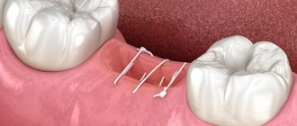
- 6. suturing with non-absorbable suture material;
- 7. The doctor will remove the stitches only after the edges of the wound have completely fused.
The duration of the procedure depends on the situation and can last from 30 minutes to 2 hours. After the operation, the doctor advises the patient regarding wound care, prescribes medications and informs the date of the next appointment.
How to restore a damaged tooth?
Treatment of a damaged tooth involves the following steps:
- examination by a dentist and assessment of further “future” damaged teeth;
- restoration of the stump area using a pin or inlay. The dentist treats the canals, strengthens the unit from the inside, thereby providing reliable support for the filling;
- The crown is built up using a composite material. If the work is done efficiently, the tooth can be visually almost indistinguishable from a natural one. The dentist repeats the shape, color and even transparency.
Reason 1: the bleeding does not stop
Heavy bleeding is the most common complication. Usually occurs in people with bleeding disorders. Before removing a tooth, no tests are required; it is for this reason that the likelihood of this particular complication is high. If you know about your problem, be sure to tell your doctor. In this case, the specialist may prescribe medications to stop the bleeding or apply sutures. If the blood does not stop or suddenly “gushes out like a fountain” shortly after the operation, immediately go to the hospital or call an ambulance.
