Diagnosing and solving problems that lie on the border of two or three medical specialties requires the most time and effort. One of these pathologies is odontogenic sinusitis, which is treated by an otolaryngologist and dentist.
Odontogenic (from the Greek odus, genus odontos - tooth, genesis - origin, development) maxillary sinusitis or sinusitis is a chronic inflammation of the mucous membrane of the maxillary sinus, provoked by pathology of the teeth of the upper jaw, namely periodontitis, tooth extraction with the formation of communication between the oral cavity and sinus, or the entry of plastic material into the sinus when filling the canals of the teeth. The share of this disease in the structure of all maxillary sinusitis ranges from 10 to 40%, and up to 75% of all unilateral sinusitis.
Reasons for the development of pathology
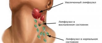
The formation of an acute purulent process in the subcutaneous fat layer near the lymph nodes can be triggered by a significant decrease in immunity. If there is inflammation of the lymph node itself, then a delay in lymph outflow and increased vascular permeability are observed.
Adenophlegmons develop due to ongoing inflammatory processes of any localization. Often this pathology is diagnosed against the background of malignant tumors, some types of dermatitis, chronic dental pathologies, and inflammation of the tonsils.
Diagnostics is aimed not only at identifying adenophlegmon, but also at establishing the cause of its occurrence - medical tactics for this pathology depend on this.
How does maxillary sinusitis manifest?
When visiting an ENT doctor, patients usually complain of heaviness, sometimes pain or swelling of the soft tissues in the area of one maxillary sinus, prolonged runny nose, hyposmia (decreased sense of smell) or, conversely, a constant unpleasant odor in the nose. Often such patients report frequent and painful sinusitis, multiple punctures, numerous courses of antibiotics, which brought only a temporary improvement in their condition or did not help at all. And only with a detailed collection of the medical history is it possible to find out about the recent or long-standing treatment of the “causal” teeth by the dentist, namely “7s”, “6s” or “8s” of the upper jaw of the corresponding side. But it happens the other way around: the patient does not make any complaints, but when preparing for dental prosthetics or sinus lifting, the dentist identifies pathology of the maxillary sinus on a computer tomogram and refers it to an ENT specialist. The reason for such careful preparation and alertness of our colleagues is the golden rule of planned surgery - the absence of inflammatory changes in the surgical area, because otherwise there is a high risk of implant rejection, scarring and a generally worse outcome of the operation.
Symptoms of adenophlegmon of different types
Symptoms depend on where adenophlegmon developed:
- With submandibular adenophlegmon, body temperature rises, swallowing may be difficult, problems with speech arise, swelling forms in the affected area, redness of the skin and pain when palpated are observed.
- With adenophlegmon of the neck, symptoms appear only when there is a critical accumulation of coccal microflora in the tissues of the anterior or lateral part of the neck. There is an increase in body temperature, pain when feeling the neck, redness of the skin, but swelling may be absent.
- With inguinal adenophlegmon, the clinic develops against the background of an inflammatory process in the genitourinary system. This type of adenophlegmon is characterized by rapid progression, while the patient experiences severe pain, due to which he cannot move freely. There are also signs of general intoxication of the body.
- With adenophlegmon of the axillary region, symptoms more often occur with open wounds of the upper extremities. Signs are pronounced: enlargement of regional lymph nodes, deterioration of general condition (including increased body temperature).
The symptoms for different types of pathology in question are similar, the differences lie only in the location of the lesion. If adenophlegmon occurs in children, then loss of appetite, increased drowsiness, lethargy, and lack of interest in games and others will be noted.
Symptoms of oral phlegmon
- General intoxication, weakness, high temperature, fever, chills;
- The tongue and lower jaw swell and become inactive;
- The tongue becomes so swollen that it may fall out of the mouth;
- Edema and swelling lead to distortion of facial shape;
- Pain at the base of the tongue, neck, painful sensations when chewing and swallowing;
- Gray-brown coating on the tongue;
- Unpleasant putrid odor from the mouth;
- Profuse salivation.
Cellulitis of the jaw develops very quickly, spreading from the affected sublingual space to neighboring areas, the floor of the mouth, and gums. With phlegmon of the lower jaw, inflammation can affect the postmaxillary and peripharyngeal space, chin, and neck.
With dental phlegmon, the symptoms are local in nature and appear against the background of untreated caries. The disease goes through several stages:
- Inflammation of dental tissue, pulpitis and periodontitis;
- Inflammation of the periosteum, swelling of the gums;
- Formation of a purulent sac on the gum.
The purulent formation grows, causes intoxication, chills, and is accompanied by bad breath. If dental phlegmon is not diagnosed in time and treatment is not started, there is a risk that the disease will spread to the entire oral cavity.
Treatment of anderophlegmon
The solution to the problem is to carry out complex treatment:
- The purulent focus is opened under local anesthesia or general anesthesia;
- it is being sanitized;
- install drainage without suturing the wound;
- apply a sterile bandage with a mandatory change once a day.
When treating adenophlegmon, antibiotics are used - not only in the form of tablets or injections, but also topically, to wash the abscess cavity. If you seek qualified medical help in a timely manner, the prognosis is favorable. But we must not forget that there is a high risk of developing sepsis.
Information about in what cases adenophlegmon of the floor of the mouth develops and what features of treatment of the disease you need to know is contained on our website https://www.dobrobut.com/.
Treatment of phlegmon
At the initial stage of the disease, drug treatment of phlegmon is possible. Anti-inflammatory therapy includes taking antibiotics and painkillers. Folk remedies, decoctions and herbal infusions are not helpful in the fight against a rapidly developing infection. Alcohol infusions are even more dangerous because... can burn the inflamed mucous membrane of the oral cavity. Do not apply heat to the affected area, because this will only provoke the development of purulent inflammation.
If it is not possible to cure phlegmon at the initial stage, the only solution is surgery. During the operation, the affected areas are excised, dead tissue is removed, and then a drain is placed at the site of suppuration. After the wound is cleared of pus, it is necessary to heal the resulting open wound.
Sutures are not used on incisions on the mucous membrane; the incisions scar quickly. But you can’t do without antibiotics and antifungal drugs in the postoperative period. If the incision was made on the outside of the jaw, sutures are placed on it after sanitation.
If the cause of phlegmon is a diseased tooth, in parallel it is necessary to treat teeth affected by caries in order to prevent the growth of foci of inflammation and the recurrence of phlegmon.
If the phlegmon caused difficulty breathing, the patient undergoes a tracheostomy: a special tube is inserted into the trachea, which supplies air directly to the lungs. After the operation, when the swelling and swelling of the larynx subsides and no longer impedes breathing, the tube is removed.
If you have a problem similar to that described in this article, be sure to contact our specialists. Don't diagnose yourself!
Why you should call us now:
- We will answer all your questions in 3 minutes
- Free consultation
- The average work experience of doctors is 12 years
- Convenient location of clinics
Single contact phone number: +7
Make an appointment
Treatment of Adenophlegmon:
Treatment of patients with adenophlegmons is based on the principles of emergency care. It is advisable to perform surgical interventions in all children under general anesthesia. If the source of infection is a decayed tooth, either its removal or opening of the tooth cavity is indicated. Then a skin incision is made and, if necessary, adipose tissue is dissected. In most cases, incision of the skin followed by pushing apart the soft tissues with the jaws of a hemostatic clamp is sufficient. Usually, the pus comes out under pressure and in large quantities. There is no need to inspect the abscess cavity. This is the main difference between intervention for adenophlegmon and that for osteophlegmon. In the case of the latter, the tissue must be cut up to the periosteum inclusive, and through this wound, the bone should be inspected and drained, with one end of the drainage installed on the bone. A bandage is applied to the wound. Dressings are carried out daily. General treatment is mandatory. The patient receives a complex of anti-inflammatory, antibacterial and, if indicated, detoxification therapy in age-specific dosages. Treatment is inpatient.
Difference between cellulitis and abscess
With an abscess, the purulent focus is clearly demarcated from the surrounding tissues without a tendency to spread.
Post-injection abscess An abscess of the leg that developed after a cat bite
With phlegmon, there is no clear demarcation, and the lesion has a “creeping” character and very quickly spreads to healthy tissue.
Phlegmon of the back of the leg
But sometimes there are cases when it is not easy to distinguish an abscess from a phlegmon, especially in the initial stage of its formation, at the very period when the contents of the purulent cavity (ichor, pus, fibrin) get out of control and just begin to spread throughout the surrounding tissues.
Abscess complicated by soft tissue phlegmon
Forecast and prospects
Depends on the severity of the infection and the area that is infected. With timely initiation of complex treatment (full surgery and adequate conservative therapy), the prospects and prognosis are favorable. In advanced cases and with inadequate treatment, the prospects and prognosis are much worse, even negative due to extremely rapidly developing severe complications (sepsis, renal-hepatic and cardiopulmonary multiple organ failure).
Prevention of the development of phlegmon
It consists of timely and complete treatment of any wounds and any suppuration of the skin, which will prevent infection from penetrating into the soft tissues. With the development of any suppuration (infected wound, boil, hidradenitis, abscess, paraproctitis, etc.), a complete and timely opening of the abscess + adequate sanitation and drainage of the purulent cavity is necessary.
Diagnosis of phlegmon
- symptoms, complaints, when they started and how long they last,
- physical examination of the patient,
- the patient's previous medical history, medications taken,
- palpation on physical examination.
Cutaneous cellulitis is always visible, while internal cellulitis is more difficult to diagnose.
Anaerobic phlegmon of the back
Examinations may include:
- blood analysis
- Analysis of urine
- Ultrasound
- x-ray
- MRI (magnetic resonance imaging)
- CT (computed tomography)
Patient management tactics
The “gold standard” for diagnosing odontogenic sinusitis is computed tomography of the paranasal sinuses, which allows not only to establish the fact of inflammation of the mucous membrane of the maxillary sinus, but also to exclude the presence of a foreign body (for example, filling material) in the sinus, and also to determine the role of dental pathology at the moment. Treatment of such patients requires teamwork between a dentist and an otolaryngologist, each of whom undertakes therapy within the scope of their professional competence. With properly selected therapy, it is often possible to solve the problem conservatively, but sometimes surgical treatment is required, which is also carried out by a dentist + ENT team.
Despite some difficulty in diagnosis, treatment of odontogenic sinusitis, as a rule, leads to complete recovery, and the patient does not require long-term medical supervision. On the other hand, periodic examination by a dentist is a necessary condition for the prevention of this pathology, even in the absence of a history of this disease.
Be healthy!
"Phlegmon of the neck"
General information about phlegmon of the neck and its ultrasound diagnosis.
Description of the disease
Diffuse purulent inflammation develops in the subcutaneous fat and is not limited to one area. The inflammatory process can spread rapidly, threatening the patient's life. Severe pathology can ultimately lead to sepsis. Therefore, when you notice the first symptoms, it is important to seek medical help.
Cellulitis most often develops as a complication of another infectious disease. In this case, the pathological microflora enters the fatty tissue from other systems of the body through the blood and lymph flow. Secondary phlegmon often develops if the patient does not adequately treat purulent processes (furunculosis, carbuncles, abscesses). In most cases, the causative agent is Staphylococcus aureus. Less commonly, other pathogenic organisms - E. coli, streptococcus, Proteus - can be detected during the diagnostic process.
Primary phlegmon develops quite rarely due to the introduction of pathogenic microorganisms into the soft tissues through wounds or cuts on the neck.
Cellulitis is severe due to diabetes mellitus. Rotting processes occur at an accelerated pace. In 10% of cases the disease is fatal.
Classification
Inflammation can occur in acute and chronic forms. In the second case, the disease is characterized by a sluggish course and the absence of acute symptoms. There are periods of remission, when any complaints are completely absent, and exacerbations, when all the symptoms characteristic of the acute form of the disease appear.
Depending on the depth of the inflammatory process, the following forms of the disease are distinguished:
- deep phlegmon (inflammation is localized under the connective membrane of the muscles);
- superficial phlegmon (pathogenic microflora develops in the subcutaneous tissue).
Depending on the location of the infection, the following types of disease occur:
- mental phlegmon;
- submandibular phlegmon (often occurs as a complication of diseases of the molars);
- phlegmon of the posterior surface of the esophagus;
- phlegmon of the sternal fossa;
- phlegmon of the anterior surface of the trachea;
Also, all phlegmons are divided into unilateral and bilateral. If we are talking about a unilateral form of the disease, there are:
- phlegmon of the back of the neck;
- phlegmon of the anterior surface of the neck;
- lateral phlegmons.
Cellulitis of the neck is a dangerous pathology that threatens life
Depending on the factors contributing to the development of the disease, the following forms of secondary phlegmon are distinguished:
- tonsillogenic (inflammatory process provoked by throat diseases);
- odontogenic (infection enters the soft tissues of the neck from diseased teeth or gums).
Based on the morphological changes occurring in the infected area, the following types of phlegmon are distinguished:
- Serous. This is the initial stage of the pathological process, which is characterized by the absence of a clear boundary between healthy and affected tissues and the formation of exudate.
- Purulent. A purulent exudate forms, the tissues swell, and a fistula may appear.
- Putrid. There is tissue destruction with the formation of gases and an unpleasant odor.
- Necrotic. In the area of inflammation, necrotic foci are formed, followed by tissue rejection. Ulcers may develop.
- Anaerobic. A secondary anaerobic infection joins the inflammatory process.
Cellulitis of newborns is another form of the disease, which is quite rare and develops at 2-3 weeks of a baby’s life. The inflammatory process is preceded by a persistent decrease in immunity. The risk of developing the disease increases if the mother experiences a bacterial infection in the last months of pregnancy.
Reasons for the development of inflammation in the neck
The disease develops as a result of penetration into the fatty tissue through a wound on the neck or through the flow of blood and lymph from other foci of infection. However, the presence of pathogenic microflora in the body does not mean that you will have to deal with phlegmon. Immunity is of great importance. If the body's defenses are working at full strength, bacteria will not be able to fully reproduce.
The following factors can become a trigger for the development of neck phlegmon:
- Diseases of the lower jaw. Foci of infection such as pulpitis, caries, periodontitis, and gingivitis can provoke infection of the soft tissues of the neck.
- Throat infections. People with chronic tracheitis, tonsillitis, pharyngitis, and laryngitis are at risk.
- Inflammation of the lymph nodes.
- Neck injuries. Through scratches and abrasions, the infection can penetrate into the deep layers of the epidermis, provoking a purulent process.
- Any infectious diseases. Pathogenic microflora enters soft tissues through the blood and lymph flow.
In some cases, phlegmon may develop not due to pathological microflora, but due to exposure to chemicals. Thus, purulent inflammation in the subcutaneous tissue of the neck will develop with the introduction of turpentine, gasoline, etc.
Symptoms
The disease in its acute form begins with a sharp increase in body temperature to 39–40 degrees. In this case, the patient has signs of general intoxication of the body:
- headache;
- reduced blood pressure;
- weakness;
- chills;
- thirst;
- sleep disturbance.
With superficial phlegmon, redness and swelling appear in the affected area. When palpating the inflamed area, the patient feels acute pain, and regional lymph nodes become enlarged. Cellulitis may be hard to the touch, and there is a local increase in body temperature (the inflamed area seems hot).
Pain at the gate of the head is one of the signs of the disease.
In deep forms of phlegmon, general symptoms are more pronounced (weakness, fever, headache, etc.). In the early stages, changes in the area of inflammation may not be noticeable. Severe pain is caused by turning the neck. The patient tries to limit his movements. As deep inflammation develops, symptoms also begin to appear in the upper layers of the epidermis. Swelling develops, the skin begins to become shiny.
If purulent masses are present inside the phlegmon, the tumor becomes loose. Pressing on the inflamed area may leave a dent. The pus forms a fistula and begins to come out or spread to neighboring tissues, aggravating the inflammation. As the affected area grows, the patient develops breathing problems due to swelling of the larynx. Body temperature can rise to 42 degrees. There is a serious risk to the patient's life.
Diagnosis of the disease
A qualified surgeon can detect superficial cellulitis during the initial examination. Deep forms of the disease require more careful differential diagnosis . In addition, it is important to identify what pathogen provokes the inflammation in order to prescribe adequate therapy.
A specialist can use the following techniques:
- Palpation of the inflamed area. Changes in the temperature of the affected area, sharp pain, absence of clear boundaries of the tumor - these symptoms indicate the development of phlegmon.
- Patient interview. The doctor clarifies when the first symptoms appeared and what preceded them. It is possible that phlegmon is a complication of a disease of the throat or oral cavity.
- General blood analysis. A change in blood composition may indicate the development of an inflammatory process.
- General urine analysis.
- Ultrasonography. The technique allows you to determine the localization of inflammation in deep forms of the disease.
- MRI of the affected area. The study also helps determine the volume and nature of inflammation. The technique is not used in all clinics due to the high cost of the equipment.
- Puncture of the inflamed area under ultrasound control. The specialist studies the obtained material and determines what pathogen provoked the inflammatory process. In accordance with the information received, medications are selected for antibacterial therapy.
Ultrasound examination of neck phlegmon.
Ultrasound examination has a number of advantages over other methods. In particular, there is the possibility of dynamic monitoring of the process, the possibility of contact between the specialist and the patient during the examination, allows for monitoring of treatment, and provides the possibility of timely controlled puncture of the object under study for the administration of medications or collection of material for research. Ultrasound does not require much time to perform and has no side effects, which emphasizes the promise of this method. Modern Doppler techniques provide the ability to assess the vascular response in the area of detected changes, while the sensitivity of the method reaches 93%. Ultrasound makes it possible to detect infiltrates and fluid accumulations that do not appear clinically in the early stages before the development of a purulent process. Ultrasound criteria for purulent tissue damage are the presence of characteristic changes in the structure of soft tissues - a decrease in their echo density with disruption of the normal anatomical structure, as well as the presence in the soft tissues of a zone of increased echo density with heterogeneous hypoechoic inclusions, as well as gas inclusions. Ultrasound makes it possible to determine the exact localization of the inflammatory focus, its depth and determination of blood flow parameters.
Ultrasound, along with high accuracy in diagnosing inflammatory changes in soft tissues, has other advantages over alternative imaging methods: it does not expose the patient and the doctor to radiation, allows monitoring the object of interest and the process of needle insertion during puncture in real time, and maintains the doctor’s contact and the patient. Ultrasound examination is most comfortable for the patient. The mobility of ultrasound devices makes it possible to use them in an operating room or intensive care unit, when it is impossible to transport the patient, for example, when performing artificial ventilation.
Intern doctor Badyukova A.A.


