A dental x-ray is a detailed image of the teeth, bones and soft tissues around them, allowing us to identify various dental and oral problems. X-rays can show cavities, hidden dental structures (such as wisdom teeth), bone loss and other abnormalities that cannot be detected during a visual examination and a routine dental consultation. Dental X-rays are usually performed annually. It is also actively used for follow-up monitoring of teeth after treatment. X-rays may be prescribed more often if your doctor monitors the development of a dental problem directly during treatment.
Types of dental x-rays
Today, there are four main types of dental x-rays according to their intended purpose:
- Bite X-ray – an image of the upper and lower rows of teeth in a closed state. This type of x-ray helps identify malocclusions, subsidence of bones, interdental caries, and the presence of hidden cavities. Also allows visualization of bone loss due to severe gum disease or dental infection.
- A peripheral x-ray of a tooth depicts the entire tooth, its surface and internal parts, that is, it is an image from the crown of the tooth to the part of the bone that supports it. These x-rays are used to identify dental problems below the gum or jaw line, such as affected teeth, abscesses, cysts, tumors, and bone changes associated with certain diseases.
- An occlusal photograph is aimed at identifying the condition of the roots of the teeth and the dental floor. It is actively used for jaw fractures, cleft palate symptoms, abscesses and suspected presence of hidden/not yet erupted teeth (including wisdom teeth). Occlusal x-rays can also be used to detect foreign objects.
- Panoramic x-ray of teeth. Allows you to obtain a detailed image of the jaws, teeth, nasal cavity and jaw joints. This type of x-ray does not allow visualization of cavities. This type of x-ray is effective in diagnosing tooth fractures, various bone pathologies, cysts and neoplasms. Actively used before orthodontic treatment.
Why does a dentist refer a patient for dental x-rays?
An x-ray gives the doctor the opportunity to most accurately assess the condition of the problem unit or all organs of the oral cavity. And also study the clinical picture in detail. In particular:
- detect foci of inflammation that have not yet manifested themselves;
- identify neoplasms in the bone and soft tissues of the dental system;
- establish the signs and stage of development of periodontitis or periodontal disease;
- establish linear dimensions, volume, structure of bone tissue before implantation;
- more accurately assess the correctness of occlusion (tight closure of the dentition);
- detect anomalies of the dental system organs;
- determine the position of erupting wisdom teeth.
As a rule, radiography is prescribed before the start of orthodontic treatment, when planning implantation and prosthetics, before and after filling the tooth canals. This study is also used to evaluate the results of conservative and surgical treatment.
Free consultation
30-40 minutes
inspection and diagnostics
treatment plan and cost
Make an appointment
What can an x-ray reveal?
Thus, using dental x-rays, the dentist can determine:
- inflammatory processes inside the tooth that are invisible during normal examination;
- presence, size and location of neoplasms (eg, cysts) or foreign bodies;
- quality of dental canal filling;
- the degree of development of caries and the presence of deep, hidden carious areas;
- the presence of an abscess or other purulent-inflammatory processes;
- cracks in teeth;
- abnormalities in bone development;
- the presence of hidden, unerupted teeth (for example, wisdom teeth).
What types of X-ray diagnostics are used in dentistry?
The main types of images obtained during x-ray photography of the dental system:
- Sighting. Provides information about the condition of one or more teeth and adjacent tissues. This helps the specialist assess the condition of dentin, root canals, periodontium, and blood vessels.
- Panoramic (OPTG, orthopantomogram). It is a detailed flat image of the jaws, teeth, joints, and sinuses. This image helps to detect carious areas, defects in fillings, impacted teeth, and cysts. And also draw up a correct plan for implantation, prosthetics, and treatment.
- Occlusal. This is the so-called bite radiography, which is often used when examining children, as well as when it is impossible to obtain clear results using targeted radiography.
In addition, photographs can be film or digital, when the resulting image is transmitted using a special sensor to a computer monitor. In the second case, the study is called radiovisiography.
Features of the appointment of dental x-rays
If you are a new patient, you will likely undergo dental x-rays so that the new dentist can get a clear picture of your dental health. This is especially important if you do not have x-rays from your previous dentist. Children may need dental x-rays more often than adults because their dentists need to monitor the growth and development of their teeth. This is important because it can help the dentist determine whether baby teeth need to be removed to prevent complications, such as adult teeth growing behind baby teeth.
Radiography
An image is taken of a small area of the jaw, more often to diagnose the condition of one tooth. It allows you to evaluate the characteristics of bone tissue, the tooth itself, the tissues around it and its root (gums, bone, ligaments).
X-rays in dentistry are used to:
- diagnose hidden caries, pulpitis, inflammation or cracks of the root canals;
- collect information about the dental system before implantation, prosthetics or monitor their results;
- assess the volume of bone tissue during sinus lift;
- check the quality of root canal treatment (must be filled without voids or pores);
- assess the structure of the dental system before and after orthodontic treatment;
- perform an accurate diagnosis if the patient’s complaints correspond to several diseases.
There are several types of X-rays used in dentistry.
Intraoral, dental. Performed to obtain an image of a single tooth or small area. The picture is taken using a radiovisiograph - a digital camera that captures an x-ray signal. The technology allows you to obtain high-resolution images: the doctor can see the condition of the canals, hard tissues, etc., and identify diseases at the initial stage. Radiation exposure with digital radiovisiography is minimal, which allows the method to be used for primary and intermediate diagnostics, when assessing the results and quality of treatment. The image is taken in a few minutes: a sensor is placed in the patient’s mouth on the side of the tooth being examined, and a pulsed radiation source is placed near the face. The image is available for viewing immediately.
Panoramic. It is performed to assess the structure and condition of the jaw as a whole, allows you to identify foci of inflammation or infection, pathology or disease of the TMJ, and assess the condition of the maxillary sinus and periodontium. Panoramic photographs are needed in preparation for implantation, prosthetics, and orthodontic treatment. They are performed using a special apparatus. The radiation dose is higher, but remains safe.
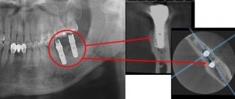
CT scan. It is used in planning sinus lifting, bone grafting, implantation, and other surgical interventions, and in diagnosing periodontal diseases. The photographs are taken in certain projections and used to build a three-dimensional model of the jaw. Computed tomography is very informative. The study provides information about the localization of inflammation (including intraosseous), the presence of tumor-like formations, their structure, size, features of the location of the root canals, etc.
Risks of Dental X-Rays
Dental X-rays involve radiation, however, the radiation levels are so low that it is considered safe for both children and adults. If your dentist uses digital X-rays instead of film X-rays, the risks from radiation exposure will be even lower.
Also, before the x-ray, the specialist will provide you with a protective apron to put on your chest, abdomen and pelvic area to prevent unnecessary radiation exposure to vital organs. In case of thyroid disease, a special thyroid collar may be used. Children and women of childbearing age may also wear it along with a lead bib.
Pregnancy is a contraindication for radiography. Women who are pregnant or think they may be pregnant should avoid all types of X-rays. Tell your dentist if you think you are pregnant because radiation exposure is not considered safe for the developing fetus.
Dental X-ray – price for 2022
The cost of one digital X-ray in Moscow will average from 250 to 350 rubles in different clinics. In addition, this price may only apply to a diagnostic initial image, while all other images taken during the treatment phase may cost less (about 150 rubles per image). Therefore, you should carefully read the clinic’s price list.
It should also be noted that there are a large number of clinics in which the cost of dental treatment is indicated on an all-inclusive basis. Accordingly, the cost of treating your tooth will already include the required number of x-rays (usually 2-4 pictures), for which you will no longer pay anything extra.
What else you need to pay attention to is that the price list of some clinics may state that the cost of a targeted x-ray indicated on the clinic’s website applies only if you are a patient of this clinic. Therefore, if an image is taken to be submitted to another clinic, its cost may be 100 rubles higher).
In addition, if you need a printout of a digital image, some clinics may also charge you about 50 rubles for this. The same applies to the description of the x-ray image: if you want to receive a written description of the image taken by a radiologist, then in some clinics they may additionally ask you for about 100-150 rubles.
Preparation and carrying out the procedure
Dental X-rays do not require special preparation. The specialist will guide you through each step of the x-ray process. He may leave the room briefly during filming. You will be required to stand or sit still for short periods of time. Spacers (film holders), if used, will move and adjust in the mouth to produce correct images.
Once the images are ready—immediately in the case of digital x-rays—your dentist will check them and determine whether or not there are any abnormalities.
Are dental x-rays safe for human health?
In accordance with SanPiN 2.6.1.1192-03, the maximum permissible annual radiation dose during diagnostic x-ray procedures is 1000 μSv (microsievert) for an adult and 300-400 μSv for a child.
To receive it, within a year you must complete:
- 500 targeted dental examinations using a radiovisiograph;
- 100 procedures of conventional x-rays with image output on film;
- 80 digital or 40 film orthopantomographies (obtaining a panoramic image).
Thus, dental X-rays for almost any purpose do not exceed the permissible doses. Therefore, it does not have a negative effect on health. Even when producing film OPTG, the radiation dose is a maximum of 30 μSv. If it is necessary to take a targeted X-ray of the teeth and use radiovisiography, you can even get several images during one appointment. Without any risk to health, since a single dose in this case is only 2-3 μSv.
Digital radiography
Today there is a new technique for dental radiography using digital technology. The new method eliminates the need to develop X-ray film in a dark room; instead, the X-rays are sent directly to a computer and can be viewed on screen, saved or printed. There are several advantages of using this technology:
- minimal radiation;
- saving time - the image is available on the screen a few seconds after shooting is completed;
- the ability to enlarge images multiple times compared to their actual size on the computer screen;
- the ability to send images by email (for example, to another specialist to get a second opinion, etc.);
- software installed on a computer can help dentists digitally compare current images with previous ones in the process of subtraction radiography. Using this technique, everything that is similar between two images is "subtracted" from them, leaving a clear, detailed image of only the part that is different. This helps dentists easily see subtle changes that might not be immediately noticeable.
Dental radiography is of fundamental importance for correct diagnosis. Thus, today radiography is an integral and extremely important part of professional dental care.
Radiology in dentistry
- Survey radiographs can be performed in three projections - direct, lateral and anterior semi-axial, which allows you to obtain an image of the entire facial and cerebral skull. The direct projection can be performed with nasofrontal or nasomental adherence to the cassette. Indications for images in the nasofrontal projection are: injuries and diseases of the brain and facial skull. This installation is also used for sialography and fistulography. Images in the nasomental projection are used: to study the bones of the middle and upper floors of the facial skull, paranasal sinuses. The condition of teeth on radiographs in direct projection is not analyzed.
- Contact radiography according to the isometric rule.
- Interproximal radiography.
- Bitewing radiography (occlusal).
- Radiography with increasing focal length using a parallel beam of rays (long-focus radiography).
Lateral photographs of the skull are taken as a mandatory addition to direct ones. However, it is quite difficult to study the condition of the bones of the facial skeleton from these photographs due to the summative effect of the right and left halves of the skull. Usually only gross, extensive bone changes are visible. Lateral photographs are often performed to examine the condition of the skull, its base, sella turcica, sphenoid and frontal sinuses, as well as to determine the location of foreign bodies.
Axial and anterior semi-axial photographs are performed if it is necessary to examine all structures of the base of the skull, bones of the midface, including the orbits, maxillary sinuses, and zygomatic bones.
Extraoral (extraoral) photographs of the jaws are performed using both dental and other X-ray machines. X-ray film and appropriate cassettes with intensifying screens are used. Extraoral radiographs are performed to study the lower jaw, zygomatic bones, temporomandibular joint (TMJ), as well as for sialography and fistulography. Indications for such images may be inflammatory, tumor, traumatic injuries of the jaws, extensive cysts, periodontal lesions of the lower jaw when it is impossible to perform intraoral radiographs. To study the condition of the TMJ, special installations according to Schüller and Parma can be used. Pictures must be taken on both sides to compare the joints.
Intraoral radiography continues to be the mainstay of radiographic evaluation for most dental and periodontal diseases. Currently, there are four methods of intraoral radiography used to study the condition of teeth, para- and periodontal tissue:
In recent years, a new branch of radiation diagnostics has emerged - digital radiography, which is not so much an independent method of X-ray diagnostics as a progressive modification of the transformation of the energy of the X-ray beam. If in classical radiography the radiation receiver was X-ray film, then in digital radiography these are highly sensitive sensors that directly form a digital image (direct digital radiography), or electro-optical converters that create an analog video signal, which is later converted into a digital signal using an analog-to-digital converter signal. The digital code is then processed by a computer and transformed again into a visible (analog) image on the monitor screen. Computer information processing allows you to improve image quality by manipulating contrast, brightness, clarity, size, eliminating technical errors, and highlighting areas of interest. The advantages of digital radiography are also a significant reduction in radiation exposure (tens of times), economic costs (since expensive X-ray film is not used), and the possibility of archiving information. The principle of digital information processing is also used in computer, magnetic resonance imaging and in some modes of ultrasound diagnostics. Currently, digital radiography has become the leading method of radiological diagnostics.
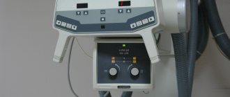
Dental clinics use a dental cone-beam tomograph GXCB-500 from GENDEX (USA) and equipment from SIRONA (Germany). Together with the Heliodent Vario dental X-ray device, the Sirona Sidexis visiograph is used here, which also allows you to significantly reduce the radiation exposure to a person: the touch sensors that radio visiographic systems are equipped with are 10 times more sensitive than X-ray film!
Also, ORTHOPHOS XG 5 DS is widely used - a digital X-ray device, which is designed for panoramic and tomographic imaging of the entire jaw, as well as the temporomandibular joints and paranasal sinuses, it is indispensable for implantology, prosthetics and orthodontics. A panoramic photograph of the teeth is taken with this device in a few seconds, and the radiation dose is so insignificant that it is recommended for use in pediatric dentistry.
Indications for use
Dental radiography is used for two main purposes: to diagnose and evaluate the quality of treatment performed.
Allows you to detect the following diseases:
- hidden carious lesion;
- pulpitis (inflammation of the neurovascular bundle of the tooth);
- cyst at the roots of the teeth;
- periodontitis (purulent inflammation of the tissue between the root of the tooth and the hole in which it is located);
- gum disease, in which bone tissue atrophies (periodontitis, periodontal disease);
- neoplasm.
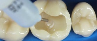
Intraoral radiography also helps evaluate the results of procedures such as:
- root canal filling;
- treatment of periodontal diseases.