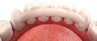
Features of dental x-rays for children
Previously (about 10-15 years ago), x-rays of baby teeth were not a common procedure. Some parents even believed in the myth that baby teeth have neither a root nor a nerve, so the causes of toothache must lie somewhere on the surface, that is, on the crown. This is absolutely not true, and childhood toothache can be caused by the same reasons as in an adult (more on this below).
When asked whether children undergo dental x-rays, we can confidently answer that yes, they do. Moreover, to accurately diagnose the problem and build the correct treatment plan, this is necessary. Another thing is that preference should be given to relatively safe types of x-ray examination, for example, digital.
There are several recommendations for parents to listen to when going for an x-ray with their child:
- You need to come to the clinic in advance so that you have time to psychologically prepare your child for the procedure. You need to tell him that the x-ray will not cause any pain, so you shouldn’t be afraid of it.
- The child's clothing should be loose, easily removable and without complex metal decorations.
- As for girls, you need to give them a simple hairstyle, without using metal pins, bobby pins, etc.
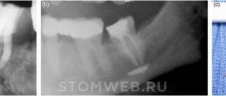
What does a dental x-ray look like?
Let's look at the most common types of x-ray diagnostics that can be used to take a picture of your teeth:
- OPTG. This is what is abbreviated as an orthopantogram, in other words, we are talking about a panoramic photograph of the teeth. When performing OPTG, you can examine the condition of both jaws at once, as well as the maxillary sinuses. This procedure is usually prescribed for dental diseases.
- TRG. Teleradiography - this technique is often used in orthodontics, for example, before installing braces. The peculiarity of the technique is that the picture is taken from a remote distance. This method allows you to get a clearer and higher quality photo.
- CT. Computed tomography allows you to obtain a 3D image of the jaw tissue and facial bone tissue. The procedure is performed using a special device - a tomograph.
Types of X-rays for children
Like adults, children may be prescribed different types of x-rays.
Sight radiograph
A targeted radiograph is performed using a special digital visiograph. A specific problematic tooth or several adjacent ones (maximum 4) are removed.
Panoramic radiograph
The panoramic image shows the entire oral cavity: the upper and lower dentition, teeth that have not yet erupted, and the jaws. It also affects the sinuses. An orthopantomogram (this is what a panoramic image is called) is often prescribed to young patients when it becomes noticeable that their teeth are erupting incorrectly: with an inclination, rotation, and so on. In this case, X-rays help to understand whether there is an anomaly in the development of the jaw bone. There are cases when, after the loss of baby teeth, permanent teeth do not appear for a long time. An x-ray will help identify the cause of this deviation.
Operating principle
The technique is based on X-ray radiation, which is combined with special programs and equipment. X-rays are a special type of radiation that can pass through the tissues of our body. These rays pass through some tissues (for example, abdominal organs) very easily, but through others (for example, muscles, ligaments or bone tissue) - worse, since they are partially or completely absorbed.
When taking a panoramic photo, the body receives a certain amount of radiation. Some of the rays are retained by tissues, while the rest passes through them. All information is sent to a special computer, which analyzes the received data and forms a contour image of the organs located in the path of radiation. As a result, the most accurate image of the oral cavity is obtained, on which the roots of all teeth, periodontal tissue, etc. are clearly visible.
Indications for testing
To determine the extent of caries damage
Children's teeth are susceptible to caries. We cannot assume that caries of baby teeth is not dangerous, since they will fall out anyway. Pathology can spread to permanent teeth even before they erupt. Therefore, it is very important to identify caries and carry out effective treatment.
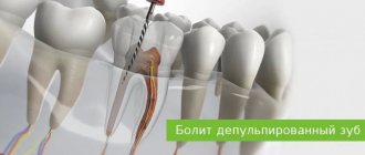
To identify periodontitis and pulpitis
Periodontitis (tooth root pathology, pulpitis) is an inflammation of the pulp where the nerve is located. It is clear that such diseases cannot be detected during the initial examination, because they are hidden in the tooth itself or inside the gums. The child’s main complaint is pain in his baby teeth, and the ability to identify its cause is through an X-ray photo.
When planning endodontic treatment
Endodontic treatment is a set of procedures aimed at preserving a tooth. When emerging caries causes complications, such as pulpitis or periodontitis, the doctor needs to perform therapeutic manipulations inside the tooth. To understand how much pathology has affected the tooth, what percentage of it is destroyed, you need to take an x-ray.
To assess the condition of the permanent tooth buds
An assessment of the condition of the permanent tooth buds is also required before endodontic treatment. It is necessary in order to understand whether the permanent teeth have been affected by pathology and how close they have already descended to the milk teeth.
To diagnose and determine treatment regimens for pathologies of occlusion or teething
At an early age, it is still relatively easy to correct an incorrectly formed bite or correct the position of erupted teeth. This can be done using braces or other special systems. However, to begin orthodontic intervention, you need to understand how serious the pathology is. X-rays help with this.
To determine the reasons for the delay in the appearance of permanent teeth
If a child does not have baby teeth before one year of age, the doctor may order an X-ray to predict their appearance. Of course, such a developmental pathology as adentia (when there are simply no rudiments of baby teeth) is extremely rare, but it is still worth excluding it completely and understanding how soon the first tooth can appear.
X-ray of baby teeth in Kharkov
An X-ray of a child’s jaw makes it possible to:
- • Diagnose carious formations at an early stage and begin timely treatment.
- • Monitor the accuracy of manipulations: during dental canal treatment;
- • Detect problems of the teeth buds: their incorrect position.
- • Most often, it is necessary to diagnose the incorrect position of the wisdom tooth, which can cause crowding of the remaining teeth and destruction of the adjacent seventh tooth.
Important!
Timely orthodontic treatment will allow teeth to erupt correctly and will also eliminate the need for expensive orthodontic treatment with braces in the future. An X-ray of a child’s jaw makes it possible to see teeth that have already erupted and those about to emerge, as well as diagnose the main cause of delayed growth of molars.
X-ray images can reveal the presence of hidden caries and diagnose its complications in bone tissue.
Types of X-rays:
- • Targeted images – can transmit images of one or two adjacent teeth, as well as the bone tissue surrounding them;
- • Panoramic X-rays – visualize the condition of the entire dentition;
- • 3D photographs allow you to obtain a three-dimensional image of the required area or the entire dentition, with the ability to view them in any section.
With the help of 3D images, the doctor is able to obtain information about the structure of the teeth, the location of fillings, the sizes and shapes of the dental canals. The need for 3D images usually arises before complex endodontic or surgical treatment.
Remember: the doctor’s ability to establish an accurate diagnosis will depend on the quality of the images! Modern digital devices allow you to save pictures on your computer for a long time. If you lose a printed original, you can always restore it in our clinic.
Is x-ray examination safe for children?
Before taking an x-ray of a child’s baby teeth, especially for the first time, every parent thinks about how safe the procedure is for the child’s health. It just so happens that since the times of the Soviet Union, the word “X-ray” has been associated with radiation harmful to the body. But the Soviet Union is long gone, and the latest medical technologies and digital equipment make it possible to conduct examinations that are completely safe for health.
The X-ray machines at our clinic carry a low radiation dose. Dental radiography is much safer for children's health than undetected maxillofacial pathologies!
Don't ignore your child's oral health screening. In our clinic, you can take an x-ray of baby teeth as comfortably and quickly as possible by making an appointment in advance. To contact us, order a call back on the website or call us yourself at the numbers listed on this page.
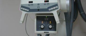
How is research conducted?
Panoramic dental x-rays are done for children, just like for adults:
- The child stands inside the orthopantomograph.
- He clamps the plastic tube between his teeth and keeps his lips closed.
- The device blade is moved as close to the patient as possible.
- A picture is taken (while the device rotates around the child’s head). This takes no more than 20-30 seconds, during which you cannot move or breathe.
A targeted photograph is taken as follows: the child sits on a chair and approaches the device.
Next, he wraps his mouth around the digital sensor and clenches his teeth. A picture is taken (takes a few seconds); during this process you cannot move or breathe. To protect the body from exposure to x-rays, a special apron is worn.
Are there any contraindications for x-rays?
Of course, any radiation poses a danger to human health. We receive different doses of radiation from household items, communications equipment, and even from the atmosphere. This cannot be avoided. For example, the natural background of the earth gives a person 2-3 mSv per year. At the same time, the power of a dental X-ray image “loses” to other types of X-rays. The radiation dose delivered by the devices is minimal – only 0.2 mSv.
There are also special schedules for X-ray procedures in children designed to control radiation exposure.
How often can a child have dental x-rays:
- babies who do not have permanent teeth can undergo an x-ray procedure once every 2 years;
- adolescents – with periapical and bitewing radiography – once every 1.5–3 years;
- adults – once every 1–1.5 years.
Contraindications
If there is bleeding
Bleeding in the mouth, caused, for example, by problem gums, can affect the quality of the X-ray image. First, the doctor must prescribe hygiene procedures that can stop the bleeding, and only then send for x-rays.
If you feel unwell
If your child is sick, has a fever, or simply feels unwell, you should not take him for an x-ray. It is better to postpone this procedure until complete recovery.
How often can you do it
How many times can an x-ray be taken? This question can be found on many forums. Including dental ones. After all, situations often occur when dental treatment is delayed for a long time, and periodically it is necessary to monitor treatment tactics using x-rays. In fact, there are no specific figures on how many times it is permissible to take an x-ray.
As mentioned above, this technique involves minimal radiation (if high-quality equipment is used), so the procedure is performed as many times as the doctor prescribes. On average, experts do not recommend taking x-rays more than 7 times a year - but this value is conditional and can be adjusted by a doctor. There are different recommendations for different shots. CT - no more than 14 images per year, OPTG - no more than 50 times, and TRG - about 10 times.