Odontogenic sinusitis is an inflammation of the maxillary sinuses that develops from infected teeth of the upper jaw. The penetration of infection is also facilitated by dental manipulations in the area of the maxillary sinuses - treatment and extraction of teeth, implantation and sinus lifting. Without identifying and eliminating the cause, standard treatment by an ENT doctor in a clinic does not bring results. Treatment of odontogenic infections must be comprehensive!
Doctor Levin Center has been specializing in the treatment of patients with combined ENT and dental pathologies for more than 20 years. Comprehensive rehabilitation programs are carried out by maxillofacial surgeons with otolaryngological training.
Causes of odontogenic sinusitis
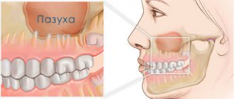
The maxillary sinuses (maxillary sinuses) are separated from the oral cavity by a thin layer of bone in which the roots of the upper chewing teeth are located. Such close proximity poses a threat of sinus infection. In 15% of cases, the apices of the roots are anatomically located under the mucous membrane of the sinus, without a bone layer, which further increases the risk of developing odontogenic sinusitis.
Conventionally, the reasons can be divided into 2 categories:
Delayed dental treatment
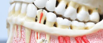
With pulpitis and periodontitis, infection from the dental canals slowly but surely spreads to the surrounding bone tissue. Without treatment, foci of infection grow, melt the bone septum, opening the “gate” for the penetration of pathogenic microflora. In advanced situations, cysts form on the roots of teeth, and when the bone barrier is destroyed, they grow into the sinus.
Medical errors
As a result of dentist errors, mechanical damage to the sinus floor is possible (perforated form of the disease). The result is the formation of a communication with the sinus, the penetration of foreign bodies with the body’s response in the form of odontogenic sinusitis.
The most common treatment errors after which patients turn to our Center:
- Unsuccessful endodontic treatment The therapist did not calculate the effort during mechanical cleaning and filling of dental canals. There was a fracture and pushing of the instrument and/or removal of the filling material beyond the root apex into the lumen of the sinus.
- Traumatic tooth extraction The surgeon, without preliminary diagnosis, having no idea about the location of the roots, damaged the bone septum. Perforation of the sinus floor occurred, often with the tooth or its fragments falling into the sinus cavity.
- Implantation without sinus lift Experimental implantation protocols without prior bone grafting result in damage to the sinus floor by the implant. If the perforation goes unnoticed during surgery, the implant flies into the sinus.
- Sinus lift without diagnostics The surgeon did not calculate the bone tissue parameters and did not assess the condition of the mucous membrane. During the preparation of the bone septum, its perforation and rupture of the membrane occurred. Bone material ended up in the sinus cavity.
How and why can a tooth end up in the maxillary sinus?
The maxillary sinus is the largest in size of all the sinuses of the nose. Its volume in an adult can reach 10 cubic centimeters. The sinus is a steam room and is located to the right and left of the wings of the nose. Visually, its projection falls on the cheek, which becomes slightly swollen and red with sinusitis.
The oral cavity and maxillary sinus are separated from each other by a layer of bone, which is lined on both sides with mucous tissue. However, both the maxillary sinus and the oral cavity are not hermetically sealed. That is why the appearance of an infection on one side of a thin bone septum (for example, pulpitis) quickly entails inflammation in the adjacent chamber.
There are several factors that significantly increase the likelihood of infection spreading:
- Anatomically, the roots of premolars and molars are located very close to the sinus. The thickness of the bone layer between them in some patients may be only 1 mm instead of the ideal 10.
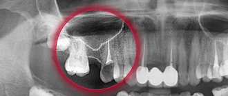
- In some lucky people, the roots of the teeth are generally located in the sinus itself, being separated from its internal space only by a thin layer of mucous membrane. What to do if your tooth roots are actually in the maxillary sinus? First of all, don't panic! And secondly, carefully monitor their health and regularly take a panoramic photo, which allows you to identify the slightest sign of trouble.
- The bony septum may become thinner over time. This occurs due to persistent inflammatory diseases - periodontitis, pulpitis and others. Therefore, if sinusitis is suspected, timely treatment and removal of teeth, including problematic wisdom teeth, is so important.
- Loose bone tissue as a result of systemic diseases, such as osteoporosis.
How to avoid
There are only two ways to prevent the development of odontogenic sinusitis:
- Treat your teeth in a timely manner, prevent infection of the dental canals and spread of inflammation beyond the apex of the tooth root.
- Contact proven clinics with experienced doctors, the opportunity to conduct a thorough diagnosis and provide for an emergency situation.
In our Center, not a single dental procedure, especially at the border with the maxillary sinuses, is performed without a thorough X-ray examination using a computed tomograph.
The study is carried out using
a high-precision Sirona device with the Galileos diagnostic software package.
Based on the results of computed tomography, we determine the location of the roots and the size of the bone septum. This makes it possible to plan treatment in such a way as to avoid risks.
Tooth extraction operations and other interventions in the area of the maxillary sinuses in our Center are performed only by maxillofacial surgeons with ENT training . Deep knowledge of the anatomy of the maxillary region allows you to avoid mistakes that a regular dentist might make. But each case is individual, and even if something goes wrong, our doctors are always ready for any turn of events and correct the situation.
Therapy for perforation of the maxillary sinus during treatment or tooth extraction
If a hole has formed in the sinus after tooth extraction, the doctor will stop the bleeding if possible by forming a blood clot in the hole. To do this, a tampon soaked in medications is inserted into the wound. The patient is advised to rest completely until the perforation heals.
If, during tooth extraction, the maxillary sinus opened, but the doctor did nothing, then inflammation may develop. In this case, if detected, an operation to restore integrity using special plates or artificial bone material is indicated.
Symptoms of odontogenic sinusitis
Inflammation from infected teeth develops gradually and in the initial stages is most often asymptomatic. If dental treatment has been carried out, symptoms may also not appear immediately - it all depends on the body’s immune system and reaction to foreign bodies. There are cases that sinusitis does not make itself felt for several years.
But sooner or later the infection will make itself known. The symptoms of odontogenic sinusitis are practically no different from rhinogenic sinusitis (when the infection enters through the nose as a result of a cold, flu or acute respiratory viral infection):
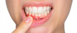
- pain and swelling in the sinus area
- nasal congestion and difficulty breathing
- purulent or mucopurulent discharge
- headaches, especially when bending forward
- decreased sense of smell
- temperature up to 39o
An unsuspecting patient first turns to an ENT specialist, but he needs to see a dentist. However, there are points that indicate the odontogenic origin of sinus inflammation:
- pain in the chewing teeth of the upper jaw, especially when chewing
- there was treatment or removal of teeth at the border with the sinuses
- implants or sinus lift were installed
The most important distinguishing feature is that only one sinus is bothering you , which is associated with bad teeth or dental treatment. In this case, first of all, you need to solve dental problems. As a rule, the symptoms of sinusitis disappear within two weeks after eliminating the source of inflammation.
Possible complications
Since inflammation of the maxillary sinuses due to teeth occurs very often, with perforation the symptoms increase significantly.
- After the removal of the upper tooth, sinusitis develops, and then osteomyelitis of the upper jaw.
- The inflammation spreads to other sinuses and paranasal sinuses.
- Teeth located in the perforation zone begin to loosen and fall out.
- A systemic infection develops. The formation of phlegmon and abscesses is possible. Meningitis and meningoencephalitis cannot be excluded.
In case of perforation of the bottom of the maxillary sinus, self-medication is unacceptable. You need to see a doctor as soon as possible!
What is the danger of the disease
Without proper treatment, a chronic inflammatory process with pathogenic microflora develops in the sinus, destroying the sinus mucosa and surrounding bone tissue. Since the maxillary sinuses are located close to anatomically important structures, severe complications can arise:
- Orbital The upper wall of the sinus is in contact with the lower wall of the eye orbit. The infection spreads into the orbit with the formation of an abscess and phlegmon. As a result, optic nerve neuritis develops, deterioration or complete loss of vision.
- Intracranial The most dangerous complications associated with the penetration and impact of infection on the brain. Meningitis, encephalitis, and abscess develop. If detected and treated untimely, irreversible consequences, including death, can occur.
Foci of infection provoked by the presence of a foreign body can cause a precancerous condition.
What symptoms may indicate a foreign body in the maxillary sinuses?
After dental treatment, a person may suspect the development of this complication if the following signs appear:
- the appearance of pain when performing light taps on the facial bone under the eyes near the nose;
- severe, aching headache;
- prolonged and persistent runny nose;
- aching pain in the upper jaw, aggravated by chewing.
One of these symptoms or a combination of several may indicate that dental medications have entered the maxillary sinus cavity. This condition is a reason for mandatory consultation with a doctor and the visit should not be postponed. If the patient does not see a doctor on time or has a weakened immune system, the disease becomes more complicated. Because of this, a person develops thick, purulent and profuse nasal discharge with a specific odor. In addition, the progression of the inflammatory process causes an increase in temperature and a deterioration in general health. In some cases, immediately after dental mixtures enter the sinuses, inflammation does not develop. However, this condition is also unsafe for his health in the future, since various negative factors (for example, hypothermia, stress, injury or hypovitaminosis) can provoke the development of inflammation and a deterioration in general well-being.
Why you should entrust treatment to the ENT department of dentistry
ENT dentistry is a symbiosis of two medical areas, a multidisciplinary approach to the treatment of inflammation of the maxillary sinuses of odontogenic origin. Only an experienced maxillofacial surgeon with ENT training can make an accurate diagnosis and create a sound rehabilitation plan.
As a rule, odontogenic causes of sinusitis are simply ignored during routine examination by an otolaryngologist. Treatment in a city clinic without high-quality diagnostics or in the absence of it at all turns into a multi-part series with monthly visits to an ENT doctor, dragging on for many years, causing inconvenience and worsening the quality of life.
Unified drug therapy or traumatic sinus punctures are prescribed, which, if they bring relief, are for a short time. Inflammation from the acute stage becomes chronic with periodic exacerbations. A person runs from one doctor to another to no avail, but without identifying and eliminating the cause, odontogenic sinusitis cannot be cured !
Diagnostics
The odontogenic form of sinusitis often remains undetected by ENT specialists due to the lack of highly accurate diagnostics and banal ignorance of the “dental” cause of the disease. Such gaps lead to the fact that even after medical and surgical treatment the person continues to suffer.
Differential diagnosis is extremely important, which allows you to determine the form of sinusitis and select the appropriate treatment.
Only computed tomography provides informative 3D images for visualizing bone and adjacent soft tissues, the condition of the teeth, the presence of foreign bodies and neoplasms in the sinus.
In our Center, CT scans are performed on a Sirona Gallileos dental tomograph with ENT mode settings.
In some cases, it will be necessary to study the microbial composition of the sinus lining to rule out a malignant process.
Stages of treatment
We strive to carry out all activities comprehensively in one day, so as not to delay the patient
- Sanitation of the oral cavity Preparation to maintain sterility during surgery to avoid secondary infection - retreatment of compromised teeth, hygienic cleaning.
- The operation is performed by a maxillofacial surgeon in a sterile operating room of the ENT department - “in your sleep”, according to the selected access protocol. A follow-up CT scan after surgery is required.
- Prosthetics If the protocol requires the removal of teeth, we install temporary orthopedic structures to close the aesthetic defect so as not to leave the patient without teeth.
Prevention
As for preventive measures regarding perforation of the maxillary sinus, they consist of following the following rules:
- A thorough and comprehensive examination of the patient before undergoing serious dental procedures, including tooth resection or implantation.
- Correct assessment of the anatomical features of the jaw structure of each patient.
- Strict compliance with all technological requirements for complex dental procedures.
Recovery after surgery
No hospitalization
A hospital stay for 2-3 days after surgery (as happens in city hospitals) is not required.
Gentle surgical protocols, the use of modern functional equipment and a microscope allow treatment to be carried out as delicately as possible .
All operations are performed in a controlled, drug-induced sleep - this is not general anesthesia ! Recovery from the state is easy, without dizziness, memory loss or clouding of consciousness. The drugs are absolutely safe for the body and are eliminated naturally 40 minutes after stopping the supply.
For patients with vascular and cardiac problems, a day hospital , where you can calmly recover and recover under the supervision of our anesthesiologist-resuscitator.
Accelerated rehabilitation
Recovery after the intervention occurs within a week. For those who want to speed up the process, our Center provides a set of procedures to reduce pain, resolve hematomas and swelling - carried out on the day of surgery.
Home care
Medications are prescribed - antibiotics, painkillers and decongestants. To prevent the patient from running to pharmacies in a postoperative state and to avoid purchasing counterfeit products “on the side,” the entire package of drugs is collected and given out free of charge .
The package contains all the necessary medications for taking and caring for the surgical area.
Please follow the recommendations and do not skip taking medications to avoid complications. The instructions are in the medicine package.
If you suspect a worsening condition, contact the clinic immediately. The telephone number for the 24-hour patient support service is listed in the memo.
Old perforations
It happens that the patient does not go to the clinic after treatment due to accidental perforation. At first the pain is sharp and pronounced, but gradually its character changes and the sensations smooth out. A fistulous tract is formed between the affected sinus and the outer part of the gum. Everything that happens is accompanied by the development of symptoms of typical sinusitis: pain from the perforated intramaxillary cavity, constant nasal congestion, outflow of pus from the nasal passages (and sometimes the fistula on the gum begins to fester). Some patients complain of swelling of the cheek on the side where the sinus is damaged.
If after treatment a person develops strange sensations, as if he feels the movement of air through the gums when sneezing, coughing, and drunk liquid enters the nose, there is a direct indication of an old perforation of the maxillary sinus.
Unfortunately, non-invasive techniques will not help here. An operation is required to remove foreign matter and foci of infection from the sinus. The fistula is cut out, and the opened defect is repaired. After the procedure, a course of antibiotics is prescribed with accompanying physiotherapeutic treatment and the use of drugs that reduce the risk of allergic reactions.