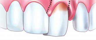
Conservative method of treating pulpitis
The simplest option used by dentists for patients suffering from focal, acute and fibrous infections. It consists of carrying out a certain list of fairly easy and quick procedures:
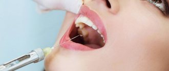
- Anesthesia using local anesthetic drugs.
- Removing affected tissue using, for example, a drill.
- Treatment of the oral cavity with any specialized antiseptics.
- Applying an insulating pad.
- Installation of a temporary filling product.
The result of such operations is a whole range of positive aspects. The patient's severe inflammation is relieved, metabolic processes are restored, dentin regeneration begins, and so on.
Surgery
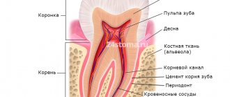
It is possible to understand what dental pulpitis is and how to treat the pathology in question, including through a detailed analysis of the techniques that require the use of any types of surgical interventions. The main indication for the use of such instructions is the presence in a person of a diffuse, focal and chronic inflammatory process of the fibrous and hypertrophic type. All activities are carried out according to strictly defined instructions:
- Anesthesia by introducing local, conduction anesthesia into the body.
- Isolate the affected area with sterile pads and pads.
- Opening the carious plane and treating the lesions with an antiseptic.
- Excision of the coronal part of the pulp extractor.
- Expanding the canals and treating them with antiseptic agents.
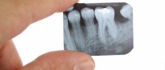
- Preliminary and final filling performed after x-rays.
It is not difficult to guess that only an experienced doctor with a variety of different skills can cope with a series of such procedures at the highest quality level. These are the specialists who work in dentistry.
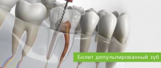
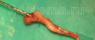
Treatment of pulpitis with removal of the dental nerve
Another event characterized by a fairly high complexity. As a rule, all medical interventions are performed in two stages. During the first of them, the specialist:
- Removes the carious plane.
- Removes damaged tissue from the pulp itself.
- Treats the excision site with an antiseptic.
- Carrying out work on installing a temporary filling.
After approximately one to two weeks, the patient necessarily comes for the second session. As part of the additional (finishing) procedure, the following actions are carried out:
- Short-term filling is removed.
- Inspection and cleaning activities are carried out.
- The oral cavity is exposed to antiseptic agents.
- A permanent material is installed to fill the voids in the tooth process.
The main advantage of all processes is the possibility of preserving a healthy dental element. In addition, the person subsequently feels positive effects due to the regeneration of vitality and sensitivity of the pulp itself.
Treatment of pulpitis of a multi-rooted tooth
A devital technique, in which the doctor initially kills the problem area using a special necrotizing paste. In the past, doctors involved in such work used ordinary arsenic, placed in the carious plane for several days.
Today, modern medicine has stepped forward, providing craftsmen with a much wider range of tools. Dental staff use the most gentle drugs that do not kill nerve tissue in the oral cavity. It is not difficult to guess that the use of such substances is a completely safe operation.
Extirpation of pulpitis
Probably the most radical treatment method, which includes a whole complex of processes to remove the inflamed neurovascular bundle, followed by sanitation and filling. The doctor can undertake its implementation only in the case when any other methods of therapy clearly do not lead to success. As in previous cases, the procedure is carried out in a vital and non-vital format: under the influence of local anesthesia or using a necrotizing paste-like drug. Depending on how complex the disease is, the specialist may need from 3 to 4 sessions, the result of which, of course, will be a complete cure of the patient from the specified range of problems.
Modeling from plastic
Plastic is another material from which you can create a beautiful tooth model. This is a fairly dense, non-sticky material that does not require special preparation for work, but it is necessary to observe the conditions for its storage: temperature changes at which the material is stored can have a negative impact on its properties. Plastic is convenient because you can work with it for an unlimited amount of time, it does not harden, which makes it possible to work out the microrelief of the future model in more detail and clearly, and make the necessary adjustments during the work.
Hardening of the material occurs when it is placed in hot water or heated to 110-120 degrees in an oven for 5-10 minutes. The possibility of long-term work and fixation of the result by heating - this is the similarity between plastic and composite. Plastic cannot be reused. Well suited for creating phantom teeth models.
The first stage of working with this material will be to warm it up in your hands and shape it into a ball, then give the overall outline of the model, determine the main surfaces of the tooth model (Fig. 16) (M - medial contact surface, D - distal contact surface, V - vestibular surface, L - lingual surface), applying markings corresponding to the first-order fissure of the F-shaped form.
Rice. 16. Giving the overall outline of the model, applying markings corresponding to the fissure of the first order, F-shaped. M - medial contact surface. D - distal contact surface, V - vestibular surface, L - lingual surface
On the chewing surface with a tool, spatula or trowel, according to the applied markings, a fissure of the first order is deepened, five main tubercles are identified (Fig. 17) (1 - anterior lingual, 2 - posterior lingual, 3 - anterior buccal, 4 - posterior buccal, 5 - distal ).
Rice. 17. Tops of the main tubercles: 1 - anterior lingual tubercle, 2 - posterior lingual tubercle, 3 - anterior buccal tubercle, 4 - posterior buccal tubercle, 5 - distal tubercle
The equator of the 36 tooth model is also formed. The formation of second-order fissures occurs by modeling the longitudinal, medial, and distal ridges of the four main tubercles (Fig. 18-19).
Rice. 18. Modeling of the longitudinal (b), distal (a), medial (c) ridges, anterior lingual tubercle (1). 1 - anterior lingual tubercle, 2 - posterior lingual tubercle, 3 - anterior buccal tubercle, 4 - posterior buccal tubercle, 5 - distal tubercle
Rice. 19. 1 - anterior lingual tubercle: (a) longitudinal ridge, (b) distal ridge, (c) medial ridge. 2 - posterior lingual tubercle. 3 - anterior buccal tubercle: (a) longitudinal, (b) medial ridge, (c) distal ridge. 4 - posterior buccal tubercle: (a) longitudinal ridge, (b) distal ridge, (c) medial ridge. 5 - distal tubercle. 6 - additional tubercle. Rice. 19. 1 - anterior lingual tubercle: (a) longitudinal ridge, (b) distal ridge, (c) medial ridge. 2 - posterior lingual tubercle. 3 - anterior buccal tubercle: (a) longitudinal, (b) medial ridge, (c) distal ridge. 4 - posterior buccal tubercle: (a) longitudinal ridge, (b) distal ridge, (c) medial ridge. 5 - distal tubercle. 6 - additional tubercle
The distal tubercle has a less differentiated surface (Fig. 20).
Rice. 20. The final result of the simulation. 1 - anterior lingual tubercle. 2 - posterior lingual tubercle. 3 - anterior buccal tubercle. 4 - posterior buccal tubercle. 5 - distal tubercle
Models made of plastic can be stored for a long time; they will remind you of the results achieved in modeling.
Rice. 21. Lingual and medial contact surfaces of the mandibular molar model. M - medial contact surface. D—distal contact surface. V - vestibular surface. L - lingual surface
Rice. 22. Vestibular and chewing surfaces of the mandibular molar model
Other treatments
In 2022, there are additional ways for doctors to save people from troubles associated with pulp inflammation:
- laser therapy;
- complex of physiotherapeutic processes.
Both mentioned operations, in principle, can also answer the question of how to cure pulpitis without removing the nerve. True, they can only be used in the early stages, when the pathology has not yet had time to develop and acquire serious symptoms. In addition, to use such funds, a person will need to go to a truly modern and advanced clinic with an experienced medical team. For example, a set of works related to the use of a laser is carried out exclusively in the presence of high-quality, proven equipment. Such installations, of course, are present in dental departments.
Is it possible to cure pulpitis and how are nerves removed: types of anesthesia
As mentioned earlier, pulp inflammation is an extremely painful pathology, which primarily affects the nerve endings inside the oral cavity. The process of its treatment necessarily includes an operation to select a high-quality antiseptic that is suitable for a particular person. Moreover, the drug is applied to the desired places using various methods:
- application;
- infiltration;
- conductive;
- stem
The anesthetics used are traditional pairs of lidocaine and novocaine, as well as more advanced and modern formulations: Ultracaine, Ubistezin, Septanest, and so on.
Is it possible to get rid of caries at home?
When medicine was not as progressive as it is now, people often got rid of caries at home using unconventional treatment methods.
Some methods of traditional medicine are still cultivated today. People are offered to prepare their own tinctures for rinsing, use propolis, soda, laundry soap, fir oil, camphor alcohol and much more.
At best, the effect will not be achieved, and at worst, a person may harm his health. Most traditional methods can damage enamel and soft tissue.
Medical representatives do not recommend treating caries at home.
If you decide to effectively fight caries, it is best to contact professionals in dental clinics in a timely manner.
Dental clinic "ORTHODONTICS" will make your teeth healthy and your smile snow-white!
Our ORTHODONTICS clinic in Krasnodar will stop caries at different stages of its progression. We offer high-quality and completely painless treatment of dental pathologies.
We employ experienced specialists who regularly improve their skills and exchange their experience with leading doctors from different countries. There is no one way for everyone to remove caries, since for each client we develop an individual strategy to make teeth healthy.
To carry out manipulations, we use advanced equipment in specialized rooms. Our clients are always satisfied with the results, delighting themselves and others with beautiful smiles. Remember that caries can be cured, the main thing is to turn to professionals in time!
Stages of treatment
Most of the operations, the main task of which is to heal the inflamed pulp, can be divided into a list of conditional stages:
- Excision of dental tissue.
- Extraction of the neurovascular bundle itself.
- Channel cleaning.
- Filling.
The question of how pulpitis is treated in 2 visits with the removal of teeth and nerves can clearly be answered by any more or less qualified dental specialist. However, not everyone, even experienced doctors, had to deal with the most severe forms of this disease. It is not difficult to guess that the key to the success of the entire therapy as a whole is a visit to a specialized specialist who knows how to handle modern equipment and the advanced range of today’s pharmacological drugs. Such professionals, of course, include dental staff.
Modeling tooth 36 from plasticine: main steps
Plasticine is perhaps the most common material; it is easily accessible and does not require special preparation for work. Having taken the required amount, just warm it up, knead it in your hands, and you can start working. Having no experience working with this material, in the first stages it is better to work without tools to feel its properties, and then create shapes using tools. After kneading, the plasticine is ready for modeling, but it does not need to be heated for long, as it becomes too soft and sticky and will not hold its shape well.
Let's consider the main stages of modeling the 36th tooth from plasticine. We give the plasticine the shape of a ball (Fig. 23).
Rice. 23. Giving the material a ball shape
Having outlined the main surfaces and tops of the tubercles of the future model, we deepen the first-order fissure of the F-shape. As a result, five tubercles are formed on the chewing surface (Fig. 24) (1 - anterior lingual, 2 - posterior lingual, 3 - anterior buccal, 4 - posterior buccal, 5 - distal).
Rice. 24. Formation of the overall outlines, the tops of the main cusps: anterior lingual (1), posterior lingual (2), anterior buccal (3), posterior buccal (4) and distal (5). Surfaces: M - mesial, D - distal, V - vestibular, L - lingual
Using the tool (Fig. 25), modeling of the distal ridge (B), longitudinal ridge (A), medial ridge (C), anterior lingual tubercle (1) and modeling (Fig. 26) of the medial ridge (B), longitudinal ridge ( A), distal ridge (C), posterior lingual tubercle (2).
Rice. 25. Modeling of the distal ridge (B), longitudinal ridge (A), medial ridge (C), anterior lingual tubercle (1). (2) posterior lingual cusp, (3) anterior buccal cusp, (4) posterior buccal cusp, (5) distal cusp
Rice. 26. Modeling of the medial ridge (B), longitudinal ridge (A), distal ridge (C), posterior lingual tubercle (2). Modeling is carried out with a spatula
Rice. 27. The final result, the model of the lower jaw molar is made of plasticine
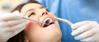
Is it painful to treat pulpitis?
Inflammation of the pulp is a disease characterized by pronounced, extremely acute symptoms, mainly in terms of pain. Almost any manipulation with a damaged element of the oral cavity causes severe discomfort to patients in medical clinics. However, such a problem can be resolved quite simply - a good dentist will easily select a high-quality, suitable anesthetic agent. That is why the process of surgical intervention itself, as a rule, takes place without third-party excesses. Much more unpleasant sensations await people who refuse to visit a doctor in a timely manner.
Features of the procedure
Wax-up for aesthetic restoration is popular among orthopedists. Wax is a malleable compound that can be molded. With its help, an accurate impression of the tooth is created, taking into account the anatomical features.
The accuracy of manipulation affects the comfort of the artificial prosthesis. Following the technology will relieve the patient from pain when talking and eating.
The Wax-up technique is safe and does not cause discomfort. This is due to the absence of violation of the integrity of the oral mucosa. There is a risk of cross-reaction in those who suffer from allergic reactions to bee products. In this case, replace the composition.