A. I. Nikolaev, L. M. Tsepov Practical therapeutic dentistry
St. Petersburg: St. Petersburg Institute of Dentistry and, 2001. - 390 p. ISBN 5-94517-001-1
The book covers at a modern level the issues of etiology and pathogenesis of one of the most common human diseases - dental caries. Its treatment using modern methods of pain relief, premedication, and psychological preparation of patients is described in detail. The characteristics of filling materials used at dental appointments are given. Medications used in endodontics are described.
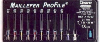
Modern methods of endodontic treatment are outlined, including a description of the endodontic instrumentation used, methods of instrumentation and root canal filling, as well as ways to improve the quality of endodontic treatment.
A separate section is devoted to the etiology, pathogenesis, clinical and morphological characteristics of chronic inflammatory periodontal diseases, and also presents the basic principles of complex therapy of chronic general and gaping periodontitis in an outpatient dental appointment. The final section provides the methodological basis for organizing periodontal care. All this gives reason to believe that the book will be useful to both an experienced dentist and a novice doctor.
The book is intended for students, interns, residents, dentists, teachers of dental faculties of medical universities, and persons studying in the system of postgraduate professional education.
Reviewers:
L. N. Maksimovskaya - Doctor of Medical Sciences, Professor, Head. Department of General Dentistry, Faculty of Advanced Training for Doctors, Moscow State Medical and Dental University, President of the Russian Academy of Aesthetic Dentistry.
V. B. Nsdossko - Doctor of Medical Sciences, Professor, Head of the Department of Therapeutic Dentistry of the Omsk State Medical Academy.
Preface.
Work in canals with pronounced curvature
The curvature of the root canals largely determines the complexity of endodontic treatment. In this article we will discuss the basic principles of working in curved canals, which a doctor can effectively implement in his practice.
Grade
It is extremely important to evaluate the location, angle and radius of curvature before starting treatment (Figures 1 and 2). Many methods have been proposed to measure root canal curvature (Figure 1), but it has not yet been established which method is best (Hartmann et al, 2018).
Photo 1: Image taken from “Methods for measurement of root canal curvature: a systematic and critical review” by Hartmann et al, 2022 (International Endodontic Journal). This image illustrates simple methods for calculating root canal curvature. These methods help determine the level of potential treatment complexity. The physician can then decide whether he is competent to perform endodontic interventions or whether the complexity of the clinical case is beyond his skill level and warrants referral to a specialist (Figure 2).
Figure 2: Tooth UR6 with sharp apical curvature at the MB root (MB1 and MB2). Apical curvature is much more difficult to manage clinically than coronal curvature. Anatomical curves associated with longer roots or calcified canals create additional problems during the treatment process.
Complications
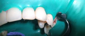
Curvatures can lead to the development of many iatrogenic complications (photo 3):
- formation of steps;
- excessive expansion of the canal and transport of curvature;
- perforations;
- apex transposition;
- file fracture.
Sometimes iatrogenic complications are quite difficult to correct; in some cases, they cannot be stopped at all.
Photo 3: Possible iatrogenic complications that may arise during the treatment of root canals with severe curvature.
Advice
Be patient: it is extremely important to adequately plan the treatment process and ensure sufficient time for its implementation (photo 4).
Photo 4: LL6 root canal treatment. A thorough analysis of the radiograph allowed us to evaluate the anatomical features and existing bends of the root canal. At first glance, LL6 does not present much complexity. However, upon closer inspection, two long mesial roots with apical curvatures were identified.
Straight access: It is important to provide direct access to curved canals. This reduces the stress on the endodontic files as they do not have to bend as they enter the root canal. The shape of the access cavity is corrected as conservatively as possible using ultrasound.
Coronal canal treatment: The quality of the coronal canal treatment reduces the degree of canal curvature and can help convert a C-curve to a J-curve (Figure 5). Rotary nickel titanium orifice files can be used for this purpose. Instruments such as Gates Gliddens on the posterior teeth can provoke a destructive effect on the hard tissues of the endodontic space and form steps.
Photo 5: Widening the coronal portion reduces the degree of canal curvature. The medial channels on the LR6 were originally C-shaped. Expansion of the coronal part of the canal transformed them into a J-shape, optimizing the treatment process.
Carpet: To avoid breaking the rotary tool, it is essential to recreate the carpet using hand files. It is better to start working in canals with pronounced curvature with files with an inactive apex. The file can also be pre-bent to the shape of the canal, since a straight tool, as a rule, cannot follow the curvature of the canal and provokes the formation of a step.
The author of the article usually starts root canal treatment with a size 8 file and manually expands it to size 10. A rotary flexible file is then used to increase the size of the endodontic space. For curvature processing, it is not recommended to use hand files larger than size 15, given their rigidity. In very curved canals, it may be necessary to gradually widen the endodontic space to the level of the canal curvature (Figure 6). In such conditions, it is much easier to work in curvature in the future.
Photo 6: UR7 with very curved channels. The canals were processed in three stages (in the coronal, middle and apical parts). You should not “forcibly” push the hand file along the curvature of the canal; the movements of the instrument must be consistent. You can process the canal with a rotary file to the depth of the manual file, and then continue working with a hand tool again until the full length of the canal is reached.
Cleaning and debridement of the endospace: Use flexible rotary instruments that allow for consideration of the anatomy of the canal when debridement of the endospace (Figure 7).
Photo 7: The use of a flexible rotary system is extremely important when working in canals with severe curvature.
Irrigation: Frequent irrigation using sodium hypochlorite helps prevent debris from clogging the root canal. However, the author does not recommend endoirrigation with EDTA during the early stages of root canal treatment because EDTA is a chelating agent that can soften dentin, causing the formation of steps.
Patency and Recapitulation: It is important to continually check for patency using small hand files (size 8-10 files). Accumulation of debris can block the patency of the endospace and cause the development of iatrogenic errors (Figure 8).
Figure 8: Endodontic treatment UR6: MB1 and MB2 are calcified and distorted. Small hand files were used to maintain patency throughout the root canal procedure. This contributed to the prevention of blockage of the endospace during treatment.
CBCT: Preoperative CBCT is extremely useful in planning the treatment of curved canals. CBCT can help identify the fusion of two canals: in such cases, the canal with less curvature is treated first, and then the canal with more curvature (Figure 9).
Figure 9: A CBCT scan was performed prior to starting root canal treatment on tooth LL7. This made it possible to verify the additional distal root. A pronounced curvature was visualized in the MB and ML, but they merged in the apical third. Taking into account all these details made it possible to plan an endodontic treatment algorithm.
Anatomical curves in the mesial-distal projection can be visualized on a targeted radiograph. However, these radiographs do not allow visualization of the curvature in the buccopalatal (or buccolingual) plane. CBCT allows clinicians to see the “third dimension”, thereby facilitating proper treatment planning and implementation (Figure 10).
Photo 10: Endodontic UR7. CBCT identified severe curvature of the MB root in the bucco-palatal plane. This feature could not be verified on a targeted radiograph.
conclusions
Endodontic treatment of canals with severe curvature is a rather complex clinical task. Before treatment, it is extremely important to carefully assess the complexity of the clinical situation in order to avoid the development of iatrogenic errors and complications. This article presents some clinical tips for creating access and treating root canals with severe curvature. CBCT is a valuable tool in planning endodontic treatment of canals with complex anatomical structures.
Posted by Creena Patel