Atheroma on the face is not just a cosmetic defect. It occurs due to blockage of the sebaceous glands. If infection penetrates inside, purulent inflammation may develop. Inflammation of atheroma on the face is very dangerous, especially if the process is located above the line of the mouth. This is due to the peculiarities of the blood supply to the head. The veins of the face drain into the venous sinuses of the skull, which leads to the rapid progression of the disease and the development of abscesses in the brain. The degree of danger of atheroma depends on other factors, including the size of the formation and the presence of underlying chronic diseases, such as diabetes. Therefore, when facial atheroma appears, treatment should only be carried out by a doctor.
Rice. 1 Facial atheroma occurs due to blockage of the sebaceous glands
Ball in mouth - what is it?
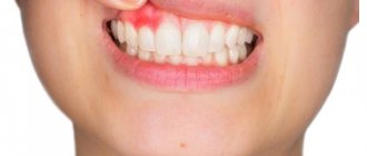
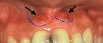
When the inflammation begins to gradually progress, a blister appears on the gum. The main reason for the formation of a lump is poor oral hygiene and prevention. But the ball can also form under the influence of other factors: inflammation occurring in the roots of the teeth, gums, mucous membranes, and periosteum.
The problem is that the disease can be diagnosed in the later stages, with the development of a purulent process. This happens because the patient does not visit the dentist. For patients who experience fear while in a chair, the doctor can give sedative anesthesia, in which the receptors are turned off and the patient falls into a shallow sleep.
Attention! It is better to diagnose the disease early in order to prevent more dangerous consequences, including blood infection and inflammation of the entire jaw.
Why does a lump appear on the gum?
The accumulation of conditionally pathogenic and pathogenic microflora due to improper oral care leads to inflammation of the mucous membrane. Other reasons why a ball appears on the gum include:
- chemical, thermal or mechanical trauma to the periodontium;
- carious formations and their complications: periodontitis, pulpitis;
- inflammation of the wisdom tooth;
- eruption of molars;
- jawbone overgrowth;
- weak immune system;
- infectious pathologies of the oral cavity: herpes, candidiasis, stomatitis.
A lump in the mouth also occurs under other conditions: epulis, exostosis, fibroma, focal fibromatosis, malignancy, cyst and flux. The main thing is to promptly establish the causes of the pathology and begin treatment.
If the ball in your mouth does not cause discomfort
The color of the bubble will help the doctor make a diagnosis. A white lump on the gum indicates the presence of exostosis or purulent exudate. A red or bloody ball indicates the development of inflammation. If the growth matches the shade of the tissue, it is the initial stage of epulis, flux, or a malignant tumor. When the tumor does not hurt, this indicates the presence of one of the pathologies:
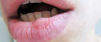
A fistula is a white ball on soft tissues that appears under or on a tooth. There is a hole on the surface for the release of pus. If, when pressed, suppuration flows out of the bladder, the patient does not feel pain. If the hole is closed with pus or bloody clots, the patient will experience discomfort with any impact.
A fistula is often formed due to advanced periodontitis, accompanied by periodontal hyperplasia. Overgrown tissue is good soil for the proliferation of microorganisms. In this case, the patient urgently needs treatment for periodontitis.
In the absence of therapy, the fistula enters a chronic stage, which can only be eliminated through surgical treatment. The progression of the disease must not be allowed, otherwise you may lose healthy molars.
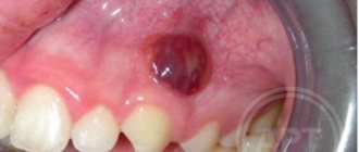
Hematoma is a round lump on the inner surface of the cheek. Sometimes it occurs in the form of a dark bluish swelling on top of the gums. Blood accumulates in or around the root of the molar. The mucous membrane grows, the patient experiences discomfort and cannot completely close the jaw. The main reasons: consequences of filling or tooth extraction, gum damage, poor blood clotting.
Hematomas are generally not dangerous. Processes take place in the body through which soft tissues are cleared of bloody clots. After some time, the bubble disappears, but if the seal remains, you need to visit a dental clinic.
Exostosis is a hard blister that is an abnormality in which the bones protrude and protrude from the jaw. Gradually, the lump increases in volume, which causes discomfort and pain. Exostosis can be provoked by various reasons:
- jaw damage;
- heredity;
- congenital disorders;
- tissue diseases after molar removal.
An examination by a dentist or an x-ray will help detect the disease. The formation will need to be removed if the development of a malignant tumor is suspected.
Epulis is a pedunculated bubble in the form of a mushroom-shaped growth on the periodontium. The tumor may be the same color as the gums or red. The reasons causing the development of pathology include:
- improper filling of the molar or too large a filling;
- dental plaque, stone;
- jaw damage;
- malocclusion;
- hormonal imbalance;
- poor prosthetic material or incorrect prosthetics.
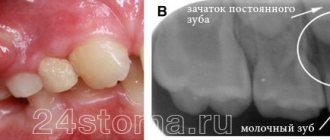
The symptoms of epulis resemble gingivitis; for diagnosis, the dentist prescribes radiography and histology to the patient. With their help, the degree of destruction of bone tissue at the site of the epulis lesion is determined. Mostly, pathology occurs in children during the growth of primary molars and in women.
Papilloma or fibroma is a bubble on the gum, sometimes a benign formation that does not pose a threat to the health or life of the patient. They are formed in people of different genders and ages. Predisposing factors to the appearance of a lump can be: damage to the mucosa, stress, systemic pathologies, heredity.
Papilloma is an enlargement of the papillary layer of skin. The bubble grows gradually, but with reduced immunity, systemic pathology, or stress, growth accelerates, but without turning into a malignant tumor. A papilloma neoplasm looks like a smooth, soft lump on the mucous membrane of a white or pink shade on a thin stalk.
Papilloma often does not create any discomfort. But after some time it may increase in size. You should consult a dentist and get tested.
The first signs of cancer and benign tumors
If a lump appears on your chin, you should visit a doctor. You won't be able to solve the problem on your own. Ignoring an obvious symptom and other characteristic signs can cost the patient’s life.
Signs of a benign neoplasm: the lump at the bottom of the chin is soft and mobile to the touch; upon palpation, slight discomfort or severe pain may be felt. In the latter case, the symptom indicates severe inflammation.
Symptoms of a compaction of a malignant nature include a hard neoplasm to the touch, it is not mobile, as if it is growing “inside” the face, there is severe pain in the absence of touching the pathological element.
Lipoma
A large pimple on the chin may be a lipoma. Lipoma is a benign formation that is formed from growing adipose tissue. In 98% of clinical pictures, there are no subjective sensations other than appearance.
If the tumor grows rapidly, pain is observed due to compression of surrounding tissues. The lump is soft and elastic to the touch, the size varies from 1 to 5 centimeters, most often it forms on the neck.
Folliculitis
It is a skin pathology. In medical practice, the disease is classified as a form of superficial pyoderma. The disease is accompanied by inflammatory processes in the upper parts of the hair follicles. It is infectious in nature.
There are several types of folliculitis, which are caused by a specific pathogen. There are staphylococcal, candidal, herpetic, acne and other varieties.
Malignant tumor
How to treat a boil on the stomach?
A tumor containing malignant cells appears as a cancer. It is difficult to describe the initial symptoms of cancer, since there are many types of them. External manifestations include swelling and compaction under the skin. They usually develop in advanced stages.
Additional symptoms: painful lymph nodes, enlarged liver, neurological phenomena (dizziness, constant headaches), joint pain, non-productive cough (sometimes mixed with blood).
The main methods of therapy include surgery, chemotherapy, radiotherapy, taking hormonal pills, and the use of powerful medications aimed at strengthening the immune status. The prognosis is determined by the type of disease and timely diagnosis.
Skin cyst
A closed cavity neoplasm of the skin and mucous membranes, which is lined with epithelial tissue or epidermis, containing contents of varying consistency - a skin cyst of true nature. The pseudocyst lacks an epithelial lining.
The compaction in the form of a cyst tends to grow quickly, which leads to subsequent rupture of the walls of the neoplasm; the contents enter the dermis, which provokes the development of a strong inflammatory process. Pain syndrome is noted.
Atheroma
The compaction is of a benign nature, contains sebum inside, and does not pose a threat to human life and health. With active growth, it can compress neighboring tissues, which leads to inflammation.
For your information, visually atheroma appears as a painful compaction that has clear boundaries and rises above the surface of the skin. Inside the element is the secretion of the sebaceous glands, epithelial cells, hairs - they look like a cheesy substance, placed in a capsule. It is the capsule that prevents the contents of the atheroma from “escaping.”
The “ball” under the skin increases relatively slowly. However, in the absence of proper therapy, it can reach up to 5 centimeters or more.
If the formation on the gum causes discomfort
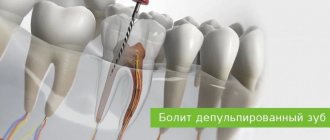
If a bubble near a molar causes pain, this indicates an infectious inflammatory process. Discomfort is also typical with gum injuries and hematomas. Painful formations can be caused by a cyst, cancer, fibroma, periostitis, periodontitis.
Gingivitis - the disease affects only one gum, and the periodontium near the molar remains uninjured. The main symptoms of the pathology are swelling, bleeding, and peeling of the epithelium. Gingivitis often occurs against the background of halitosis. Sometimes pathology develops as a result of endocrine and metabolic disorders.
When treating, it is necessary to eliminate the cause of gingivitis. Professional oral hygiene, diagnosis and treatment of metabolic disorders are performed. To eliminate inflammation and prevent further spread of bacteria, antibiotic therapy is administered. For pain and severe discomfort, analgesics are prescribed.
Periodontitis - purulent bumps on the gums appear as a result of degeneration of the alveolar process, through which the tooth root is held in the alveolus. Tissues can be destroyed due to poor oral hygiene and internal pathologies.
In the initial stages, periodontitis is treated by teeth cleaning at the dental clinic and further proper care. An advanced form of the disease, in which the teeth become loose, pus accumulates, requires surgical intervention - restoration of the alveolar processes or removal of molars.
Periostitis or flux is a dense formation near a problematic molar with carious lesions. Patients complain of pain radiating to the temple, chest, neck, ear. The condition gradually worsens and the temperature can rise to 38 degrees. In the oral cavity, lesions due to periostitis are hyperemic. A purulent fistula forms. When the discharge is removed, the discomfort decreases.
Causes of periostitis: trauma, periodontitis, osteomyelitis, weak immune system, infectious pathologies occurring in the body and vitamin deficiency.
Lump under the chin
An internal lump on the chin under the skin appears as a neoplasm that may appear suddenly. In the evening a person falls asleep without having problems with the skin, and in the morning he discovers a cosmetic defect.
Most often, the location of the pathological element is under the lower jaw or in the occipital region. It is there that the lymph nodes are located, which tend to increase due to existing inflammatory processes.
In case of viral, infectious and bacterial pathologies, activation of these nodes is observed, as a result of which they become larger. The lymphatic system in the human body acts as a kind of filter that prevents inflammation from spreading throughout the body.
Advice: When a lump in the chin area feels hard, stiff and extremely painful, it is recommended to immediately consult a doctor. These signs indicate a developing tumor, which, unfortunately, is not always benign.
Note that if the lump appears within a few days or even a couple of hours, then there is no need to panic, it is definitely not cancer. Oncological compactions develop gradually and relatively slowly, and other negative signs are always observed.
Treatment of neoplasms in the oral cavity
A lump under the jaw or on the gum can be detected upon examination. For an accurate diagnosis, a biopsy or x-ray is prescribed. Treatment is selected taking into account the cause of the formation of a bubble in the oral cavity. It will be necessary to stop the further spread of infection and eliminate discomfort.
The doctor will determine how to remove the lump after a complete diagnosis, but the main measures include:
- fistula - pus is removed by rinsing with a disinfectant solution;
- epulis - surgical intervention (removal using diathermocoagulation, cryodestruction or a scalpel);
- periodontitis - removing fillings, cleaning canals, removing pus, rinsing with herbal decoctions and soda solution;
- periostitis - placement of special drugs under a temporary filling (if there is no result, the tooth will have to be removed);
- gingivitis - cleaning of periodontal tubules, antiseptic and antibacterial therapy, removal of lumps.
When a formation appears on the gum, you can rinse your mouth with vodka, diluted alcohol or juice from fresh Kalanchoe leaves.
Timely diagnosis allows you to prevent further spread of infection and save teeth.
Only an experienced dentist will help you get rid of a lump in your mouth, minimizing the risk of developing serious complications. At the first signs of pathology, you should immediately consult a doctor. The doctor's consultation
Treatment
Most lumps are removed through surgery. However, there are formations that are treatable.
For example, boils are treated with antimicrobial and disinfectant drugs.
If the doctor expresses concerns about the identified lump, then it is worth starting treatment as quickly as possible.
After all, benign tumors eventually develop into malignant tumors, the treatment of which takes much more effort, time and money.