Mucosal melanoma is a relatively rare disease and accounts for less than 1% of all melanomas.
These formations have a much more aggressive growth compared to cutaneous forms, are prone to active metastasis to regional and distant sites, and often recur, which causes high mortality rates. The prognosis for mucosal melanomas is poor, with a five-year survival rate of 10–15%.
Melanomas of the mucous membranes of the head and neck region account for half of all mucosal melanomas. They are mainly localized in the projection of the upper respiratory tract, oral cavity and pharynx. Other forms of mucous melanomas belong to the urogenital area. The distribution of tumors by location is presented in the table.
| Localization of melanoma | Prevalence |
| Mucous membrane of the head and neck | 50% |
| Rectal mucosa | 25% |
| Mucous membrane of the female genital area | 20% |
| Mucosa of the digestive tract, conjunctiva and urethra | 5% |
According to scientists, unlike other dermatological cancers, mucosal melanoma does not depend on exposure to ultraviolet radiation. In addition, there are no obvious risk factors for this type of tumor, including family history.
Melanoma of the mucous membranes affects the following organs:
- oral and nasal cavity;
- paranasal sinuses;
- trachea and bronchi;
- lips;
- pharynx;
- esophagus;
- stomach;
- intestines;
- gallbladder;
- anorectal area;
- vulva and vagina;
- urethra and bladder;
- conjunctiva of the eye.
For convenience, mucosal melanomas are sometimes divided into three subgroups:
- melanoma of the gastrointestinal mucosa;
- respiratory;
- genitourinary melanomas.
Given the tendency to early lymphogenous and hematogenous metastasis, it is sometimes difficult to determine whether a mucosal tumor is primary or metastatic. Depending on the location, the tumor will have certain characteristics. For example, primary melanomas of the oral cavity, nose, pharynx, as well as the anorectal and genital areas first develop in a radial direction, increase in area, taking the form of a spot; only then do they gain volume, rising above the surface of the mucosa, and begin to infiltrate the underlying base.
Some mucosal melanomas develop from melanocyte cells that are present in the tissue structure of the organ (lips, nose, oral cavity, anorectal area, etc.). The development of primary melanomas on the mucous membrane of organs where pigment cells are initially absent (trachea, bronchi) can be explained by disorders of tissue embryonic development.
Types and classification
In accordance with the international classification of cancer stages (TNM) from 2022, several types of mucosal melanoma are distinguished.
By tumor prevalence:
| T3 | Neoplasms limited to the mucous membrane and lying directly under the soft tissues, regardless of the largest size or thickness |
| T4a | Neoplasms spread to bone structures, deep-lying soft tissues, skin, cartilage |
| Т4b | Tumors spread to the hard muscle, zygomatic bone, cranial nerves, carotid artery |
Based on the presence of regional metastases, tumors are divided into three types:
| Nx | The presence of regional metastases cannot be assessed |
| N0 | There are no regional metastases |
| N1 | Regional metastases are present |
Based on the presence of distant metastases, melanoma of the mucous membranes can be of three types:
| cm0 | No distant metastases were found |
| sM1 | Distant metastases are present |
| рМ1 | Histologically confirmed distant metastases are present |
What are the causes of melanoma?
DNA is the genetic material in our cells. It passes on genetic information to the next generation, making children, for example, look like their parents. In addition to information regarding hair color, facial features, and other aspects of appearance, DNA also contains information for a cell in the body about how to grow and perform activities necessary for life.
Ultraviolet radiation can damage DNA. Most melanomas have abnormalities in the chromosomes, where the DNA is located. This damage makes the DNA less able to control cell growth and division. In some situations, this leads to the onset of cancer development. Most ultraviolet radiation comes from sunlight, but some comes from artificial sources such as ultraviolet booths. Some such exposures may occur several years before cancer occurs. However, the tumor develops as a result of exposures that occurred many years ago. Children and young adults often receive multiple intense sun exposures that may not become apparent for years or even decades.
The relationship between DNA and melanoma is currently being studied. It has been discovered that the DNA of certain genes is often damaged in melanoma cells. Most of these DNA changes are not inherited and may be the result of sun exposure. It is believed that some people are better able to repair damaged DNA than others and are less likely to develop melanoma. By understanding the causes of DNA changes that lead to melanoma, it will be possible to use gene therapy to repair the resulting DNA damage.
In some melanomas, DNA changes can be inherited. Inheriting certain mutant genes from one parent may increase the risk of developing melanoma. Research is currently underway to identify altered genes using blood tests.
Although most nevi (moles) never develop into melanoma, in some cases they do. Some changes in benign nevus cells can lead to their transformation into melanoma cells. However, it is not yet known exactly why some moles become cancerous and why having multiple moles (nevi) or atypical moles increases the risk of melanoma.
Symptoms
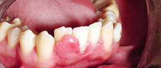
The disease is most dangerous because in about a third of cases there are no signs, that is, melanoma is asymptomatic. The other two-thirds of patients note the following first signs of the disease:
- ulceration of the mucous membrane;
- bleeding in the area affected by the tumor;
- nodular or mocular neoplasm, which can have various shades - reddish, brown, purple, black, gray, white;
- first noted pain caused by wearing orthopedic devices in the oral cavity.
Diagnostic methods
In order not to miss this disease and to identify it at the earliest possible stage, you need to regularly visit an otolaryngologist and dentist for routine preventive examinations of the mucous membranes of the oral cavity and ENT organs.
The following laboratory and hardware techniques are also used to detect pathology:
- MRI, CT, ultrasound (examination of secondary damage to areas in which regional and distant metastases are located is carried out);
- biopsy, after which the resulting biomaterial is sent to the laboratory for cytological and histological examination;
- determination of the presence of mutations in the BRAF and C-KIT genes.
Morbidity statistics
Detection rate
Melanoma is quite rare - from 3% to 6% of the total number of malignant tumors. It is considered the most aggressive type of skin cancer, characterized by rapid growth and early metastasis. Of all deaths due to malignant skin tumors, it accounts for 80%. The average annual increase in the incidence of the disease in Russia is 3.9%.
Age and gender of patients
The tumor is mainly detected between the ages of 30 and 60 years, and also occurs in older people. Recently, the disease has become “younger” and is increasingly diagnosed in patients aged 24 to 40 years. The average age of patients is 52 years, more than a quarter of cases are detected in people under the age of 45.
Under the age of 40, neoplasms are more common in women. After 40 years, the incidence is higher in men and, as age increases, this trend becomes more pronounced - at the age of 65, men get sick twice as often, and at 80, three times more often than women.
Dependence on race and skin color
The risk of melanoma is highest in Caucasian people with fair skin. Hispanics, Asians and African Americans are less susceptible to the disease. At the same time, African Americans have the lowest survival rate.
Survival
The average mortality rate from melanoma in Russia is 2.13-2.52 per 100 thousand population. The average annual increase in the mortality rate is about 1.5%. Detecting a tumor in the early stages allows you to achieve almost 100% survival within 5-10 years.
Treatment
When treating such a disease, a multidisciplinary, that is, an integrated approach is used. The following treatment methods are used in various combinations:
- surgical intervention in which the primary lesion is removed, and then the issue of the need for cervical lymph node dissection (removal of lymph nodes in the neck) is decided;
- adjuvant radiation therapy;
- adjuvant chemotherapy;
- immunotherapy (if the presence of a mutation in the BRAF and C-KIT genes is confirmed).
Can melanoma be prevented?
The most important way to reduce your risk of melanoma is to reduce your exposure to intense sunlight.
Stay in the shade. The simplest and most effective way to limit your exposure to ultraviolet rays is to minimize your exposure to sunlight. This is especially important from 10 a.m. to 4 p.m., when the effects of ultraviolet rays are most pronounced. Remember that the sun's rays can be reflected from water, clouds, sand, cement and snow.
Protect your skin with clothing. You can protect most of your skin with clothing, such as long-sleeve shirts and a wide-brimmed hat. Thick, dark-colored fabric usually works well to provide the best protection for the skin.
Use of protective creams. Use protective creams, especially in cases where sunlight is intense. Use creams even on cloudy and cloudy days because ultraviolet rays penetrate clouds and fog.
Sunscreens should be applied to unprotected areas of the skin 20-30 minutes before going outside so that the skin absorbs the cream. Apply a thick layer to face, ears, arms, legs and neck. Remember that skin treatment must be repeated every 2 hours. It is also recommended to treat the lips.
Protective creams and products are not used to keep you in the sun for a longer period of time. These products do not prevent melanoma, they only reduce the intensity of exposure to ultraviolet rays.
Wearing sunglasses. Sunglasses provide 99-100% protection to the eyes and the skin around them from exposure to ultraviolet rays.
Avoid other sources of ultraviolet radiation. The use of ultraviolet lamps is hazardous to health, as their light can damage the skin, so their use is not recommended. These lamps increase the risk of melanoma.
Protecting children from the sun. Children deserve special attention as they spend a lot of time outdoors and get sunburned quickly. Older children should be aware of the dangers of prolonged sun exposure and the possibility of melanoma. In high mountain areas and areas with intense sun exposure, the use of protective equipment should become a habit for you and your children.
Identification of altered moles (nevi) and their removal. The presence of certain types of moles (nevi) is accompanied by an increased risk of melanomas. Depending on the appearance of these moles, your doctor may recommend careful monitoring or removal if malignant degeneration is suspected. Routine removal of multiple moles as a preventive measure for melanoma is not recommended. If you have multiple moles, regular monitoring by a dermatologist is recommended, as well as monthly self-examination. If you discover an unusual mole or its change, you should immediately contact a specialist.
Genetic counseling. If more than one member of your family has had melanoma, if you have had multiple melanomas, or if you had melanoma at a young age or dysplastic nevi, you may have a gene mutation(s). In this regard, genetic counseling is necessary. In some families with a high frequency of melanomas, a mutation of the CDKN2A gene has been found.
Stages of the disease
We suggest you familiarize yourself with the stages of the disease. The staging table is based on the International Cancer Staging (TNM) classification. Each stage is assigned to certain types of melanoma, which we presented above in the tables.
| Stage | T | N | M |
| III | T3 | N0 | M0 |
| IVa | T4a T3-T4a | N0 N1 | M0 |
| IVb | T4b | Any N | M0 |
| IVc | Any T | Any N | M1 |
Forecast
The survival rate of patients who received combination treatment is 4.5-49.2%.
Factors that significantly influence survival prognosis:
- are there regional metastases;
- are there distant metastases;
- what stage – IVa, IVb, IVc;
- invasion (germination) of the neoplasm into the underlying tissues;
- non-radical (incomplete) surgical removal of the primary focus of the disease;
- localization of the tumor (for example, in patients with MSO of the oral cavity, regional metastases appeared in 36.4% of cases, in patients with MSO of the nasal cavity and paranasal sinuses - in 7.8% of cases).
Provoking factors
The direct causes that influence the formation of gingival melanoma have not been established. However, there are various factors that have a negative impact on soft tissue, which creates a favorable environment for the development of tumor cells on the gums.
Diseases
Chronic and inflammatory gum diseases (periodontitis, papilloma and others) can give rise to malignant neoplasms if not treated in a timely manner.
Damage
People whose gums are regularly exposed to mechanical damage are more likely to develop melanoma. Crowns, dentures and piercings often lead to such damage.
In addition, unprofessional tooth extraction also damages the gums and can lead to inflammation and further development of cancer.
Wrong lifestyle
Smoking, drug addiction and alcoholism are considered provocateurs of various diseases, including gum melanoma.
Poor nutrition
The soft tissue of the gums is very sensitive. Eating too hot or spicy food makes them more vulnerable over time.
If a person has a combination of the above reasons, then the risk of developing gum melanoma increases at least twice.