MAKE AN APPOINTMENT
- Removal cost
- Indications
- Preparation
- Methods and steps
- Complications
- What to do after the procedure
- Our doctors
- Reviews
- To make an appointment with a doctor
In practice, specialists at Plomba dentistry periodically encounter cases of complete destruction of the crown of a tooth and infection of the root system, in which a purulent-inflammatory process occurs. Most often, this problem can be solved only by removing the root and remains of the tooth. But sometimes the root system can be preserved for subsequent prosthetics. How the removal will be carried out, what measures and tools will be used, the doctor will be able to decide after familiarizing himself with the clinical picture.
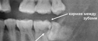
Indications for tooth root removal
- Complete destruction of the coronal (supra-gingival) part, affecting the root system;
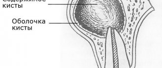
- extensive purulent-inflammatory process at the root: cyst, abscess;
- longitudinal axial fracture;
- previous incorrect extraction - during removal, fragments remained in the hole, causing an inflammatory process that affected nearby tissues.
The damaged area of the tooth is easily identified visually. Additional symptoms of the need for urgent medical intervention and even possible removal of a diseased tooth are:
- twitching, throbbing pain;
- acute pain due to mechanical action - pressing, biting, chewing food;
- unpleasant odor;
- gum hyperemia;
- a purulent process is a direct indication of the need to remove the root of a diseased tooth;
- elevated body temperature.
If one or more symptoms are present, the destroyed units are removed. In some cases, incomplete removal is performed - resection of the tooth roots. This usually occurs when the root apex is affected by periodontitis, a small cyst, or granuloma. Often in such diseases the coronal part is preserved. In this case, the damaged part is removed through an incision in the gum. Subsequently, installing a crown solves the problem of restoring the chewing unit.
Preparation
Before the procedure, the doctor conducts a consultation, a detailed examination and an x-ray of the tooth to be extracted: the degree of damage to the dental crown and the presence of anomalies are revealed. Based on the diagnostic results and radiographs, a clinical picture is formed, the number, size, shape of the roots, their relationship with the mandibular canal and maxillary sinus, and the size of bone resorption are established.
When removing 8 units, be sure to take into account its position, the width of the retromolar space, and the depth of immersion. Based on these data, the doctor draws up a surgical procedure and selects surgical instruments.
If the patient has an inflammatory focus in the periodontium, antibiotics are prescribed 24 hours before the planned surgery. Patients who are terrified of the dental chair are given sedative preparation.
Preparing for tooth extraction
Removing a tooth or its roots is a rather complex surgical dental procedure, but it will not be difficult for the patient to prepare for it. If local anesthesia is planned to be used for pain relief, the patient should eat a large meal before visiting the dentist because:
- After removing a tooth or root, it is forbidden to eat for several hours;
- salivation after eating is significantly reduced, which will make the dentist’s work easier;
- after eating, blood glucose levels are normalized and the risk of loss of consciousness under the influence of local anesthesia is reduced
In the case of general anesthesia, on the contrary, it is necessary to abstain from eating for several hours before the removal procedure begins. Drinking alcohol before visiting the dentist is prohibited. Alcohol affects the structure of the blood and does not combine well with anesthetics, not to mention the negative impact on the human psyche and behavior.
Inflammatory and infectious diseases of any nature must be cured before surgery to remove a tooth or its roots. The dentist must be warned about the presence of allergies to certain medications, in particular to anesthesia drugs.
A normal pregnancy in general is not a contraindication to dental procedures. However, during this period the use of a number of drugs used in dentistry is prohibited, so information about pregnancy is entered into the patient’s dental record. Also, detailed information about the patient’s chronic diseases, especially heart pathologies, is recorded in the dental record.
Preventive measures
In order to avoid serious complications after molar tooth extraction, you need to follow a number of recommendations:
- 20 minutes after the end of medical procedures, the gauze swab should be removed from the socket of the extracted tooth;
- In order for healing to occur faster, you should not drink or eat for the first 2-3 hours after surgery;
- for several days you should avoid eating solid foods, as well as spicy and hot foods and drinks;
- hot water procedures and physical activity are contraindicated until the hole is completely healed;
- You cannot wash the hole yourself or try to clean it with foreign objects that have not been disinfected;
- During the first 24 hours after surgery, you should not rinse your mouth;
- A cold compress on the right side of the cheek will help prevent swelling and reduce the likelihood of bleeding.
If the hole does not heal for a long time or it seems that there are remains of a tooth in it, then you should contact your dentist again and under no circumstances try to solve these problems yourself.
Methods and stages of removing teeth or their roots
Most often in modern dentistry, only two methods of removing teeth or their roots are practiced:
- removing a tooth from the gum using forceps;
- rocking of the tooth and its rotation around its axis by elevators.
In cases where the roots are deep, the gum tissue can be cut with a scalpel. In general, the process of removing teeth or their roots is divided into the following stages:
- separation of the round ligament from the neck of the tooth (ligamentotomy);
- applying (installing) forceps to the tooth;
- advancing the fixing elements of the forceps under the gum;
- final fixation of the forceps;
- rotation (rotation) or luxation (swaying) of the tooth;
- extracting a tooth or its roots from the socket.
Removal techniques
The dental surgeon determines the extraction tactics individually for each patient. Takes into account the location of the tooth, the type of pathological process, and the degree of spread. Before extraction, the doctor sends the patient for an x-ray. The goal is to obtain accurate information about the roots and structural features of adjacent teeth and the jaw apparatus in which the pathological unit is located.
Simple operation
In the case of a conventional location, removal occurs in a simple way, which takes no more than 10 minutes, taking into account the effect of anesthesia. The dental surgeon performs the operation according to the following scheme:
- a preliminary survey of the patient about allergies to anesthesia drugs, chronic pathologies and the medications he uses;
- administration of a drug for superficial sedation in order to normalize the mental state and ensure a comfortable extraction process (at the request of the patient or according to indications);
- injection of a local anesthetic into nearby tooth tissues (gums, cheek), usually using ultracaine, which is considered hypoallergenic;
- after 4-5 minutes, the surgeon begins the removal process by loosening the tooth in the surrounding tissues;
- after ensuring mobility, the specialist separates the tooth from the gum and with a sharp movement turns it out of the alveolar socket;
- the wound is treated with an antiseptic solution;
- a tampon with a medicinal substance is applied to the wound, after which the bleeding stops for several minutes;
- if after tooth extraction the wound surface is too large, the doctor will apply several stitches, which will prevent the possibility of postoperative infection and bleeding in the future;
- the patient receives recommendations and goes home after stabilizing his physical and emotional condition.
After removal using a simple method, the patient feels well. If the wound bothers you after the anesthesia wears off, it can be easily eliminated by following your doctor's recommendations.
The hard way
When the doctor sees abnormalities on the x-ray, removal will be carried out in a complex way. The experience and skill of the doctor matters.
Stages in complex removal:
- taking anamnesis;
- choice of anesthesia;
- mandatory administration of sedatives to relieve tension in the patient;
- injection of local anesthesia into 5-6 areas surrounding the tooth to be removed;
- the doctor makes an incision in the gum to access the tooth and roots;
- drilling into several parts using a drill for easy removal and prevention of injury;
- the tooth is separated from the gum in parts and removed;
- a mandatory revision of the resulting hole is performed;
- the wound is treated with an antiseptic and sutured;
- To prevent infection and bleeding, the doctor places a drug or PRP membrane in the wound. They dissolve along with the sutures.
Extraction during the normal course of the process takes 20-30 minutes. If the operation is complicated, the procedure may take up to 2 hours. After a complex removal, the wound is wider, the tissue is more traumatized, the pain is more pronounced and lasts longer.
It is important that removal is carried out carefully, taking into account future implantation. Therefore, it is better to entrust the operation to an experienced maxillofacial surgeon who will not damage the alveolar ridge. In this case, you will not have to build up bone tissue before installing the implant and eliminate possible complications. Average surgeons usually do not stand on ceremony during operations. The result is broken bone structures of the jaw, wandering remnants of roots, unremoved cysts growing into the maxillary sinus, perforations, fistulas, osteomyelitis and much more. Not to mention the shocking post-operative pain that overtakes the patient after such punitive surgery. Only the maxillofacial surgeon has enough theoretical and practical skills to perform tooth extraction without complications.
Common complications after tooth extraction
Tooth extraction is essentially a full-fledged surgical intervention. Symptoms such as pain and inflammation in the surgical area are considered normal for the rehabilitation period, unless they are too severe and are not eliminated 3-4 days after tooth (root) extraction. The rehabilitation period may also be characterized by increased body temperature and enlarged lymph nodes.
More serious clinical complications include:
- renewed bleeding from the socket after tooth (root) removal - methods for eliminating minor bleeding can be discussed by the dentist; in case of intense bleeding, it is necessary to urgently consult a specialist;
- incomplete removal of the tooth root - the presence of residues is diagnosed by x-ray and follow-up and quickly eliminated;
- alveolitis is a dangerous, but easily eliminated by antibiotics, inflammatory process in bone tissue, characterized by a significant increase in body temperature, swelling, severe pain, and requires immediate treatment, as it can lead to sepsis.
In the first hours after removal, the patient should not eat. Cotton swabs from the surgical area can be removed 30 minutes after completion of all manipulations. In the first days after surgery, it is not recommended to eat sour, sweet, salty, very chilled or hot foods. Short-term cold compresses can relieve pain in the first days after tooth extraction.
Tooth extraction is a last resort in modern dentistry. A qualified specialist must strive by all means to save the tooth even in the most difficult cases.
The final stage of restoring oral health is not the removal, but the replacement of teeth (except for wisdom teeth).
Impacted wisdom tooth
These are usually the hardest eights to remove out of all the others. We have already numbed the surgical field. What's next?
It's under the gum! So, we take a scalpel in our hands and make a delicate incision in the area of the tooth being removed. This creates access to the wisdom tooth being removed. It is isolated from the surrounding tissues using special instruments, and now we can visually assess its position and choose a removal technique.
If the tooth does not erupt, it means that something is preventing it. This “something” will also interfere with its removal, and this “something” could be a neighboring tooth, a bony protrusion, etc. However, you won’t also remove the seven to get to the wisdom tooth, right?
Special surgical tip for removing wisdom teeth. Rotates at the right frequency, provides the right torque, does not burn tissue or inflate emphysema. In surgery, we use only such devices.
Therefore, we divide the tooth into parts. Using a special tip with a cutter speed of 150,000 rpm - this is no longer a simple angle cutter, but not yet a turbine cutter. The latter, by the way, is highly undesirable to use for removing teeth, because at 500,000 rpm it is easy to burn everything with a hellish flame, and with air from the cooling nozzle you can also inflate emphysema over half your face. In general, for removal you need to choose the right tools; there are no trifles or compromises here and cannot be. And you should think a hundred times before removing such problematic teeth in a one-chair dental office at a rural club on the “Half-Empty Bins” collective farm.
Impacted teeth are removed mainly with an elevator, and not with forceps, as many are accustomed to thinking
So, we divide the tooth into 2-3 parts in order to remove it carefully and with little trauma to the surrounding tissues. And teeth are usually removed using an “elevator” (in the picture on the left). Forceps, which everyone associates with removal, are actually used extremely rarely.
Well, the tooth has been removed. Next, we clean the tooth socket from “sawdust” and small tooth fragments that might remain. Using a curette.
When removing wisdom teeth, no biomaterials are used; the hole is filled with a blood clot on its own, this is quite enough for normal healing.
Moreover, “pushing” biomaterials into the hole can complicate the healing process, so let the regeneration process take place naturally and simply, and not fancy, as some doctors suggest.
After removal, resorbable (absorbable) sutures are placed on the hole; most often they do not need to be removed.
The clot is in place. Next, we bring the edges of the wound together and put stitches so that food doesn’t get stuck in the wound, it doesn’t bleed too much, and it heals faster. But at the same time, the sutures should not be tight, because the wound may bleed significantly during the first 24 hours. And if you don’t create an outflow, edema often develops.
What to do after tooth extraction
The operation cannot be prescribed without an X-ray examination, which makes it possible to determine the amount and degree of root destruction and the extent of the inflammatory process. Before root removal, the patient is given anesthesia, after which the gum is separated from the neck of the tooth. The surgeon's further actions are determined by the clinical picture. Dentists call the most difficult operation the removal of the deep and often twisted roots of the “figure eight” – the eighth tooth in the dentition. But the specialists of the Plomba clinic successfully cope with this too.
When is molar tooth extraction indicated?
Only an experienced doctor can make an adequate decision about whether it is necessary or not to remove a molar tooth after a thorough examination. For detailed diagnostics, not only a visual examination is used, but also an analysis of the results of radiography or visiography. Indications for the removal of permanent teeth can be divided into absolute (in which the tooth requires mandatory removal) and relative (in these cases, the opinions of doctors may be divided).
Mandatory removal of a molar tooth is indicated in the following cases:
- there is a high probability that odontogenic osteomyelitis will develop;
- there is a risk of developing phlegmon or abscess.
- tooth root fracture.
Among the relative indications for the removal of molars are the following:
- if due to a dental disease, inflammation has developed in the area of a specific tooth (sinusitis);
- with longitudinal fractures of the tooth root;
- if the root and coronal part are so badly destroyed that therapeutic treatment is fraught with serious difficulties;
- if during the treatment of root canals their walls were damaged;
- tooth mobility is in the third or fourth degree;
- a fracture of the alveolar process, on the line of which the tooth is located, was diagnosed;
- in the oral cavity there are pathologies occurring around dystopic or impacted teeth;
- if orthopedic treatment is being carried out and certain molars cannot be used as support;
- if an orthopedic dentist or orthodontist has prescribed removal of molars and premolars.
As for the relative indications for molar tooth extraction, if you disagree with the opinion of one specialist, you should consult with another. This will help you choose the most gentle treatment and, possibly, save teeth that are at risk of removal.
Prices for complex removal of teeth of the upper and lower jaw
Tooth extraction is a difficult operation that requires special knowledge and skills from the surgeon. Our clinic employs experienced specialists who use modern anesthesia, the latest technologies and professional equipment, so the manipulations are performed efficiently and as painlessly as possible. The price for the service in Moscow varies between 5,000-8,000 rubles.
To find out the cost of complex tooth extraction, book a consultation at our clinic in Moscow by calling: 8 (495) 380-01-38.
| Code no. | Name of procedures | Unit of measurement | Cost, rub. |
| 601 | Application anesthesia | 150,00 | |
| 602 | Infiltration anesthesia, conduction | 550,00 | |
| 605 | Removal is difficult | 1 tooth | 3 500,00 |
* The prices indicated on the website are not a public offer. The exact cost of treatment can only be determined at an appointment with a doctor.
Prices for treatment in Moscow full price list
Share on social media networks:
Article Expert:
Lavrentyev Sergey Sergeevich
Doctor - dentist, surgeon, implantologist. He regularly improves his professional level and studies modern methods of dental implantation and surgical dentistry at international seminars and conferences of outstanding implant surgeons in the world.
Work experience 25 years
Operation stages
- Pain relief
. General or local anesthesia is used, the anesthetic is selected depending on the health status, age, individual characteristics, and allergic status of the patient. - Impacted tooth
. Removing an impacted tooth is made difficult by limited access to the problem tooth - the crown part closed by mucous/bone tissue. After anesthesia, an incision is made into the mucous membrane, the unit is divided with a bur, and it is removed in fragments. - "Eight"
. The eruption of a wisdom tooth in 75-80% of cases occurs against the background of complications and pain, so it is better to remove it in order to avoid problems with the gums and healthy neighboring units in the future. The operation is performed under anesthesia: the doctor dissects the gum and extracts the tooth. The edges of the wound are connected and sutures are applied. - Root
. The complexity of the manipulation depends on the presence/absence of inflammation, the degree of neglect of the diseased tooth, the depth of the branches: the root is cut, the crown is destroyed into individual elements, and all tooth fragments are removed.
Methods and tools for difficult removal
Special instruments are used with soft periodontal tissues:
- tongs/elevators
. They are used to remove the roots of multi-rooted teeth and severely damaged units: the dentist loosens the tooth with the root using an elevator, dislocates it with forceps, and removes it from the gums. For individual groups, there are various forceps, the shape of which is determined by their purpose: for roots, units with a crown, for teeth of the upper/lower jaw; - chisel
_ The device is used when removing the wall (outer) of the alveolar ridge, dense bone structure - when it is impossible to place an elevator in the area between the root and the alveolus: the surgeon places the chisel in the working segment, performs 2-3 blows, dislocates and removes the tooth; - drill
_ Helps the doctor when sawing a unit, dividing roots, removing bone tissue. During the manipulation, the mucous membranes and tissues are damaged to a minimum extent.