Lip cancer is a malignant neoplasm that consists of elements of the integumentary epithelium of the red border of the lips. A tumor on the upper lip appears rarely. Over time, lip cancer spreads to the bones of the lower jaw. Atypical cells are transported with lymph to the lymph nodes, resulting in the appearance of new malignant foci.
Doctors at the Yusupov Hospital carry out early diagnosis of lip cancer using modern research methods. Oncologists provide radiation therapy and perform gentle surgical interventions. In the later stages of lip cancer, complex therapy is used. Early diagnosis of a malignant lip tumor and adequate therapy can not only save the patient’s life, but also avoid cosmetic defects after surgery.
Lip cancer - what is it?
Depending on the type of tumor growth, the following forms of lip cancer are distinguished:
- Papillary;
- warty;
- Ulcerative;
- Ulcerative-infiltrative.
A malignant tumor of the lip in 95% of cases is represented by keratinizing squamous cell carcinoma. In 5% of cases, histologists have squamous cell non-keratinizing cancer of the lip mucosa. A tumor of the lip, which from the inside, on the mucous membrane, is characterized by a malignant course (infiltrative growth and early metastasis to regional lymph nodes).
A tumor of the lower lip most often affects the border of the lower lip. A crack, ulcer or swelling forms on it, which looks like a wart. The disease is predominantly diagnosed in people over 60 years of age.
Cancer of the upper lip occurs less frequently than the lower lip, but the tumor is more aggressive. Upper lip cancer has a high risk of metastasis and spreads quickly. This is explained by the fact that the malignant tumor is located close to the nasal cavity, where the blood supply system is developed. A tumor on the inside of the lip is dangerous because it quickly grows into the soft tissue.
Melanoma on the lip is 10 times less common than squamous cell carcinoma of the lip, but is characterized by a high degree of malignancy. Basically, the tumor is located on the red border of the lower lip. It penetrates deeply into tissues, quickly transfers to nearby tissues and gives metastases.
At the Yusupov Hospital, oncologists make a diagnosis of lip cancer using the following methods:
- Examination with the naked eye and using stomatoscopy;
- Cytological examination of impression smears from a tumor ulcer;
- Histological examination of lymph node punctate.
After establishing the primary diagnosis, a comprehensive examination of the patient is carried out.
Option 2. Labia too large
In the case of the labia majora, the consequences of dysfunction are similar:
- Excess tissue rubs, gets injured, and in some patients even forms folds where secretions accumulate, which are an excellent habitat for bacteria.
- Playing sports or any other serious physical exercise leads to microtraumas, wounds, tissue hypoxia and, as a result, susceptibility to various infections. Irritated tissue is easily permeable to microbes, which leads to diseases, papillary and even cancerous lesions.
- Another big problem for patients with long labia is difficulty during sexual intercourse, during which the lips are retracted deep into the vagina.
Such pathologies are a medical indication for reducing enlarged labia using labiaplasty. This operation involves giving the correct shape to the labia minora (labiominoroplasty) or labia majora (labiomyoroplasty).
Labiaplasty
Causes of lip cancer
Lip cancer develops for many reasons:
- From smoking;
- After a long stay in the sun;
- As a result of inflammatory processes of an infectious and non-infectious nature;
- Under the influence of high temperatures;
- In the presence of microtraumas;
- Due to prolonged exposure to chemicals.
Lip cancer often develops from smoking. Men get lip cancer much more often than women. Currently, the trend is changing, because many women suffer from tobacco addiction. Doctors believe that lip cancer develops due to smoking strong cigarettes for a long time.
The average smoker smokes at least ten cigarettes a day. In this case, the paper surface is constantly in contact with the lips. The skin here is especially delicate and sensitive. Microcracks appear on its surface. They are invisible to others and do not cause problems for the smoker. Damaged areas of the epithelium are affected by tobacco smoke. It contains a lot of harmful substances. Skin cells begin to degenerate.
The cause of mechanical trauma that causes lip cancer can be improperly made dentures, the habit of holding various objects with the lips (nails, the mouthpiece of a smoking pipe), or biting the lower lip. The mechanism of development of lip cancer is as follows: a long-term non-healing crack, wound, inflammation on the lip, papilloma develops into leukoplakia, Manganotti cheilitis, keratoacanthoma, warty form of dyskeratosis or other precancerous diseases. Against this background, lip cancer occurs.
Precancerous diseases of the lips
Lip cancer does not occur on healthy mucous membranes. Malignant neoplasms develop against the background of obligate or facultative precancerous diseases. Obligate precancerous diseases include:
- Abrasive precancerous cheilitis Manganotti;
- Warty precancer of the red border;
- Limited precancerous hyperkeratosis of the red border.
Facultative precancerous diseases with greater potential for malignancy are:
- Erosive and verrucous leukoplakia of the lip;
- Papilloma;
- Keratoacanthoma;
- Cutaneous horn.
Malignant neoplasms of the lip can develop against the background of optional precancerous diseases with less potential malignancy:
- Flat leukoplakia of the lip;
- Chronic ulcers;
- Ulcerative and hyperkeratotic forms of lupus erythematosus and lichen planus;
- Chronic cracked lips;
- Post-X-ray cheilitis;
- Meteorological and actinic cheilitis.
Background conditions that are precursors to lip cancer include scars after burns, trauma, surgery, and benign neoplasms.
Heilith Manganotti
Abrasive precancerous Manganotti cheilitis occurs in older people. Small, round erosions appear on the lips, which do not heal for a long time. They have a smooth surface of yellow-red or bright red color. In some cases, a bloody or serous crust appears on the surfaces of erosions. If you remove it, the opened wound bleeds a little. Touching erosions does not cause pain. Once erosion appears, it does not heal for several weeks or months. After disappearing, soon enough new erosions appear in their place or nearby.
Manganotti cheilitis is diagnosed by external examination and questioning of the patient. This disease has symptoms similar to those of herpes, leukoplakia, lichen planus or lupus erythematosus. For differential diagnosis, oncologists scrape the affected area of the lip and send it for a thorough histological examination. This scraping makes it possible to detect emerging cancer cells in a timely manner and prevent the development of a malignant neoplasm of the lip.
Leukoplakia and lip hyperkeratosis
Leukoplakia of the lip is a lesion of the mucous membranes with keratinization of the epithelial tissues. Unfavorable factors that contribute to the development of leukoplakia may be alcohol abuse, smoking and eating very spicy foods. Leukoplakias of the lower lip most often develop in the mucous membranes at the corners of the mouth.
Hyperkeratosis of the lips appears as a limited area from 0.2 to 1 cm in diameter. Its surface is smooth, covered with thin, tightly packed grayish-white scales. Scraping cannot remove them.
Lip tumors
Of all the anatomical parts of the oral cavity, the red border of the lower lip is most often affected. Lower lip cancer accounts for approximately 12 - 25% of the total number of tumors in this location. For reasons that are not entirely clear, tumor lesions of the red border of the upper lip are an extremely rare occurrence. The disease most often occurs in people over 60 years of age. Men are more often affected, the male/female ratio approaches 13/1. Predisposing factors are solar radiation, smoking, and alcohol abuse.
Clinically, the disease manifests itself as an exophytic (i.e., protruding) tumor or, much more often, an ulcerative bleeding surface. Since it is very difficult not to notice this lesion, the vast majority of patients seek medical help at an early stage of the disease. This circumstance (treatment of the early stages of any malignant tumors always brings better results) allows us to count on high effectiveness of treatment. In addition, lower lip cancer rarely metastasizes to regional (cervical) lymph nodes. At the time of initial treatment, involvement of the cervical lymph nodes is diagnosed in only 2–12% of patients. Damage to regional lymph nodes significantly worsens the prognosis of the disease. Thus, the 5-year survival rate in the presence of this lesion drops from 90 to 50% and forces one to resort to more aggressive methods of treatment.
Treatment for lower lip cancer is determined by the stage of the disease. Early stages can be cured with surgery or radiation treatment. There are 2 types of radiation treatment: remote and contact therapy (so-called brachytherapy).
The lip has a rather complex anatomical structure, which includes muscles with different directions of muscle fibers, skin, salivary glands, and subcutaneous tissue. Naturally, the goal of surgical treatment is not only radical removal of the tumor, but also restoration of the structure and function of the lower lip. Modern methods and techniques of plastic surgery can achieve good cosmetic and functional results. This is illustrated by the photographs below of patients who underwent surgical treatment for cancer of the lower lip.
Develops against the background of obligate (warty precancer, limited dyskeratosis, Manganotti cheilitis, Bowen's disease) and facultative (leukoplakia verrucous, keratoacanthoma, cutaneous horn, papilloma with malignancy, erosive-ulcerative and hyperkeratotic form of lupus erythematosus and lichen planus, post-radiation cheilitis) precancerous processes .
Warty precancer is characterized by the appearance on the red border of the lip of a sharply demarcated hemispherical lesion with a diameter of 4 mm to 1 cm, which protrudes above the surrounding tissues and has a dense consistency. Its color varies from an almost normal red border to a stagnant red. The nodule may be covered with thin scales and resemble a wart or keratinizing papilloma. Malignancy can occur 1-2 months after the onset of the disease.
The productive form of focal dyskeratosis is characterized by excessive keratinization of the epithelium and looks like a white spot with flat protrusions on a red border or an area of hyperkeratosis with stylized horny protrusions. The destructive form (erythroplakia) is distinguished by the fact that limited defects of the epithelial cover appear on the red border of the lip - erosions, ulcers, cracks. Malignancy can occur 6 months after the onset of the disease.
Abrasive pre-cancer cheilitis Manga-Notty is characterized by the formation of an oval or irregularly shaped erosion on the lip, often with a smooth, polished surface of a deep red color. Crusts may form on the surface of the erosion, which may cause slight bleeding when removed. There is no tissue compaction at the base and around the erosion. Spontaneous epithelization with subsequent recurrence is possible. Malignancy occurs 3 months to 30 years after the onset of the disease.
Lip papilloma is a clearly circumscribed tumor from 1-2 mm to 2 cm in diameter on a wide base with a villous or rough surface. As keratinization increases, the tumor acquires a whitish tint. Lip papilloma is considered a progressive precancerous disease. Compaction of the base of the papilloma and the appearance of pain are signs of its degeneration.
Treatment of diffuse lesions of the lower lip (cheilitis, leukoplakia, etc.) is predominantly conservative. Focal lesions are treated surgically, laser and cryodestruction.
Lip cancer
The term “lip cancer” refers to malignant tumors that arise in the mucous membrane of the red border of the lip. Neoplasms that have developed on the skin near the lip or mucous membrane of the vestibule of the mouth are not included in this group of tumors.
In the structure of cancer diseases in Russia (1997), lower lip cancer accounted for 1.4% of the total number of malignant neoplasms with an incidence of 2.8 per 100,000 population. The disease is most often observed in men over 70 years of age. In rural areas, lip cancer occurs 1.5-2 times more often than in urban populations. In recent decades, there has been a gradual decline in incidence. Approximately 90% of tumors of the lower lip develop due to exposure to known carcinogens, the main of which are tobacco and tobacco-containing substances. Alcohol is comparable in action to tobacco and is part of a group of agents that can potentiate tobacco-induced carcinogenesis. Other important etiological factors are chronic injuries and inflammation of the lip, ultraviolet radiation, changes in weather conditions, and occupation (work in the oil refining, coal, textile industries).
International classification according to the TNM system
Anatomical areas and parts:
1.Upper lip, red border. 2.Lower lip, red border. 3. Angles of the lips (commissures). T - primary tumor: Tx - insufficient data to evaluate the primary tumor, T0 - primary tumor not determined, Tis - preinvasive carcinoma (carcinoma in situ), T1 - tumor up to 2 cm in greatest dimension, T2 - tumor up to 4 cm in greatest dimension , T3 - tumor more than 4 cm in greatest dimension, T4 - tumor spreads to adjacent structures - bone, tongue, facial skin.
N/pN - regional lymph nodes. Regional lymph nodes for all parts of the head and neck, with the exception of the nasopharynx and thyroid gland, are the lymph nodes of the neck, including: submental, submandibular, nodes at the base of the skull near the great vessels (deep cervical), in the bifurcation zone of the common carotid artery (deep cervical) , posterior cervical (superficial cervical), located along the accessory nerve, supraclavicular, preglottic and paratracheal, retropharyngeal, parotid, buccal, postauricular and occipital.
N/pNx - there is insufficient data to assess the condition of the regional lymph nodes, N/pN0 - there are no signs of metastatic lesions of the regional lymph nodes. pN0 - histological examination of material from a selected area of neck tissue includes 6 or more lymph nodes; histological examination of the material obtained using cervical lymphadenectomy includes 10 or more lymph nodes, N/pN1 - metastases in one lymph node on the affected side, up to 3 cm or less in the greatest dimension, N/pN2 - metastases in one or more lymph nodes on the affected side, up to 6 cm in greatest dimension; or metastases in the lymph nodes of the neck on both sides or the opposite side, up to 6 cm in greatest dimension, N/pN2a - metastases in one lymph node on the affected side, up to 6 cm in greatest dimension, N/pN2b - metastases in several lymph nodes on the affected side, up to 6 cm in the greatest dimension, N/pN2c - metastases in the lymph nodes on both sides or on the opposite side, up to 6 cm in the greatest dimension, N/pN3 - metastasis in the lymph node more than 6 cm in the greatest dimension.
Note. The lymph nodes on the affected side include the lymph nodes located in the midline.
M - distant metastases: Mx - the presence of distant metastases cannot be assessed, M0 - no distant metastases, M1 - distant metastases.
The requirements for determining the рТ category correspond to those for determining the T category.
Grouping by stages
Stage 0 TisN0М0 Stage I Т1N0М0 Stage II Т2 N0М0 Stage III Т3N0М0 Т1-3N1М0 Stage IVA Т4N0-1М0 Any ТN2М0 Stage IVB Any ТN3М0 Stage IVC Any Т Any NM1
Clinic . Cancer most often affects the lower lip midway between the midline and the commissure. The main morphological form is squamous cell carcinoma (80-95%). Metastasis later, mainly to regional lymph nodes (5-9.2%). In the clinical course of cancer of the lower lip, two groups are distinguished - exophytic and endophytic. There are transitional forms between them. Exophytic cancers of the lower lip include papillary and warty forms. The papillary form develops from papilloma or against the background of focal productive dyskeratosis. Exophytic cancer visually represents a tumor of irregular shape (round, lumpy, flat), rising above the surface of the red border. There is infiltration of the underlying tissues without clear boundaries. The warty (fungous) form develops against the background of productive dyskeratosis and is characterized by the fusion of multiple small growths on the lip (“cauliflower appearance”), a gradual increase in infiltration of the underlying tissues and tumor disintegration. Endophytic cancer of the lower lip is represented by ulcerative and ulcerative-infiltrative forms. The ulcerative form is characterized by the formation of an ulcerative defect on the lip with a gradual deepening of the ulcerative surface, its bottom becomes uneven, its shape is irregular, the edges rise above the level of the red border, everted. The ulcerative-infiltrative form is characterized by infiltrative growth, which makes it difficult to determine the boundaries of the tumor. Endophytic forms of cancer are more malignant.
Diagnosis is based on anamnesis, examination, palpation of regional lymph nodes (submental and submandibular lymph nodes are examined bimanually), cytological and/or histological examination data. When making a differential diagnosis, it is necessary to keep in mind tuberculosis and syphilitic lesions.
Treatment . In stages I and II, the standard treatment method is radiation therapy (short-focus radiotherapy, electron therapy, contact radiation, interstitial or combined radiation therapy) with a total focal dose of 60 Gy.
The treatment of choice for stage I lip cancer is cryodestruction. Block resection of the lip with simultaneous plastic surgery according to Bruns is rarely used due to the poor cosmetic effect. Surgery is more indicated in the presence of residual tumor after completion of a radical course of radiation therapy; it is performed within 3-6 weeks after the end of irradiation.
For stage II tumors, 2-3 weeks after completion of treatment of the primary lesion, prophylactic upper fascial-sheath excision of the neck tissue on both sides is performed. The block of tissue removed includes the tissue of the submental and submandibular region and the lymph nodes of the fork of the common carotid artery, the lower pole of the parotid salivary gland.
For stage III lower lip cancer, combined radiation therapy is used (external gamma or electron therapy + interstitial or short-focus radiotherapy). The presence of clinically detectable metastases in the lymph nodes is an indication for preoperative irradiation of the cervical lymphatic collector on the affected side. Preventive or therapeutic fascial-sheath removal of cervical tissue (on both sides) is carried out after complete subsidence of radiation reactions and completion of regression of the primary tumor focus. The method of choice for treatment of stage III lower lip cancer, especially when the tumor has spread to the corner of the mouth, is a combined one. In this case, lymphadenectomy is performed simultaneously with removal of the primary tumor. To eliminate lip tissue defects formed after treatment, primary reconstruction is performed using local or regional flaps.
Treatment for stage IV lower lip cancer is combined. For multiple metastases in the lymph nodes of the neck, treatment begins with preoperative external beam radiation therapy. 2-3 weeks after completion of the first stage of treatment, surgery is performed on the primary lesion with simultaneous cervical lymphadenectomy. Fascial-sheath excision of the cervical tissue is performed for single, dislocated metastatic lymph nodes that are not fused with adjacent anatomical structures of the neck. The presence of multiple displaced metastases in regional lymph nodes or single, but limitedly displaced, fused with the internal jugular vein and the sternocleidomastoid muscle is an indication for performing the Crail operation. In case of bilateral metastatic lesions of the lymph nodes, cervical lymphadenectomy is performed simultaneously on both sides. In elderly and weakened patients, this operation can be done first on one side, and after 2-3 weeks on the other.
In case of unresectable tumors and the presence of contraindications to combined treatment, a course of palliative remote gamma therapy is carried out according to a radical program (60-70 Gy). Chemotherapy is used as part of palliative chemoradiation treatment (see Cancer of the mucous membrane of the floor of the mouth).
Recurrent lower lip cancer is treated surgically and in combination. For late limited relapses (developing 1 year or more after completion of short-focus radiotherapy), a course of radiation therapy (60-70 Gy) can be repeated.
Forecast . According to the summary data of various authors, the five-year survival rate for stage I is 91.2-96.9%, for stage II - 82.8-83.6%, for stage III - 63.5-75.4%, for stage IV - no more thirty%.
Prevention of lip cancer consists of timely treatment of precancerous diseases, quitting smoking, eliminating unfavorable external factors (increased insolation, etc.), and using protective lip creams.
SIGN UP FOR A CONSULTATION by phone +7 (921) 951 - 7 - 951
Share link:
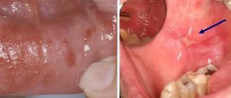
Symptoms of lip cancer
There are local and general signs of lip cancer. Local symptoms of a malignant neoplasm can often be seen on the lower lip. When the pathological process is located on the mucous membrane of the lips, facing the vestibule of the mouth, the tumor has a pronounced malignancy. General signs of lip cancer can develop if the tumor is not detected in a timely manner and treated inadequately in the later stages of cancer.
First symptoms
The first signs of lip cancer usually go unnoticed. First, you can determine the enlargement of the mental lymph nodes. You can notice this by feeling the lower jaw. The next early sign of lip cancer is a swelling of a dense consistency. Itching occurs in it. This neoplasm is usually mistaken for a herpetic rash.
A small ulcer with a crust forms in the center of the swelling, which does not cause pain. If it is removed, the patient feels quite severe pain, and upon closer inspection, he may find a bleeding base, which is formed by tubercles.
Local signs of tumor
Symptoms of lip cancer are:
- Dyskeratosis of the lips;
- Papilloma;
- Erosion;
- Cheilitis.
In most cases, dyskateriosis looks like cracks and ulcers. The erosions are covered with a crust and resemble herpes in appearance, but, unlike it, they do not heal after a certain period of time. Some patients have no ulcers or erosions. Instead, a small compaction appears, which over time grows and becomes covered with a crust.
On the red border of the lower lip, away from the midline, a patch or formation may appear that protrudes above the surface. An erosion or ulcer with a granular surface and a roll-like edge forms in the center of the tumor. The formation has a dense consistency and gradually increases in size, eventually acquiring an irregular shape. Its boundaries are unclear.
Exophytic lip cancer predominantly develops from a warty form of productive diffuse dyskeratosis of papilloma. With exophytic growth, the tumor has a dense consistency, often covered with flat scales. Endophytic growth of a cancerous tumor is characterized by the formation of an ulcer with uneven, dense edges. It often appears against the background of destructive dyskeratosis, quickly infiltrates the soft tissues of the lip and is prone to metastasis.
Symptoms of the disease should be a signal to immediately contact oncologists at the Yusupov Hospital. Lip cancer, treatment of which is started on time, is completely cured in 90% of cases.
Symptoms and first signs
It is known that cancer begins to manifest itself at a time when it is too late to treat it. But if we are talking about lip cancer, then its first signs appear long before the formation of metastases.
Most often, patients complain:
- For the appearance of a lump in the area of the damaged part of the lip.
- The seal causes some discomfort as it increases in size.
- The top of the tumor is covered with a dense film, and when you try to remove it, severe pain occurs.
- Gradually, the compaction grows and increases in size, involving new tissues in the pathological process.
When the process of metastasis begins, a person exhibits other, specific signs:
- lymph nodes enlarge, they lose their mobility;
- their pain on palpation is noted.
The process of eating is gradually disrupted. A person cannot chew food and takes it in liquid form, resulting in exhaustion. The patient loses weight, his body cannot fully absorb nutrients and vitamins.
initial stage
At the first stage of disease development, scanty manifestations of cancer may be present. The person is not bothered by anything at all or the symptoms are mild.
It is worth paying attention:
- The appearance of a non-healing wound in the lip area. This could be a deep crack or ulcer that does not heal over a long period of time.
- The edges of the wound become thicker and painful. Gradually the size of the damage decreases.
- And the compaction grows, involving other tissues in the process.
Since tumor growth can be slow, it is recommended to pay attention to a wound that does not heal. It can bleed and secrete.
Stages
Currently, the generally accepted classification of lip cancer is TNM (T-size of the tumor, N-damage to the lymph nodes, M-metastases). Based on the size of the tumor, there are 4 stages of lip cancer.
Table 1. Stages of lip cancer
| Stage | Tumor size |
| T1 | Less than or equal to 2cm |
| T2 | More than 2-4cm |
| T3 | More than 4cm |
| T4a | The tumor grows into the cortical layer of the bone, tongue muscles, maxillary sinus and skin |
| Т4в | The tumor grows in the bed of the masseter muscle, the pterygoid process, the internal carotid artery and the base of the skull |
If on the affected side there are single enlarged lymph nodes, the size of which is less than 3 cm, this is stage N1 lip cancer. At stage N2, enlarged lymph nodes are detected on the affected side, the diameter of which is more than 3 cm. If the patient has single enlarged lymph nodes on the affected side measuring 3-6 cm in size, this is stage N2a of lip cancer. At stage N2, oncologists determine multiple metastases to the lymph nodes. Their size is equal to or greater than 6cm. In the presence of bilateral metastases in the lymph nodes measuring 6 centimeters, they speak of the N2c stage of lip cancer. If the diameter of the lymph nodes exceeds 6 cm, this is stage N3 of the disease.
In the absence of distant metastases, oncologists determine stage M0 of lip cancer, if there are distant metastases - M1, in the case of distant metastases that cannot be assessed - MX. The diagnosis of “early stage lip cancer” is made in the presence of a tumor less than or equal to 2 cm, the presence of single enlarged lymph nodes less than 3 cm on the affected side and the absence of distant metastases. This is T1 N1 M0.
Diagnosis of a tumor by a doctor in a hospital
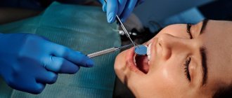
What does lip cancer look like? It could be a small ulcer or a widespread tumor. Doctors at the Oncology Clinic of the Yusupov Hospital make a diagnosis of lip cancer based on the patient’s complaints, the results of an external examination and additional studies. The oncologist carefully examines and palpates the lips, cheeks, gums and regional lymph nodes. When examining the red border of the lips, skin and mucous membrane, use a magnifying glass.
How to identify lip cancer? Further examination is carried out using instrumental and laboratory diagnostic methods:
- Ultrasound examination;
- X-rays of the lower jaw;
- Panoramic tomography;
- Cytological examination of material obtained by taking fingerprint smears from the surface of the ulcer or histological examination of tissue obtained during a biopsy.
For lip cancer with lymphatic metastasis, a biopsy of the lymph nodes is performed. To exclude hematogenous metastases, chest X-ray and ultrasound examination of the abdominal organs are used. When the diagnosis of lip cancer is confirmed, an X-ray examination of the chest organs, general clinical and laboratory examination (electrocardiography, blood and urine tests) are performed.
A PET-CT study (positron emission computed tomography) is prescribed for the following purposes:
- Determining the stage of lip cancer;
- Assessment of response to treatment;
- Detection of disease relapse during the observation period.
PET-CT is an innovative method that combines the capabilities of computer technology and radiology. At the Yusupov Hospital, it is used not only to diagnose lip cancer, but also to monitor tumor development and evaluate the outcome of treatment. Thanks to PET-CT, radiologists have the opportunity to very accurately carry out radiation treatment, significantly reduce the irradiation area, and minimize the impact on healthy organs and tissues.
In many cases, the use of PET-CT excludes a number of additional studies in the future. This allows patients of the Yusupov Hospital to save time and money. The issue of the need to use this method is decided individually for a specific patient at a meeting of the expert council with the participation of professors and doctors of the highest category. A comprehensive examination of patients using the latest diagnostic equipment, the use of modern methods of performing laboratory tests using high-quality reagents allows oncologists at the Yusupov Hospital to obtain reliable results and provide adequate therapy for lip cancer at an early stage.
How to care for your labia?
When the labia are in good condition, normal hygiene procedures are sufficient. Good conditions for the functioning of this genital organ will be provided by: a daily shower, the use of special cosmetics for intimate hygiene and the choice of cotton underwear.
If the labia are deformed and enlarged, secretions can accumulate in the folds of the skin, which is an excellent breeding ground for various bacteria. In this case, an important task is careful care agreed with the gynecologist.
It is very important to properly moisturize delicate intimate areas. To do this, you should use preparations containing hyaluronic acid, which makes the skin smoother and more elastic, and reduces the friction of the lips against each other.
Moisturizing the intimate area
Proper care of the labia not only prevents bacteria and viruses from entering the reproductive system, but also makes the vulva healthy.
Treatment of lip cancer
How to treat lip cancer? When treating the disease, oncologists at the Yusupov Hospital take into account many different factors: the patient’s age, histological type of tumor, features of the spread of the tumor. Doctors at the Oncology Clinic provide multidisciplinary treatment for lip cancer:
- Surgical interventions;
- Radiotherapy;
- Chemotherapy.
Regardless of the chosen technique, they affect the lesion or tumor, areas of regional metastasis. For grades I and II of lip cancer, radiation treatment is performed, which includes external radiotherapy and surgery.
Treatment in the initial stages
Squamous cell carcinoma of the lower lip is a common complex tumor process. Its treatment consists of two stages: removal of the main focus and the fight against metastases that have spread to neighboring tissues and organs. Radiation treatment can suppress the development of a tumor focus. The radiologist selects the method of radiation therapy individually, depending on the size of the tumor, stage of the disease, and age of the patient.
Removal using the Krail method
In the initial stage of the tumor, surgeons perform a wedge-shaped excision of the entire thickness of the lip under local anesthesia. In order to remove a defect on the lip, plastic surgery is performed - skin flaps are transferred from the patient's cheek. Removal of the lesion is performed using the Krail technique. The operation is performed at the first and second stages of lip cancer, when the tumor has not spread to nearby tissues. Surgical excision of carcinoma makes sense in the absence of metastases.
The decision to remove a lymph node is made by an oncologist surgeon if the primary tumor process has already been cured or if the lymph node is removed along with the removal of the main lesion. In some cases, surgeons remove the primary cancerous tumor, the affected lymph nodes and the vessels that connect them. The scope of the operation is determined collectively by the clinic's oncologists.
Due to the fact that metastasis can occur in a cross way, the lymphatic apparatus of the suprahyoid region on the left and right is removed. If the oncologist sees that there are cancer metastases in certain lymph nodes, he decides to remove them, and those that are located along the lymph outflow.
Treatment tactics for lip cancer depend on the stage of the disease:
- Preventive surgery at stages I and II of lip cancer is carried out exclusively in cases where it is not possible to control the dynamics of the disease and there are unfavorable prognoses regarding the spread of cancer;
- At stage III, in the absence of metastases, treatment is carried out using a combined effect on the lesion and adjacent areas;
- In case of spread of cancer cells and the presence of single metastases in the lymph nodes, oncologists at the Yusupov Hospital perform combined treatment of lip cancer with subsequent surgery, plastic surgery and surgical correction of the lips;
- In stage IVC, palliative chemoradiotherapy is performed.
Candidates and doctors of medical sciences use innovative methods of treating lip cancer. The presence of modern equipment and competent medical personnel who are attentive to all the wishes of patients allows us to achieve good results in the treatment of lip cancer for many years. Lip cancer, treated in the early stages, is cured in 97-100% of cases. At stage III, 67-80% of patients can be cured. In the fourth stage of cancer and repeated relapses, complete recovery occurs in 55% of cases.
Option 1. Labia too small
Such cases occur in several cases:
- in elderly patients, which is associated with changes in hormonal levels and general aging of the body;
- in women with a congenital or acquired (due to illness) tendency to tissue atrophy;
- The labia may be underdeveloped from birth.
It happens that the labia have become too small due to the fault of the surgeon, who severely trimmed them during labiaplasty.
When this reproductive organ is too small, the patient may feel pain during intercourse because there is not enough tissue to limit penetration too deep. This leads to injury and even pain in deeper organs.
In addition, if the labia are too small, the vagina is always open to microorganisms, which leads to inflammation of the female genital tract. The function of maintaining moisture is also impaired: the vagina quickly dries out, the mucous membrane ages prematurely, and muscles and other structures weaken.
One of the solutions to restore labia insufficiency is augmentation, which consists of filling the tissue with hyaluronic acid. After injection of hyaluronic acid, they increase in size, look more attractive and close the vagina as it should normally.
The second option for restoring the labia is autotransplantation of adipose tissue.
Unfortunately, these are not permanent procedures - after some time, the hyaluronic acid and fat are absorbed, so injections should be repeated from time to time.
Prevention
There are primary and secondary prevention of lip cancer. Oncologists recommend:
- Take precautions when exposed to the sun (wear a wide-brimmed hat);
- Stop smoking pipes and cigarettes;
- Maintain oral hygiene;
- Change working conditions;
- Do not drink strong alcoholic drinks.
For persons prone to dyskeratosis of the lips and cheilitis, oncologists at the Yusupov Hospital conduct an annual preventive examination. Secondary prevention of lip cancer consists of regular dental treatment at the dentist, surgical intervention in the presence of dyskeratosis and cheilitis.
Vascular anatomy of the lips: which arteries supply blood to the lips
The lips receive most of their blood from the labial arteries. Thanks to their various branches, the labial arteries can effectively supply blood to the lips.
In addition to providing blood supply to the lips, the labial arteries provide adequate blood supply necessary for breathing, eating and facial activity.
The labial arteries are direct branches from the facial arteries .
Co-supply of the upper lip from the facial artery occurs in 97.3% of cases, from the artery arising from the transverse artery of the face in 1.8%, and from both at the same time in 0.9%.
Lip cancer: treatment at Yusupov Hospital
Effective treatment of lip cancer in Moscow is carried out by doctors from the oncology clinic of the Yusupov Hospital. Oncologists have many years of experience in treating malignant neoplasms of the lip. Surgeons are fluent in surgical techniques. The cost of surgery is lower than in other oncology clinics in Moscow, despite the high level of qualifications of doctors and modern equipment.
Radiotherapy is carried out using modern radiation installations from leading European and American manufacturers. In order to undergo a course of effective treatment for lip cancer at an affordable price, call the Yusupov Hospital. After the operation, surgeons at the Oncology Clinic perform lip plastic surgery using innovative surgical techniques.