Modern dentistry cannot be imagined without accurate diagnostics. A set of measures to establish a diagnosis is indispensable in the treatment of teeth and gums; it plays a key role in implantation and prosthetics, and is also important during orthodontic treatment.
Diagnostics is carried out at all stages of preventive and therapeutic tasks. The dentist-therapist resorts to full diagnostic measures when the patient comes to the clinic. The specialist also uses operational diagnostic measures as an intermediate stage of treatment after installing a filling. Upon completion of prosthetic procedures, full diagnostics allows you to evaluate the results and correct them in a timely manner.
Dental diagnostics is, without exaggeration, 50% of the success of treatment and prevention.
Diagnostics involves the following activities:
- History - information about the problem from the patient himself. Mandatory primary stage of diagnosis. The patient indicates the area of concern - which tooth hurts, where there is increased sensitivity, etc.
- Introduction to medical history . Modern technologies make it possible to provide a specialist with images and examination results taken earlier. The information in the medical history card can greatly facilitate the doctor’s work when developing the optimal treatment strategy, so it is important to have it with you whenever possible.
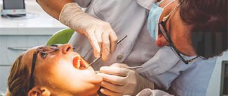
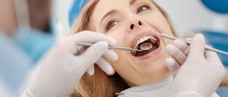
- A thorough visual examination and examination of the condition of the teeth and gums. The specialist determines the problem by the appearance of the tissue.
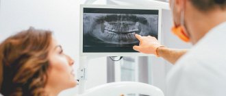
- Instrumental (X-ray) examination is a study using special equipment. A key element of modern dental diagnostics.
X-rays of teeth
Milk, mixed, permanent dentition. Their characteristics.
Milk: 20 teeth, 1 increase in bite during teething 1 mol. molar, shape of dental arches - semicircular, physiological trema/diastema, physiological abrasion of teeth, distal surfaces of second piers. molars form the mesial stage, resorption of the roots of the molars. teeth, reducing the depth of the incisal overlap to a direct ratio, the formation of the TMJ ends.
Replaceable: root resorption, physiological abrasion, trema, eruption of permanent teeth (timing, sequence, pairing), 2nd stage of increasing the bite during eruption 1st post. molars
Constant: 3 increase in bite during the eruption of canines and 2 molars, 32 teeth, upper z.r. - semi-ellipse, lower - parabola, each tooth has two antagonists except (3.1, 4.1, 1.8, 2.8), the upper tooth is in contact with the same and behind the lower one , normally - med.cheek tubercle of 1st molar. located in the fissure between honey. and Wednesday cheeks with cusps of 1 molar of the lower part, and the upper canines are located between the lower canines and premolars, the sagittal gap between the incisors is 1-2 mm, the upper incisors overlap the lower ones by 1/3 (incisal-tubercle contact between them), the upper lateral teeth overlap the lower ones by 1/3 the depth of the longitudinal fissure, the palatal cusps of the upper molars and premolars lie in the longitudinal fissures of the lower ones, the midlines coincide, tight contacts, between the most protruding cusp of the second molar of the mandible and the cutting edge of the lower central incisor there should not be a distance of more than 1.5 mm. (Curve of Spee).
X-ray diagnostics
The use of X-rays and other high-precision equipment in diagnostics allows us to obtain, in real time, accurate images of the desired areas of the oral cavity in optimal resolution.
What equipment is used for instrumental diagnostics:
- Radiovisiograph (visiograph, videograph, dental imaging apparatus) is a device that converts X-ray data into a digital image on a computer in real time. The dentist, using a visiograph, controls the treatment procedure without leaving the chair with the patient. The device allows you to quickly take a photo and obtain an image of the problem tooth.
- Digital 3D orthopantomograph - allows you to take a panoramic image of the desired area of the skull or an individual tooth. The device is indispensable for prosthetics, treatment, implantation, bite correction (orthodontics), etc.
- Rheodentograph is a device for accurately assessing the functional state of the pulp (tissue containing nerve endings) of a tooth according to hemodynamic indications. Allows you to quickly, painlessly and accurately determine the extent of tissue damage. The device is important in the gentle treatment of pulpitis.
- A tool for diagnosing early and latent caries. Pathological processes of hard tissues in the early stages are often difficult to determine. At the same time, treating caries in the early stages allows you to solve the problem with minimal impact on the tissue. The device allows you to effectively identify crisis areas on the tooth.
The capabilities of the equipment allow you to print the results or transfer them to electronic media.
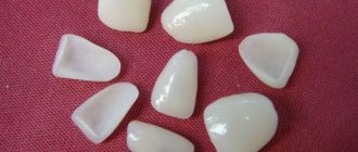
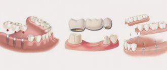
Diagnostic measures include control modeling methods, as well as taking impressions. These methods can significantly increase the effectiveness of orthodontic treatment and prosthetics.
The main task of the dentist is to maintain the health of teeth and gums, maximize tissue preservation, and ensure functionality and aesthetics. All these problems cannot be solved without competent dental diagnostics.
Gnathology
What is gnathology?
Gnathology is a science
that studies the effectiveness of the dental system in normal and pathological conditions.
Chewing is a vital function of processing and swallowing food. Gnathology - as a field of dentistry
, deals with the identification and treatment of pathology of the temporomandibular joint.
What does a gnathologist treat?
The most important element of the human masticatory apparatus is the paired
temporomandibular joint (TMJ). Movements of the lower jaw occur as a result of the complex interaction of the masticatory muscles, temporomandibular joints and teeth and are regulated by the neuromuscular system. The temporomandibular joint coordinates the movement of the lower jaw in three directions, and any movement occurs with simultaneous sliding and rotation of the heads of the lower jaw. During normal operation of the joint, painless opening and closing of the mouth is ensured by the intra-articular disc, which is a cartilage pad. Dysfunction of the temporomandibular joint (TMJ) interferes with proper chewing of food, speech formation,
and all this is accompanied by pain and a “clicking” of the joint when opening or closing the mouth.
The position of the jaw is disrupted, which leads to improper closure of the dentition. Any deviations in the functioning of the elements of the mandibular joint require competent consultation with a gnathologist. IMPORTANT: A gnathologist-dentist is engaged in functional diagnostics and treatment of dysfunctions of the temporomandibular joint (TMJ).
TMJ dysfunction leads to difficulties in diagnosis, as it has a variety of clinical manifestations. It is important to emphasize that a gnathologist as a specialist must have extensive practical experience in therapeutic and orthopedic dentistry. Incorrectly placed fillings, crowns and orthopedic structures in most cases are the root cause of pathology and a trigger for the malfunction of the dentofacial apparatus. As a specialist, a dentist orthopedic gnathologist will not only determine the cause and degree of dysfunction of the dentoalveolar system, but also propose an optimal treatment plan to eliminate them.
IMPORTANT:
If you need a highly professional consultation with a gnathologist, contact the Dentist dental clinic. Specialists with more than 17 years of experience will explain to you in detail and clearly the cause of your illness and offer a reasonable treatment plan.
Symptoms for which you should contact a gnathologist.
It is no secret that patients mostly turn to the dentist complaining of pain in the teeth. But how do you understand that pain and discomfort in the teeth and jaw are related to the joint? The first signs of dysfunction that you should pay attention to are:
- Rapid fatigue and muscle hypertonicity when chewing.
- Involuntary clenching of the jaws.
- Local or diffuse pain in the masticatory muscles.
- Discomfort when opening and closing the mouth.
- Bruxism (teeth grinding) Unconscious muscle activity not associated with chewing or speaking
Signs indicating the presence of TMJ dysfunction in the later stages:
- Clicking in the joint that occurs when opening the mouth, chewing, or yawning.
- Joint blocking and displacement. The mouth opens in a zigzag or S shape.
- Inability to chew solid food due to pain
- Limited mouth opening.
Patients report severe headaches, migraines of unknown etiology, congestion and tinnitus. Pain and muscle spasms not only in the masticatory muscles, but also in the muscles of the neck and occipital region. With joint dysfunction, dizziness, dry mouth, sleep disturbance, snoring, sleep apnea (breathing stops for 10 seconds) may also occur. IMPORTANT:
When such symptoms appear, treatment of TMJ by a gnathologist is mandatory. Otherwise, the incorrect functioning of the joint will eventually lead to the development of irreversible degenerative processes and immobilization (arthrosis).
Causes of TMJ dysfunction.
Overload of the masticatory apparatus is the basis of dysfunction, but there can be many reasons.
- Occlusal-articulatory
(closing of the dentition) the problem includes:
incorrect prosthetics, placement of flat fillings, single and multiple defects in the dentition,
pathological abrasion of teeth
jaw injuries, malocclusion. All this is accompanied by a decrease (decrease) in the height of the bite. - Another problem is related to mechanical overload of the jaw muscles
and their spasm. Unilateral chewing, bruxism, and heavy speech load ultimately lead to chronic microtrauma of the elements of the TMJ. - And the third important factor is neuropsychic and physical stress
associated with changes in the activity of the central nervous system (CNS).
How does a gnathologist consultation work?
Consultation with a gnathologist-dentist is an important stage in the treatment of TMJ dysfunction and
begins with collecting complaints and anamnesis. Then the doctor proceeds to examine the oral cavity, assesses the closure of the teeth, the functioning of the muscular system, the position of the head, as well as the severity of facial asymmetry. Particular attention is paid to the temporomandibular joint, assessing it in a static position and when performing movements.
A gnathologist, who is maximally focused on obtaining the most effective treatment result, will study all the elements of the muscular-ligamentous apparatus of the neck area, the tension of the back muscles, and the patient’s posture. These elementary examination methods allow a competent specialist to quickly make a preliminary diagnosis and determine the scope of the examination and treatment tactics.
An obligatory part of diagnostics is computer and hardware research. From the variety of studies, the gantologist will select those necessary for treatment.
Often used in the practice of a gnathologist are:
- Orthopantomogram and plain radiograph of the TMJ, allowing to detect
abnormalities in the development of bone tissue of the ligamentous apparatus. - Computed tomography, which provides a layer-by-layer study of the condition of the joint and dystrophic changes.
- MRI of the joint has the greatest diagnostic value for studying the condition of the periarticular soft tissues and articular disc.
- Graphic research, axiography, method of recording movements of the lower jaw using a special apparatus;
- Analysis of occlusal contacts using an articulator device - a device that reproduces the movements of the lower jaw.
- Diagnostic plaster models of jaws.
Among functional diagnostics, myography is especially in demand - a method for studying the masticatory muscles, the quality of static and dynamic closure of teeth. There are other techniques: electromyography, phonoarthrography, gnathodynamometry. Modern computer programs help speed up the diagnostic process for TMJ lesions, as well as form a more accurate opinion about the mechanism of development of the pathological process. With their help, it is possible to offer optimal correction methods. All of the above methods evaluate the functioning of muscles and joints, determine exactly how the teeth close and help identify the cause of joint dysfunction.
IMPORTANT:
To ensure high quality diagnostics, all these methods can be used by gnathologists at the Dentist clinic.
What treatment methods are used.
Treatment carried out by a gnathologist usually involves an integrated approach. In the treatment of gnathological disorders, it involves the use of therapeutic and diagnostic devices.
- To restore the correct position of the jaw, use special
joint splints
. The technique is effective in combating bruxism, pathological tooth wear, and for relieving muscle tension in the maxillofacial and cervical region. - To stimulate the masticatory muscles of the face and neck, a high-tech system is used - a deprogrammer
. Designed specifically for use in neuromuscular dentistry and gnathology. This unique stimulator allows you to erase (deprogram) the masticatory muscle dysfunction reflex and restore normal tone. - If the dysfunction of the dentofacial apparatus is caused by poorly performed orthopedic treatment, with a serious decrease in the bite, then before repeated prosthetics, an individually made occlusal splint (splint)
, which has a lasting memory effect. Can be installed on one or two jaws at once. Splin not only corrects occlusion (closing), but also solves several functional problems. Evenly distributes the load when chewing, reduces the activity of the muscular system and thereby corrects the direction of movement of the lower jaw.
Dentist orthodontist-gnathologist.
Orthodontists recognize that most malocclusions are caused by problems other than dental problems. Most often, the development of orthodontic problems is caused by such anomalies as violation of the dimensions of individual elements of the jaw apparatus, deviations in the position of the jaws relative to the base of the skull, pathology of the temporomandibular joints or
chewing muscles. Cases of the development of malocclusion due to orthopedic problems of the spinal column are no exception. Malocclusion in 90% of cases is accompanied by pathological abrasion
, and consequently, a decrease in the height of the lower half of the face. Treatment with braces and aligners in such situations is useless and ineffective.
IMPORTANT: Orthodontic treatment cannot restore the previous volume of dental tissue in case of pathological abrasion, it cannot restore single and multiple defects in the dentition, and all this is directly related to dysfunction of the mandibular joint.
When, during a consultation, an orthodontist encounters existing disorders in the functioning of the maxillofacial apparatus, he is obliged to consult with a gnathologist-orthopedist. This will help to avoid mistakes and negative results after orthodontic treatment. If we objectively approach the problems of dysfunction, then an orthodontist-gnathologist
is not the best combination of specializations
. Since orthodontists, as specialized specialists, are very far from therapeutic and orthopedic dentistry and do not have sufficient knowledge and experience necessary for the effective treatment of TMJ dysfunction. The orthodontist corrects the abnormal position of the teeth in the jaw, improves the appearance of the dentition, and restores the correct closure of the dentition, but this is not enough to treat such a complex pathology.
IMPORTANT:
The specialists of the Dentist clinic are well aware that an interdisciplinary approach is very important and the orthodontist acts only as a specialist whose work is necessary in the intermediate stage of treatment of joint dysfunction.
Dentist orthopedist-gnathologist.
The gold standard or standard of a gnathologist is an orthopedic gnathologist.
In a broad sense,
If we consider the specialty of an orthopedist in dentistry, then this particular doctor is in charge of
diagnosing
and correcting defects of the musculoskeletal system. Having extensive experience in eliminating problems associated with the masticatory apparatus during prosthetics and deep knowledge in restoring its functions, this is an ideal candidate for a gnathologist.
The success of orthopedic treatment directly depends on the design of the prosthesis and its ability to evenly distribute the load during chewing. In this case, the condition of the temporomandibular joint and masticatory muscles is taken into account. Uniform distribution of the load and restoration of the correct bite height is the most important stage in the treatment of TMJ dysfunction.
Advantages of contacting specialists at the Dentist clinic.
By contacting the Dentist clinic on Chistye Prudy,
Each patient can count on highly qualified
consultation
with an experienced gnathologist-orthopedist. The center will be able to undergo examination using modern diagnostic methods, as well as carry out all stages of this treatment.
The advantages of the Dentist clinic are:
- High professional level of doctors in this profile.
- The ability to use advanced diagnostic and treatment methods, which will completely eliminate existing symptoms
- At the highest level, carry out orthopedic or orthodontic treatment for TMJ dysfunction.
The price for a consultation with a gnathologist in Moscow varies widely, and often does not correspond to the professional level of the specialist or the diagnostic capabilities of the center.
If you decide to make an appointment with a gnathologist-orthopedist at the Dentist clinic, you will be pleased to discover that, despite the high level of service, the consultation is free .
Initial examination
Diagnosis of diseases in dentistry begins with a patient interview and visual examination. It is very important to tell your doctor the symptoms of the disease in detail. These may include: pain, discomfort when eating hot or cold food, sweet or sour taste. If you have allergies to any medications, hereditary or genetic diseases, be sure to tell your doctor. Smoking can also affect the condition of your teeth and gums.
The dentist will examine your mouth using a mirror and probe. For a detailed examination, all Dental Guru clinics have microscopes. If the problem and its solution are obvious, then if you agree, treatment can begin immediately. To clarify the diagnosis, the doctor will conduct the necessary functional diagnostics.
Diagnocat artificial intelligence: fast CBCT analysis, accurate diagnosis
Only in our clinic, Diagnocat artificial intelligence is used for CBCT analysis! The program uses 3D images to determine the condition of the teeth, finds problems and suggests how to treat them. In just a few minutes, Diagnocat will examine your CT scan, identify problem areas, and generate a report for each tooth. And the doctor will make an accurate diagnosis! More about Diagnocat here.
Are there any disadvantages to digital diagnostics?
The disadvantages of high-precision digital solutions include their low prevalence due to the high cost of the equipment, as well as the time spent by the doctor. The technologies are relatively new and are just beginning to come into practice. There are still few doctors who have a full range of equipment and can carry out the analysis inside and out. Most are just learning new products and using only individual devices - for example, myographs or face arcs. That's why you won't see them in every clinic.
Smile-at-Once is so far the only clinic in Moscow with a full diagnostic range of devices and programs for planning orthopedic treatment and creating prosthetics.
Content
- X-ray
- Panoramic shot
- Computer radiovisiography
- Tomography
PROMOTION
Dental restoration, installation of fillings
from 2200 rub.
The importance of correct diagnosis in dental treatment is difficult to overestimate. The effectiveness of treatment, its duration, and long-term results directly depend on how correct the doctor’s assumption regarding your clinical picture is. Diagnostic methods in dentistry depend on the level of equipment of the clinic you choose.
Practical significance of the photo protocol
Creating a photo protocol is necessary for a more detailed assessment of the condition of the dental system. When diagnosing a disease, photography is an integral part of the procedure.
The manipulation is carried out in accordance with the protocol, so even the smallest details are subject to analysis and assessment by a specialist.
According to the object, the photo protocol can be extraoral and intraoral. An important nuance of the procedure is the position of the patient’s head; when creating a series of images, it must remain unchanged.
What information can be obtained as a result of creating an extraoral protocol:
- indicators of tension in the muscular system of the neck and head;
- data on the symmetry (asymmetry) of the dental system;
- information about the individual characteristics of the patient’s appearance.
The intraoral protocol allows:
- analyze the condition of the jaws;
- identify the presence of phenomena such as tooth abrasion or wedge-shaped defect;
- assess the position of the teeth.
The photologging technique is used to evaluate both intermediate and final results.
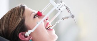
The importance of condylography in identifying dysfunction of the dentoalveolar system
Condylography is a method used to analyze the position of the temporomandibular joint. The main task of the manipulation is to establish the trajectory or, more simply put, the path of movement of the jaw. The technology significantly simplifies the diagnostic procedure and helps identify pathological processes.
Note! The information obtained as a result of condylography can be used not only when making a diagnosis, but also to implement the prosthetic procedure. Thus, the availability of data on the individual indicators of the patient’s TMJ movement trajectory allows the orthodontist to make the most suitable design.
Scanning facial parameters using the Proface scanner
The final stage with the participation of the patient is scanning of facial parameters. There are two options here - use a facial scanner and/or a special application for Mac OS on the iPad.
The Proface scanner or program creates a three-dimensional model of the patient's appearance. For this purpose, a specially developed craniometric analysis algorithm is used (craniometry is a technique for measuring the skull). That is, many points of the skull are calculated - due to this, a copy of the patient’s face and all the bones of the head is transferred to the computer.
In addition, photometry is carried out - photographing the face is necessary to evaluate the result “live” and maintain a digital photo record.
An example of photometry is photographing the face from all angles before and after dental implantation with the installation of a fixed prosthesis immediately
We approach diagnostics wisely! We use the latest developments and artificial intelligence! We make an accurate diagnosis and find the best solution to your dental problems.
Sign up for a consultation
Optical strip scanning
This technology shows better results compared to the above. A special grid or stripes are projected onto the object. Based on their distortions, the relief of the scanned surface is determined. In this case, it is enough to determine the coordinates of the object’s surface using one camera and additional structured lighting. This approach is most often used in modern orthopedic dentistry.
Structured light can be different - laser, infrared, visible. The main condition is automatic recognition of these lines by software. The known distance between the projected stripes allows you to calculate the coordinates of the surface points. The more lines, the more accurate the result will be. The more cameras are used, the higher the scanning speed will be.
Hardware methods of functional diagnostics
Detection of dental ailments in the initial stages is carried out using devices such as radiovisiograph and orthopantomograph. The radiation exposure of these devices is within normal limits, so the risk of adverse effects is excluded.
Hardware methods include:
- tomography;
- rheodentography;
- electroodontodiagnostics.
When making a final diagnosis, dentists adhere to the principle of complexity. It is possible to obtain an overall picture of the condition of the patient’s dental system only through a series of diagnostic procedures and their subsequent analysis.