Errors in dental prosthetics - classification
The most common mistakes made by dentists when making dental prosthetics:
- Purpose for installation of a bridge prosthesis. Such an error may be associated with the patient’s desire to install this particular type of prosthesis for existing problems in the dentition. Dentists often follow the lead of their clients and this leads to sad consequences: the bridge is either not clearly fixed or has mobility, teeth are injured, and periodontitis develops.
- Choosing porcelain crowns in the absence of indications. Porcelain crowns are considered the highest quality, most expensive and “prestigious” - it is not surprising that clients of dental clinics insist on installing them. But the doctor should not expand the list of indications for such a procedure - for example, installing porcelain crowns for malocclusion (deep-set upper teeth) will lead to their rapid wear and certain inconvenience when chewing.
- Ignoring the need to treat gums and bone tissue when installing a complete removable denture. Under no circumstances should removable dentures be made, let alone prescribed for wearing, if there are existing inflammatory/infectious diseases of the oral mucosa! Such an unprofessional approach will lead to thinning of the bone tissue, jaw deformation, and the development of osteomyelitis.
- Violation of dental treatment rules. Depulpation of a tooth before prosthetics is a mandatory procedure, which, if ignored, can lead to the development of pulpitis, tooth removal, and the development of severe inflammatory processes in bone tissue. And this, in turn, is fraught with surgical intervention and plastic surgery.
Installation of low quality prosthesis
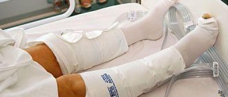
Initially, in the practice of endoprosthetics, low-quality products were used, which had a number of significant disadvantages. When installing them, repeated operations were required in 70–80% of cases after the first 2 years of use due to:
- periprosthetic infection;
- frequent dislocations;
- prosthesis fractures;
- instability of fixation of implant components;
- displacement of the head of the knee endoprosthesis or hip endoprosthesis;
- destruction of components (primarily the liner).
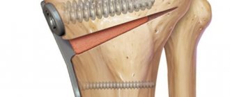
Today in Russia, knee endoprostheses have been replaced by analogues of a more advanced configuration made from modern materials of high reliability. They solved many problems. Although Russian practice has still not reached the level of prosthetics results in countries with developed medicine (Germany, Israel, USA). For effective surgical treatment, high-quality prosthetic structures are not enough. A set of clear, understandable standards for endoprosthetics and a revision of rehabilitation protocols are also required.
Solution
Errors in dental prosthetics usually occur due to improper work of the dentist. Any neglect of the necessary procedures preceding the actual prosthetics leads to irreversible complications. It will be impossible to correct/correct anything - you will have to remove the prosthesis (even permanent crowns and onlays), prescribe full treatment for the problem, and then repeat the prosthetic procedure again.
The dentist should never make concessions to patients - they often ask to install a prosthesis for carious teeth, not to pay attention to swelling of the gums (and this is a sign of inflammation), to choose the most expensive crowns. Such rashness has no place - the doctor must explain the situation to the patient in detail and inform the patient about the possible consequences. And patients themselves should be guided by the recommendations of professionals - this will be the key to obtaining the desired effect.
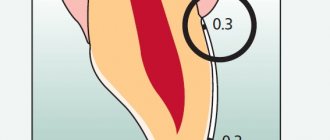
Different leg lengths after endoprosthetics
Changes in limb length after endoprosthetics are more often observed when a hip joint endoprosthesis is installed. As a rule, the leg in which the joint was replaced becomes longer, less often - shorter, than the second limb.
Reasons for changes in leg length:
- lack of radiological control during surgery;
- selection of an implant without preliminary collection of parameter measurements (offset, SDU). This error is typical for cases of installation of a knee joint endoprosthesis or hip replacement with cementless monoblock legs.
In some cases, a change in the length of the limbs can also occur with the correct installation of a knee endoprosthesis . A common reason is the lack of quality rehabilitation or its negligent implementation.
Measuring the lateral inclination of the acetabulum. The right minor trochanter is located lower than the left, indicating a leg length discrepancy. Normal horizontal center of rotation (red line).
What errors occur during implantation and prosthetics on an implant?
All possible errors during surgery can be divided into two groups: without using scientific medical terminology, they can be called surgeon errors and orthodontist errors.
Surgeon mistakes
Errors of a purely surgical nature include the following incorrect actions:
- neglect of the rules of antisepsis and asepsis (that is, working in unsterile conditions, unclean instruments, etc.);
- incorrect selection of anesthetics for a particular patient (after all, before administering anesthesia, the operating surgeon must find out whether this person has specific reactions to the proposed anesthetic, whether there are additional diagnoses, chronic diseases, etc., which may affect susceptibility to certain painkillers) ;
- incorrect administration of the anesthetic (to the wrong point, in the wrong volume, etc.);
- inattention to the presence of anatomical features in the patient’s operated area;
- carelessness when handling alveolar ridge tissues;
- incorrect selection of the diameter of the dental implant - if it exceeds the diameter of the mucotome;
- the drills do not cool down while working with them, their rotation speed is incorrectly selected;
- selection and use of drills in the wrong sequence (according to the rules, you should select from smaller to larger diameter - although this is not always relevant);
- the distance between the implants and the roots of adjacent teeth is not maintained;
- the dental implant is inserted into the prepared socket at the wrong speed (it cannot be quickly and easily inserted like a designer part - the doctor must constantly monitor the insertion so as not to damage the tissue and cause complications during healing);
- the whole operation is carried out too hastily or too slowly (both have their downsides and their risks).
Orthodontist mistakes
Basically, these are those inaccuracies due to which the implant is ultimately not ideal for installation in a particular patient, as well as errors in the installation of the implant. Mistakes made by an orthodontist when installing dental implants include:
- the supporting parts are prepared inaccurately;
- the axes of the implant support elements are not parallel;
- fewer supports than necessary;
- the height of the lower facial region is incorrectly determined;
- the neck of the implant does not fit tightly with the edges of the crown - that is, these elements do not fit well;
- the length of the implant and the height of the tooth crown are in the wrong proportion;
- the width of the crown is significantly greater than the diameter of the implant;
- the crown attached to the implant contains a part of artificial gum made of plastic;
- the axis at the crown and the axis of the dental implant have an angle between themselves exceeding 27 degrees;
- the configuration of the tooth crown is incorrect, which creates a risk of the abutment unwinding or breaking;
- a gap between the abutment and the implant itself, which leads to poor fixation;
- the prosthesis on a dental implant is poorly fixed, which can lead to decementing or the screw that secures the crown may come loose;
- fissure-tubercle contacts, which should be between the implanted artificial tooth and the “native” antagonist teeth, are formed incorrectly, which can ultimately lead to traumatic occlusion;
- the garland of the implant crown is poorly polished and has an insufficiently smooth surface;
- the denture is fixed in a rigid way on the implant and conditionally “movable” teeth;
- the intercoronal spaces are too narrow, which will create difficulties for the patient in cleaning them and performing hygiene procedures;
- gingival risk factors and the risks of periodontitis are not taken into account;
- the dimensions of the console and the crown do not correspond (i.e., the implant will be permanently overloaded on one side).
Complications during the second stage of the operation
- Removing the intraosseous element of the implant along with the plug.
- Penetration of the implant into the maxillary sinus.
- Formation of a section of bone tissue over the intraosseous element.
The intraosseous element may become unscrewed if the process of reparative bone regeneration is disrupted and implant integration is lacking. In this case, the implant can simply be returned to its original place, the patient can be prescribed calcium supplements, and after a month and a half, the second stage of the operation can be repeated.
Cases of pushing the intraosseous part of the implant into the cavity of the maxillary sinus, as a rule, are the result of subantral implantation and a slowdown or disruption of the course of reparative bone regeneration. In this situation, surgery is required to remove the implant from the sinus cavity.
If bone tissue has formed over the intraosseous implant, this phenomenon is not considered a complication. You just need to make an incision in the periosteum and mucous membrane, remove the bone formation using a saw, and during the installation of the former and the gingival cuff of the implant, make sure that nothing else prevents their correct screwing.
II. Learning objectives of the lesson:
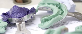
- Have an idea of the complications that may arise at the clinical and laboratory stages of manufacturing bridges and obtaining working prints (α - I);
— Know the features of preparing teeth for bridges (α — II);
- Know the principles of casting (α - II);
- Know the principles of designing bridges (α - II);
— Be able to perform the clinical stages of manufacturing stamped-soldered and solid-cast bridges.
What to do if there is inflammation under the prosthesis?
The first thing to do is contact your doctor or, better yet, go to a good dental clinic that will offer several treatment options.
If prosthetics involves installing a pin, treatment becomes more complicated, since removing it from the root canal is quite difficult and can result in a root fracture. After this, the tooth will have to be completely removed. The issue of repeated prosthetics is quite complicated, since a root fracture can damage the bone plate and the implant cannot be installed, just like any pin.
When the cause of a complication after installing a crown is improper treatment of the canals, again the refilling is difficult and may result in perforation.
When it is possible to unseal the canal or remove the pin, anti-inflammatory therapy is carried out. After this, the canals are re-filled and a new prosthesis is installed.
We can conclude that it is better to carry out painstaking preparation for prosthetics and take into account all possible complications before installing the prosthesis, rather than treating the resulting inflammation.
Oral hygiene after dentures
To maintain a healthy and beautiful smile, it is not enough to simply insert a tooth. You also need to take care of careful care of the oral cavity and the dentures themselves. Hygiene is the basis of dental health. After installing a denture, hygiene requirements increase, since food particles can get stuck in the elements of the orthopedic structure, plaque accumulates on the walls of the denture, and thus creates a favorable environment for bacteria - the main culprits of all diseases of the teeth and gums.
Care of fixed dentures:
- 1
regular brushing of teeth (recommended not only in the morning and evening, but also after each meal);
- 2
rinsing with special antiseptic preparations;
- 3
professional dental cleaning every 3 months.
Care will become much easier if you purchase an irrigator - a device that effectively washes away food debris in the most inaccessible areas of the oral cavity and is completely safe for dentures.
Rules for caring for removable dentures:
- after each full meal or snack, the denture should be removed and washed under running warm water;
- In the morning and evening, dentures, just like teeth, need to be cleaned (for this, use a toothbrush with soft bristles and a special paste for dentures);
- in order to prevent the accumulation of plaque, it is recommended to soak the denture in a special solution for 10-20 minutes after each cleaning;
- From time to time it is necessary to take your dentures to the dentist for cleaning;
- Removable orthopedic structures should be stored in clean water or a special solution for storing prostheses.
New generation removable dentures, made of high-quality polymers, are so comfortable that they can be left on at night.
Prosthetic stomatitis
Denture stomatitis is a consequence of the inflammatory process that develops in the gum tissue under the denture.
The cause of inflammation may be:
- improper fixation, excessive pressure of the structure on the gum, due to which blood circulation in the tissues is disrupted, bedsores appear, ulcers form, and the necrotic process begins
- diabetes;
- long-term use of steroids and some antibacterial drugs;
- low quality of oral hygiene.
In response to inflammation of the mucous membrane, the lymph nodes may become inflamed. The treatment program for prosthetic stomatitis will depend on its causes. If necessary, the installation of the prosthesis is corrected. As part of preventative treatment, proper, thorough hygiene is key. Additionally, the oral cavity can be sanitized with antiseptic drugs.
Why does inflammation begin under the prosthesis?
At the present stage of orthopedic treatment, all materials are so carefully selected that the risk of complications is reduced to a minimum. But everything has its exceptions.
Poor canal filling before prosthetics is a mistake not of the orthopedist, but of the therapist. Incomplete filling of the tooth canal leads to the entry of unfavorable microflora into it, which multiplies and provokes inflammation of periodontal tissue. The patient feels pain, swelling at the site of the installed prosthesis, and pulsation. In this case, it is extremely necessary to re-treat and reinstall the prosthesis.
Inflammation may occur under a fixed prosthesis or crown if an instrument is left in the canal during treatment or the canal wall is perforated. In any case, 95% of complications are associated with improper root canal treatment.
A carious cavity under the prosthesis is a complication of prosthetics and is also associated with improper preparation for prosthetics. Caries under a denture develops in two cases: incomplete removal of carious tissues and non-compliance with the rules of sealants during prosthetics. When there are holes for food debris to penetrate, it always leads to the formation of plaque and caries.
Complications during implantation
- Fracture of the pilot drill or bur.
- Damage to the bottom of the maxillary sinus or penetration of the drill into the nasal cavity.
- Violation of the integrity of the wall of the mandibular canal and damage to the inferior ventricular nerve.
- Damage by bur to the lower and lateral compact layers of the lower jaw.
- Partial or complete absence of primary fixation of the implant.
- Violation of the integrity of the wall of the alveolar process.
Damage to instruments can have various causes: excessive pressure on the fissure bur at the time of longitudinal drilling of the implant bed, violation of temperature conditions for sterilization of equipment, or the implant exhausting its service life in 30 sterilization cycles.
Damage to the floor of the maxillary sinus may be the result of an erroneous determination of the height of the alveolar process or excessive pressure on the instrument. If such a situation arises, you should refrain from installing the implant in this place and, if possible, install it in close proximity to the already formed bed. Another possible option is to install an implant, the length of the intraosseous part of which is two millimeters less than the depth of the finished bed. In this case, the bed must first be filled with bone chips or hydroxyapatite removed from the instrument. The recommended method of implantation in this case is a two-stage one, and it is better to choose a screw or combined intraosseous element.
Damage to the inferior ventricular nerve and injuries to the wall of the mandibular canal can be caused by negligence in preparing the bone bed or incorrect determination of the size of the implant due to possible distortion of the vertical size of the mandible on the orthopantomogram. If the preparation of the canal wall results in the occurrence of an intracanal hematoma and subsequent compression of the nerve, then restoration of sensitivity in the area of innervation can be expected in two to three weeks. In cases of osteoporosis, the wall of the mandibular canal may be defective or absent altogether; in this case, the effect on the lower ventricular nerve can be explained by hemorrhage in the area of the medullary spaces, as well as swelling of the reticular tissue of the bone marrow. Partial loss of sensation (or parasthesia) in the lower lip area may be felt the day after surgery and disappear completely after five to seven days. If the decrease in sensitivity of the lower lip, caused by a violation of the integrity of the wall of the mandibular canal and the inferior alveolar nerve, persists for one to two weeks, then the implant must be removed and the necessary symptomatic treatment performed.
Violation of the integrity of the lower or lateral compact layer of the lower jaw, by and large, is not a complication, but if during control radiographs it turns out that part of the implant extends beyond the jawbone by more than two millimeters, it is necessary to replace the installed implant with another one, whose height of the intraosseous part is less.
A fracture of the alveolar process wall very often occurs as a result of the installation of a plate implant if the bone bed under it was formed of a smaller size than necessary. Another possible reason for this complication is the narrowness of the alveolar process. In this case, you need to press the broken part to the appendage and stitch the wound.
If the implant in the bone bed is mobile and there is no fixation, the reason for this may be either improper preparation of the bone bed or osteoporosis. If the preparation of the bone bed was performed incorrectly, the installed implant can be replaced with a similar one, but of a slightly larger diameter (if this is allowed by the existing anatomical conditions), or the installed implant can be kept in the existing bed, filling the gaps in its upper part with bone chips. If the cause of implant mobility is osteoporosis, it can be fixed by filling the bed with osteoconductive or osteoinductive material. There is another option: replacing the existing implant with an implant of a different design, for example, a cylindrical one with a screw one, without cutting threads in the bed, which was prepared for installing a cylindrical implant.
Galvanic syndrome
If metal is present in the denture structure, it is very important that all subsequent dental prostheses are made from this metal. The fact is that different metals have different electro-mechanical characteristics, which can lead to the occurrence of galvanic currents upon contact with an electrolyte (saliva plays the role of an electrolyte in the oral cavity).
Symptoms of galvanic syndrome:
- 1
the appearance of a sour or metallic taste in the mouth, a feeling of soreness;
- 2
changes in taste sensations (for example, a feeling of bitterness when eating sweets);
- 3
burning sensation on mucous tissue, tongue;
- 4
increased saliva viscosity, dryness;
- 5
pathological changes in mucous tissue - the appearance of swelling, redness, numbness;
- 6
sensation of electric current flowing in the gum tissue, along the teeth, tongue or in the palate;
- 7
hypersensitivity of the gums and tongue;
- 8
change in color of dentures (darkening).
Galvanic syndrome causes not only local discomfort in the oral cavity, but can also cause a general deterioration in the patient’s well-being.
Chips in the lining of orthopedic structures
The outer layer of orthopedic products is made of ceramics. Thanks to the veneer, dentures and crowns look like real teeth – natural and aesthetically pleasing. The facing layer is quite durable, and subject to high-quality prosthetics and proper operation, orthopedic structures retain their original appearance and integrity for many years.
Chips of the aesthetic layer are possible for two reasons - errors at the planning stage, manufacturing of the orthopedic structure, and the patient’s violation of the ban on cracking nuts and gnawing bones.
In case of a chip, depending on the nature and size of the damage, you can correct the situation in two ways:
- 1
Replace the ortho construction.
- 2
Restore the facing layer without removing the prosthesis.
IV. Interdisciplinary integration
| Disciplines | Know | Be able to |
| Normal anatomy | Structure of the oral cavity | Graphically depict the anatomical shape of teeth |
| Histology | Features of the structure of oral tissues | Graphically depict the histological structures of the tooth |
| Physics chemistry | Physical and chemical properties of materials used for the manufacture of bridges | |
| Orthopedic dentistry | Classifications of dentition defects, principles of bridge design prostheses, biomechanics. Methods of preparation when turning and tilting abutment teeth. |