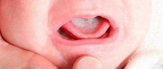
Milium (horny cyst, whitehead, “millet”) is an epidermal cystic formation at the mouth of the hair follicle, looking like a small white grain. Typically, milia are localized on the face in areas with relatively thin skin, very rarely on the body, and exist for a long period without any changes. Milia are a fairly common cosmetic problem and can occur at any age. Whiteheads appear especially often during puberty; also often found in newborn babies (neonatal acne). Female representatives are more susceptible to the appearance of milia.
Causes
Milia occur in patients with a thick form of oily seborrhea, characterized by hyperfunction of the sebaceous glands and changes in the chemical composition of sebum and creating a favorable background for the development of acne.
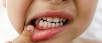
There are closed comedones - whiteheads (milia) and open comedones - blackheads (blackheads). Thick seborrhea is also characterized by the appearance of deep cysts of the sebaceous glands - atheromas and acne vulgaris. Milia have the appearance of small rounded dense nodules of a white-yellowish hue, 0.5-3 mm in size with clear boundaries, protruding above the surface of the skin and resembling millet grains (popular name - “millet”). Milium is characterized by an uninflamed head, since the whitehead does not have a natural outlet and contact with the environment. Milia do not change in size for a long time. When microorganisms enter the milium, inflammation may develop with the formation of an abscess. Milia (milia) can often be found on the wings of the nose in recently born babies; they are considered borderline conditions in newborns and soon disappear spontaneously without any treatment.
Milia are recognized based on an examination of the skin by a dermatologist-cosmetologist based on characteristic external signs.
Features of children's skin
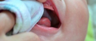
At birth, babies have very thin skin. The skin of a newborn is almost half as thick as the skin of an adult. The outer layer thickens with age.
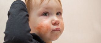
The skin of newborns is red or purple in color due to the close proximity to the upper layer of blood vessels and an insufficient layer of subcutaneous cellular tissue, as a result, the skin appears “transparent”. This phenomenon is especially pronounced if the baby is frozen - the appearance of a marbled vascular network on the body is observed.
Moisture evaporates faster from the skin of newborns. Children are more susceptible to bacteria, viruses, fungi and mechanical influences. Thickening of the skin begins at 2-3 years of age and ends by 7 years.
Treatment of milia
Milia do not go away on their own; it is almost impossible to get rid of them with the help of cosmetics. Under no circumstances should you squeeze out milia yourself, as this will damage the hair follicle and sebaceous gland. Such self-medication often leads to the subsequent formation of a larger acne or the addition of an infection, which can cause the formation of a rough scar.
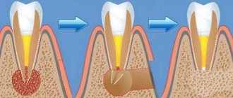
Treatment of milia is carried out by a dermatologist and consists of opening and removing the cyst capsule with its contents. The choice of removal method depends on the location, number, size and depth of the milia. For single milia, mechanical removal is carried out (using a sterile needle and curettage) with preliminary and subsequent antiseptic treatment of the skin. Small wounds left after such a procedure heal on their own without leaving a trace.
For multiple whiteheads, modern methods are used - removal of milia with a laser, radio wave method or electrocoagulation. The crusts formed at the cauterization site disappear on their own within 10-14 days. Removing more than 10 elements at one time is not recommended to avoid significant trauma to the skin and disruption of the sebaceous glands.
Symptoms of atheroma in children
In 80-85% of cases, apart from the visual defect, children do not present any other complaints. Uncomplicated atheroma is characterized by:
- painlessness;
- mobility;
- elasticity upon palpation.
The skin over the surface of the tumor is smooth and does not fold. The clinical picture may change when the contents of the atheroma become inflamed against the background of the addition of bacterial flora. In this case, patients may note:
- pain when pressed;
- redness and increase in size of the tumor itself;
- local increase in body temperature.
If the above symptoms occur, you should immediately seek help.
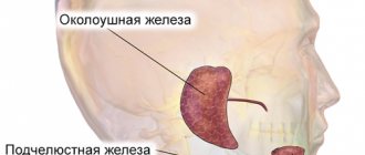
This situation threatens the penetration of infection into nearby tissues and blood with the spread of microorganisms to distant areas of the body and can cause a deterioration in the general condition of the child. Atheromas occur in areas of the skin that are rich in sebaceous glands. Therefore, they especially often have the following localization:
- scalp;
- face and neck area;
- shoulder girdle, armpit area;
- back.
Neoplasms occur less frequently in the groin area and scrotum (in boys). In these areas they have the appearance of a skin growth and require differential diagnosis with other skin tumors.
Prevention of milia
To prevent the appearance of milia, careful facial skin care is required using cleansers and moisturizers appropriate to the skin type, scrubs, peels, cosmetic masks that normalize the activity of the sebaceous glands; elimination of provoking factors, proper and rational nutrition, healthy lifestyle. Salon cosmetic procedures, including mechanical, ultrasonic, vacuum facial cleansing, peelings (chemical, mechanical - microdermabrasion), laser resurfacing, are quite effective in preventing the appearance of milia.
Diagnosis of atheroma in children
It is relatively easy to identify atheroma.
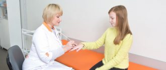
Even at the stage of the initial conversation with the patient or his parents, the surgeon assesses the general well-being of the child, collects anamnesis and analyzes complaints. When examining a neoplasm, the doctor pays attention to its size, location, pain, and the presence of redness. Before surgery, the surgeon prescribes a number of tests to comprehensively assess the child’s condition:
- general and biochemical blood test;
- general urine analysis;
- blood test for infections (HIV, syphilis, viral hepatitis B, C);
- ECG, fluorography.
The child is also examined by an anesthesiologist and pediatrician before the operation. A mandatory diagnostic method is histological examination of tumor tissue after its removal. At this stage, it is possible to determine whether it was an atheroma or a lipoma (a tumor similar in appearance).
Questions
- Which doctor treats atheroma in children?
A pediatric surgeon is involved in identifying and treating atheroma in children. - Is it possible to cure atheroma without surgery?
Sometimes in the early stages of the disease the surgeon chooses a wait-and-see approach. In 20-35% of cases, atheroma can resolve on its own. However, if the capsule is formed and the tumor is large, it will not be possible to do without surgical intervention. - How dangerous is atheroma in childhood?
Atheroma is a relatively safe tumor. It does not become malignant, rarely becomes complicated and is not accompanied by the development of permanent defects. However, this does not eliminate the need to consult a pediatric surgeon. Only a doctor can establish the correct diagnosis and select treatment. In addition, atheroma can sometimes become inflamed due to the addition of bacterial flora and cause a deterioration in the child’s well-being. - Is it possible to play sports after surgery?
After surgical removal of atheroma, it is recommended to refrain from active activities for 2-3 weeks. This will allow the tissues to fully heal and recover.
Treatment of atheroma in children
The optimal method of treating atheroma in children is its surgical removal.
However, the age of the patient decides a lot here. Thus, most pediatric surgeons, in the absence of severe clinical symptoms, recommend a wait-and-see approach. Sometimes the swelling may resolve on its own. This is especially true for newborns and infants. In such cases, the development of the neoplasm is observed until the age of three, and if it persists, only then is it removed. Surgical intervention can be carried out traditionally using a scalpel or minimally invasively using a laser. In the first case, the surgeon excises the tumor with a capsule, which eliminates the risk of relapse. After this, cosmetic stitches are applied to the tissue to achieve the best aesthetic result.
Laser removal in children is used for small tumors. Using this technique, it is possible to avoid incisions and the formation of postoperative scars. In both cases, a comprehensive examination of the child is first carried out with the selection of the optimal treatment option.
In the postoperative period, the child may be prescribed the following groups of medications:
- painkillers;
- anti-inflammatory;
- antibacterial.
Drug therapy is aimed at eliminating discomfort and preventing infection of the postoperative wound.
Signs of mastitis in newborns
- when touching the mammary glands, the baby cries;
- as a rule, the infection penetrates into one of the glands (much less often it affects both), due to which the mammary gland becomes denser, the baby develops severe pain when touched;
- in the area of infection the skin turns red;
- The baby’s body temperature rises to 39 °C;
- the child cries when he is swaddled or changed;
- the baby refuses to breastfeed and becomes lethargic;
- later, pus accumulates at the site of infection; when pressure is applied to this area, the child screams very loudly and experiences pain;
- with purulent mastitis, pus may spontaneously (without pressure) be released from the nipples of a newborn.
This condition is extremely dangerous for a child. Therefore, at the first symptoms of mastitis, you should consult a pediatrician or call an ambulance. If help is not provided in time, the pus can melt the tissues near the mammary gland and penetrate into its other parts. This condition can lead to a diagnosis of phlegmon of the chest wall - purulent inflammation of fatty tissue. More serious conditions that mastitis in a newborn can lead to are sepsis and generalized infection (spread of infection throughout the body), which threatens the life of the child.
For a newborn girl, mastitis is more dangerous than for a boy: if the acini (components of the mammary gland) die during the disease, connective tissue appears in their place. In this case, when the girl grows up, her breasts will most likely develop asymmetrically. And during breastfeeding, an adult woman runs the risk of serious lactostasis (milk stagnation), which will be difficult to cure without surgery.
How does physiological mastopathy differ from mastitis in newborns?
What happens to the baby's mammary glands after birth? The endocrine system is activated, so the baby’s body needs maternal hormones less and less. The pituitary gland of a small organism begins to produce estrogens, resulting in the release of prolactin: this causes engorgement of the mammary glands in the child, from which a milky fluid can be released.
Within a week after birth, the baby experiences a condition called a hormonal crisis. Nature is designed in such a way that it is the hormonal crisis that forces the body to adapt to the world: having gone through it, the child receives certain protection from problems with the immune and neurological system.
How is mastitis treated in newborns?
Clinical guidelines for the treatment of neonatal mastitis state that before prescribing therapy, it is necessary to establish the stage of development of the disease.
If this is the initial (infiltrative) stage, then treatment of mastitis in a newborn is recommended:
- special ointments applied to the bandage;
- ultraviolet irradiation or UHF, which has a detrimental effect on bacteria;
- compresses with magnesium to relieve swelling and pain;
- compresses of dimexide with saline as an antiseptic;
- alcohol compresses;
- Vishnevsky ointment;
- suppositories or syrup with paracetamol to relieve pain and fever;
- Treatment of neonatal mastitis can be enhanced with broad-spectrum antibiotics.
However, all of the above actions can be effective until a purulent focus has formed in the gland. If it occurs, the newborn will most likely require surgery, during which the pus will be removed and the cavity will be washed. After surgery, a bandage with a hypertonic solution will be applied to the wound. In addition, the doctor will prescribe a course of antibiotics.
Neonatal mastitis
Mastitis is an acute or chronic inflammation of the mammary gland. It is usually customary to talk about mastitis in a woman who is breastfeeding, but children also have mastitis , especially in the newborn period, when the baby experiences a sexual crisis with swelling of the glands and parents try to “treat” this crisis with various warm-ups, ointments, tinctures and squeezing out milk. from pieces of iron. Usually, all these attempts lead the child and his parents to the surgeon, at best for an appointment, at worst - to the operating table with a purulent abscess.
Mastitis can also develop due to defects in care, when prickly heat with pustules appears on the skin, the baby is rarely washed or his immunity is reduced and the infection penetrates through the nipple area when they are injured.
Features of breast swelling
Physiological mastopathy or engorgement of the mammary glands is the physiological condition of a child’s breasts, in which they increase in size. Usually the mammary glands are enlarged evenly, occasionally there is unilateral enlargement. An increase of up to 3 cm in diameter is considered normal if there is no redness or changes under the skin or on its surface.
Sometimes grayish or milky-white contents may be released from the mammary gland ducts; its composition is comparable to that of colostrum.
Typically, breasts begin to enlarge on the second day after birth, and gradually decrease from the end of the first week, but they completely disappear by the month. Such mastopathy does not require any treatment; you should not press the breasts, trying to remove milk from them, apply compresses with ointments, especially like Vishnevsky ointment, camphor and others, which is widely recommended on the Internet.
Swollen breasts do not cause any discomfort to the child, they are not treated in any special way, only with strong magnification can a clean, dry and sterile bandage be applied. It is necessary to protect the breasts from friction with clothing.
However, many parents are worried about breast enlargement and begin using bandages, squeezing out milk, secretions and fiddling with the swollen breasts, unwittingly introducing microbes into the cracks of the papillae. They penetrate deep into the chest and cause the development of a serious complication - neonatal mastitis. If its onset is neglected, the course of the disease can be severe, even fatal. How to distinguish the onset of mastitis from physiology?