Dental implantation is a complex procedure that requires intervention in the soft tissues of the oral cavity. To avoid all sorts of complications, it is very important to follow the dentist’s recommendations. Swelling and redness may be observed for several days after surgery, which is considered normal. If other unpleasant symptoms appear, you should consult a specialist to prevent rejection of the prosthesis and other pathological conditions.
Functions of the gums during implantation
The purpose of implantation is to restore not only the chewing, but also the aesthetic functions of the dentition. This problem can be solved only by having a sufficient volume of bone tissue (to hold the implant, distribute the chewing load) and healthy mucous membrane (to protect the bone, provide access to nutrients, and protect the implantation area from infection).
Therefore, the gum around the implant must have the desired histological structure and in sufficient volume to provide:
- Aesthetics Recreate a gingival contour that does not differ from the area of neighboring teeth.
- Functionality Provide nutrition to the bone around the implant, as well as support for the crown or prosthesis.
- Prevention of rejection Protect the implant from plaque and bacteria, reduce the risk of peri-implantitis.
BozhkovRoman Andreevich
Implant surgeon, 13 years of experience
Nobel Biocare Certified Specialist
Leading specialist in bone reconstruction and ultrasonic sinus lifting, high-complexity gum surgery
More about the doctor
WHAT ARE THE SIGNS OF GUM RECESSION?
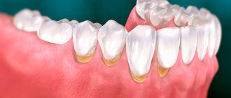
Recession (recession) of the gums is accompanied by various symptoms. During recession, the gums become shorter, gradually exposing the roots of the teeth. Therefore, one of the first symptoms of gum recession is increased tooth sensitivity. At the same time, the teeth look longer, and there is a small groove at the edge of the gum, where it comes into contact with the tooth.
Even with these signs, you can't be sure if you have gum recession. To ensure early detection and treatment of gum disease, visit your dentist regularly. It is especially important to detect gum recession in the early stages, since timely treatment is the only way to prevent further development of the disease.
When is soft tissue augmentation necessary?
An operation to replenish gum volume is performed when there is a deficiency of gum, which can arise due to a number of factors.
Initial gingival deficiency
To achieve good aesthetics, the thickness of the gum tissue must be at least 5 mm. But such ideal conditions are extremely rare for reasons:
- Long-term absence of a tooth - without chewing loads, the bone atrophies, the gums also shrink and decrease
- Wearing removable dentures or adhesive bridges - mechanical pressure leads to soft tissue loss
- Thin mucosal biotype is a physiological feature of the body in which the gum thickness is only 2-3 mm
In such cases, there is a high risk of the formation of an unaesthetic gingival contour and the absence of gingival papillae around the implant.
Even when planning treatment, our specialists analyze the volume and condition of the gums in order to, in case of deficiency, provide for surgery to build up soft tissue at one of the stages of treatment.
Errors during implantation
Loss of gum volume is also possible during implantation if the operating surgeon does not know how to work correctly with mucous tissues. Common mistakes made by novice doctors:
- Excessive, thoughtless mechanical damage to gingival tissue at one of the stages of the operation
- Excessive tension when suturing - healing occurs with pronounced defects
Not only the surgeon, but also the orthopedist must understand the importance of maintaining the volume and quality of soft tissue. Temporary structures during the healing stage should not interfere with the regeneration process. Incorrect placement and pressure of the temporary prosthesis on the mucous membranes leads to their slow growth and trauma.
If the operation and prosthetics were carried out negligently, plastic surgery on the gums after implantation cannot be avoided in 99% of cases.
Therefore, we recommend that you carefully choose a clinic and doctors. Exposure of the implant
The formed gum around the implant (as well as around the teeth) is of two types:
- Attached - motionless, tightly fused with the periosteum
- Movable - moves when lips and cheeks move
If there is a lack of attached gum or the patient does not comply with the rules of care (aggressive brushing of teeth or, conversely, lack of oral hygiene), the implant neck is gradually exposed - soft tissues are torn off, the gums recede, and periodontal pockets are formed.
To prevent such situations, it is necessary to replenish the gum volume even before implantation and follow the doctor’s recommendations during the recovery period. If the moment was missed and the implant was exposed, gingivoplasty is performed, but in such cases a number of procedures are first performed to eliminate inflammation.
If you ignore the problem, inflammatory processes can spread to surrounding tissues and provoke serious complications:
- Mucositis - inflammation of the soft tissue around the artificial root without involving bone structures
- Peri-implantitis is a more severe condition where bone tissue detaches from the implant.
In our Center, implantation is performed by experienced implantologists and maxillofacial surgeons who are fluent not only in implantation protocols, but also in various techniques for carefully working with soft tissues. We do not strive to quickly “screw in” implants at any cost. Our task is to take care of the condition of the mucous tissues, since the success of implantation depends on this, which we carry out in the format of a lifetime guarantee.
Materials and manufacturers
In the manufacture of implantation systems and auxiliary products, medical titanium, stainless steel, and other metal alloys are used. For installation in the frontal zone, there are models made of zirconium that are not visible through the gum tissue. Manufacturers offer different models of shapers. The doctor selects an option according to the clinical situation. The classification is based on the shape, size and type of part. The line includes standard, narrow, and wide models.
Standard ones are used in cases involving a translingual method of intervention, when correction of the mucous membrane is carried out immediately before installation of the implant. Markers are applied to the surface of the element, allowing you to control the level of fouling of soft tissue. If the diameter of the prosthetic structure is more than 5 mm, wide models are used, if less than 5 mm, narrow models are used.
The most famous manufacturers of implantation systems and components for them:
- NSK (Japan)
- Nobel Biocare (USA)
- Astra Tesh (Sweden)
- Trate AG (Switzerland)
- Bicon (USA)
- Niro (Germany)
- Semantos (Germany)
- Impro (Germany)
- Adin (Israel)
- Alpha Bio (Israel)
- NeoBiotech (South Korea)
Of course, the list is not limited to the brands listed. The cost of placing a gum former depends on the material, brand, technical parameters, and size of the superstructure. For good results, it is recommended to install implants and auxiliary components from the same manufacturer.
The network of RUTT clinics in Moscow uses Swiss implantation systems and ROOTT superstructures produced by Trate AG. All components are made of the same grade of titanium and fit perfectly due to their exact size. This is a universal, reliable implantation system, the effectiveness of which is scientifically proven.
At what stage of implantation is gum plastic performed?
Depending on the causes of the gum deficiency and the period of manifestation of the defect, the procedure is performed during one of the stages:
- Before or simultaneously with implantation In case of initial gum deficiency or thin biotype, operations are best performed before implantation. But if the implantologist is skilled in perioplasty, the procedure is also possible during implant installation.
- After implantation before prosthetics This stage is often chosen after osseointegration of the implant - during its opening in preparation for the installation of an orthopedic structure. At this stage, errors after implantation are corrected through gingivoplasty and the immediate installation of a gum former.
- In the delayed period of operation This is carried out if the parameters of the mucous membranes were not taken into account in the early stages, and over time the implant neck became exposed. It is recommended to carry out the procedure as soon as possible to reduce the risk of bone inflammation and prevent rejection of the artificial root.
Gumplasty is a necessary solution for successful implantation
By replenishing the missing volume of soft tissue, the aesthetic gum contour is recreated, and the risk of infection of bone tissue and implant rejection is reduced.
Levin Dmitry Valerievich Chief physician and founder of the Doctor Levin center
How long to wear?
Typically, the process of forming the gingival contour lasts from 2 to 4 weeks; this all happens individually for each person. Therefore, to answer the question: how long can you walk with a gum former? – we can answer that it must be used until the formation of uniform soft tissue edges is completely completed. Your healthcare provider will let you know when to replace the element and secure the abutment. Under no circumstances should you ignore the specialist’s request and continue to wear the gum former.
Operation stages
The operation in the area of one implant lasts 30-60 minutes. Absolutely painless, because... performed under local anesthesia. For patients with fears, it is possible to perform the operation “in their sleep”.
- Anesthesia Infiltration or conduction anesthesia is used. If desired by the patient or according to indications, sedation (controlled sleep).
- Plastic Depending on the clinical picture, local tissues are used to form the gums, a flap of the required size or matrix is applied.
- Suturing is carried out under the control of a dental microscope, using the finest threads. Neat sutures without displacement or tissue tension improve healing.
Why is it better in sedation
Sedation

