Hygienic index
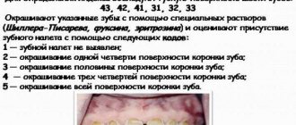
The Green-Vermilion hygienic index allows you to evaluate the amount of tartar and plaque separately. To determine it, six teeth are studied: 31, 11, 16, 26 - vestibular planes, and 36, 46 - lingual. Plaque can be assessed using dye solutions (Fuchsin, Schiller-Pisarev, Erythrosin) or visually.
The following codes and criteria for dental plaque exist:
- 0 – no layers;
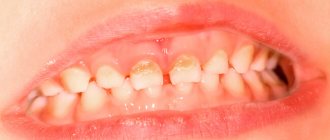
- 1 - soft plaque covering no more than 1/3 of the tooth plane, or the presence of any number of colored deposits (brown, green and others).
- 2 – thin layer located on less than 2/3, but more than 1/3 of the surface of the molar;
- 3 – soft plaque, occupying more than 2/3 of the tooth plane.
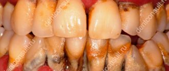
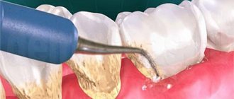
Determination of sub- and supragingival molar stone is carried out using a dental probe.
What else is good about the Green-Vermilion index? Assessment of dental calculus (criteria and codes) is as follows:
- 0 – no stones present;
- 1 – supragingival deposit covering no more than 1/3 of the tooth plane;
- 2 – formation located above the gum, covering less than 2/3, but more than 1/3 of the plane of the tooth, or the presence of individual growths in its cervical region;
- 3 – supragingival layer covering more than 2/3 of the tooth plane, or large deposits of stone located near its neck.
The Green-Vermilion index is calculated by adding the values produced for each of its elements, dividing by the number of planes studied and adding both values.
12.4. Determination of gingival index - gi (Loe, Silness, 1963)
Intended to determine the location and severity of gingivitis. Used for clinical and epidemiological studies.
Each tooth is examined in four areas:
- gingival papilla from the medial and distal surfaces of the tooth;
- marginal gums from the vestibular and lingual surfaces of the tooth.
To determine bleeding, the gums are probed with a blunt instrument.
Codes Criteria
0 - Normal gum.
1 - Mild inflammation, slight discoloration, slight swelling, no bleeding on probing.
2 - Moderate inflammation, redness, swelling, bleeding on probing.
3 - Severe inflammation with noticeable redness and swelling, ulceration, and a tendency to spontaneous bleeding.
The gums are examined in all teeth or selectively by segments, sextants. Key teeth are 16, 21, 24, 36, 41, 44. The GI value for a site is determined by summing the codes around the examined tooth. The sum of the site codes divided by 4 indicates the GI of the tooth. To obtain the GI values of an individual examined, all GI values of the teeth must be summed and divided by the number of teeth examined.
Formulas for calculating the index:
| GI tooth = | ∑ points |
| 4 |
Interpretation:
0.1-1.0 - mild gingivitis;
1.1–2.0-moderate gingivitis;
2.1 – 3.0 — severe gingivitis;
| Individual's GI = | G.I. teeth |
| n teeth |
Inspection and subsequent calculation of the index require certain knowledge and manual skills. GI is the most accurate in assessing the effectiveness of the anti-inflammatory effects of drugs.
Cliche
The calculation formula is attached as follows:
IGR-u = sum of plaque values/number of planes + sum of stone values/number of surfaces.
The interpretation of the index (the IGR value at the level of medicine) is proposed as follows:
- 0.0-1.2 - flawless;
- 1.3-3.0 – acceptable;
- 3.1-6.0 – low.
The Green-Vermilion index has the following values for dental plaque standards:
- 0.0-0.6 – impeccable;
- 0.7-1.8 – tolerable;
- 1.9-3.0 – bad.
KPU indices
What do oral hygiene indices express? One of the basic dental coefficients (BDC) demonstrates the intensity of decay. The letter “K” means the number of damaged teeth, “P” - the number of filled teeth, “U” - the number of teeth to be removed or liquidated. The sum of these values gives an idea of the development of the decay process in a particular person.
There are three types of KPU coefficient:
- KPUz – the number of carious and treated teeth in the subject;
- KPU of planes (KPUpov) – the number of destroyed faces;
- KPUpol – the sum of fillings and carious cavities.
For non-permanent teeth, the following indicators are used:
- KP – the number of damaged and healed teeth of short-term occlusion;
- KP – sum of rotten planes;
- CPR – number of carious cavities and fillings.
Teeth lost as a result of physical change or extracted are not taken into account in an unstable dentition. In children, when changing teeth, two coefficients are used simultaneously: KPU and KP. To identify the overall intensity of the disease, both degrees are summed up. KPI from 6 to 10 confirms a high intensity of decay, 3-5 – moderate, 1-2 – low.
These standards do not show the real picture, as they have the following shortcomings:
- both extracted and cured teeth are taken into account;
- can only increase over time and begin to reproduce past carious damage with age;
- do not allow for initial damage to be taken into account.
Serious shortcomings
Significant flaws in the indicators of KPUz and KPUpov include their uncertainty with increasing decay due to the formation of new depressions in healed teeth, loss of fillings, the occurrence of secondary caries and similar factors.
The multiplicity of caries is shown as a percentage. To do this, the composition of people in whom this disease was found (except for focal demineralization) is divided by the number of those studied in this team and multiplied by one hundred.
In order to assess the prevalence of tooth decay in a particular region, the following estimated conditions for the level of prevalence among twelve-year-old children are used:
- low intensity level – 0-30%;
- relative – 31-80%
- large – 81-100%.
Evaluation of indicators
There are three main directions for assessing caries lesion indicators;
- Extent of caries spread. This indicator is the percentage of the number of patients diagnosed with caries to the total number of people examined.
This technique is often used to compare oral health status in different regions of the country.The levels of caries prevalence among 12-year-old patients are used as assessment criteria: a low level corresponds to an indicator from 0 to 30%, an average level from 31 to 80%, and a high level from 81 to 100%.
- The intensity of spread is an indicator that determines the degree of caries damage to the elements of the jaw row of a particular patient. For children under 12 years of age, the following assessment criteria apply:
- very low intensity – up to 1.1;
- low – up to 2.6;
- moderate – from 2.7 to 4.4;
- high – from 4.5 to 6.5;
- very high – more than 6.5.
For adults, this breakdown changes slightly and looks like this:
- very low – up to 1.5;
- low – from 1.6 to 6.2;
- moderate – from 6.2 to 12.7;
- high – 12.8-16.2;
- very high – more than 16.2.
- Increase in caries infection. This indicator allows you to assess the dynamics of the development of the carious process in an individual patient.
To determine it, the intensity of caries over a certain period of time is revealed.A change in the value of the indicator up or down indicates the effectiveness or ineffectiveness of the therapeutic process.
Components of the Opalescence home teeth whitening system and how it works.
Come here to find out if dental teeth whitening is harmful.
At this address https://zubovv.ru/krasota-i-uxod/otbelivanie/domashnee/limonom-i-kak.html we offer several effective recipes for teeth whitening with lemon.
CPITN Index
The hygienic state of the oral cavity is assessed using different indices. Let's consider the CPITN coefficient. It is used in clinical practice to monitor and examine periodontal health. Using this index, only those signs that can begin to develop in the opposite direction are recorded (tartar, gum inflammation, which is judged by bleeding), and does not take into account irreversible changes (tooth mobility, gum recession, loss of epithelial attachment).
CPITN does not record process activity. This coefficient is not used for treatment planning. Its most important advantage is the speed of identification, information content, simplicity and the ability to compare results. The need for treatment is determined based on the following signs:
- code X or 0 means that there is no need to treat the patient;
- 1 indicates that a person should take better care of his or her mouth;
- 2 means that it is necessary to eliminate the factors influencing the retention of plaque and carry out professional hygiene;
- Code 3 indicates constant oral hygiene and curettage, which usually reduces inflammation and reduces pocket depth to less than or equal to 3 mm;
- 4 means that adequate hygiene of the oral mucosa is necessary, as well as deep curettage. In this case, combined treatment is required.
General overview
In dentistry, indices are a quantitative reflection of the progress of the pathological process that has developed in the periodontium.
When compared with clinical diagnostic methods, index assessment is considered relative, since it does not reflect the absolute accuracy of the ongoing process.
But periodontal indexing has some advantages that distinguish it from clinical diagnosis. With its help you can:
- measure the dynamics of tissue infection;
- monitor the spread and development of periodontal diseases;
- monitor the progress of treatment;
- evaluate its success and the results of preventive measures;
- perform epidemiological screening of the population;
- make comparisons of the results of different studies.
There are several dozen assessment indices for studying the condition of periodontium. All of them are divided into 3 groups:
- Reversible . With their help, the dynamics of diseases and the effectiveness of treatment are assessed. Reflect the severity of the inflammatory process, bleeding gums, loose teeth, the size of periodontal and gum pockets.
- Irreversible. They convey the manifestation of such signs as resorption (absorption) of bone tissue in the alveolar process, amyotrophy of the gums.
- Complex. They help to give a comprehensive description of the condition of periodontal tissues, taking into account the existing symptoms and the degree of development of pathological processes.
Children's hygiene
What is the Fedorov-Volodkina index? It can be used to determine whether the patient takes good care of his teeth. This indicator should be used to assess the oral health of children under 5-6 years of age. To establish it, the labial edge of six teeth is studied.
Using special solutions, teeth are stained and the presence of plaque on them is assessed. Determination of sub- and supragingival calculus is carried out using a dental probe. The calculation of the coefficient consists of the numbers obtained for each of its elements, divided by the number of planes studied, followed by the addition of both values.
Norm
The Fedorov-Volodkina index (1968) is still used in our country today.
First, the labial surface of the six front lower teeth is stained with a potassium-iodine-iodide solution. The hygienic index is determined by the intensity of the resulting color, then it is assessed using the five-point method and calculated using the formula Kcp=(∑Ku)/n, where:
- Ksr – general hygienic cleaning coefficient;
- Ku – healthy indicator of cleaning one tooth;
- n – number of teeth.
Coloring the entire plane of the crown means 5 points; 3/4 – 4; 1/2 – 3; 1/4 – 2 points; lack of color – 1. Normally, a healthy indicator should not exceed 1.
Assessment of tooth mobility using the Miller scale as modified by Flesar;
Determination of tooth mobility according to D.A. Entinu.
Algorithm for determining tooth mobility.
Algorithm for determining the dental plaque index according to Silness-Loey.
Algorithms for determining indexes.
The index evaluates the amount of soft plaque in the gingival area. The assessment is carried out visually and with a probe without staining, 4 surfaces are examined, for better diagnosis, the area of the tooth neck is pre-dried with an air jet.
Plaque intensity, evaluation criteria:
0 – there is no plaque on the tip of the probe;
1 - a small amount of plaque;
2 – a thin layer of plaque near the neck, a significant amount at the tip of the probe;
3 – a significant amount of plaque in the gingival area and in the interdental spaces.
The index is calculated using the formula:
General index = (sum of points) / (number of teeth examined).
Mullemann bleeding index (modified by Cowell).
Determines the degree of bleeding of the gingival sulcus during probing or pressure on the gingival papilla.
In the area of “Ramfjord's teeth” (16,21,24,36,41,44) from the buccal and lingual (palatal) surfaces, the tip of the periodontal probe, without pressure, is guided from the medial to the distal surface of the tooth.
0 — after the study there is no bleeding;
1 - bleeding appears no earlier than after 30s;
2 — bleeding occurs either immediately after the test or within 30 s;
3 - bleeding occurs when eating or brushing teeth.
Index value = (sum of indicators of all teeth) / (number of teeth).
The generally accepted classification of pathological tooth mobility according to D.A. is based on Entin (Entin D.A. 1954) is the direction of the visually determined displacement of the tooth relative to its axis.
I degree – tooth displacement only in the vestibulo-oral direction;
II degree – visible displacement of the tooth both in the vestibulo-oral and medio-distal
III degree – tooth displacement in the vestibulo-oral, medio-distal and vertical
directions: when pressing, the tooth is immersed in the hole, and then
it returns to its original position again.
The method used to assess pathological mobility according to the Miller scale (Miller SC 1938) modified by Fleszar (Flezar et al., 1980):
0 - stable tooth, there is only physiological mobility;
1 - tooth displacement relative to the vertical axis is slightly greater, but does not exceed 1 mm;
2 - the tooth moves 1-2 mm in the buccal-lingual direction, the function is not impaired;
3 - mobility is pronounced, while the tooth moves not only in the buccal-lingual direction, but also vertically, its function is impaired.
Algorithm for determining the class of furcation defect.
Furcation defect of the alveolar bone is a defect in the bone tissue of the interradicular septum in the area of furcation of multi-rooted teeth. To determine the state of the furcation defect, a furcation probe is used. Probing of the furcation is carried out in the horizontal direction. Based on the magnitude of the horizontal spread of the process, the class of furcation lesions is distinguished. There are several classifications of furcation defects.
Classification by I. Glickman (1958):
Class I - alveolar bone resorption, which exposes the root furcation area, but is not accompanied by destruction of the interradicular bone.
Class II—interradicular bone is partially lost, but there is no through defect.
Class III - a through defect in the furcation area is detected during probing, but is hidden by the gum.
Class IV - a through defect of the interradicular septum, the furcation area can be examined in the oral cavity and is not hidden by the gum.
Classification by PJ Heins, SP Canter (1968):
Class I - the apex of the alveolar ridge exposes the arch of the root furcation; horizontal probing of the bone defect can be accompanied by immersion of a graduated probe up to 2 mm in the direction of the interradicular septum.
Class II - in addition to signs of a class I furcation defect, a horizontal immersion of a graduated probe in the direction of the interradicular septum by more than 2 mm is possible, but the instrument does not penetrate to the opposite side.
Class III - corresponds to the free penetration of a graduated probe to the opposite side when it moves in the horizontal direction.
Classification by J. Lindhe (1983):
Initial (grade 1) destruction of the interradicular septum by 1/3 of its cross section or less.
Partial (class 2) destruction of the interradicular septum exceeds 1/3 of its cross section, but does not form a through defect.
Total (grade 3) destruction of the interradicular bone in the horizontal direction with the formation of a through defect.
Inspection
What are the indicators for assessing the results of medical examination by a dentist? It is known that a comprehensive examination of residents involves a method of protecting their health, consisting of providing conditions for their impeccable physical development, preventing illnesses through the implementation of proper sanitary, hygienic, preventive, therapeutic and social measures.
The purpose of medical examination is to strengthen and preserve people's health and increase their life length.
A medical examination is designed to solve the following problems:
- annual analysis of a person’s well-being;
- comprehensive monitoring of patients;
- combating bad habits, identifying and eliminating the causes of tooth decay;
- active and timely implementation of health-improving and therapeutic measures;
- increasing the efficiency and quality of medical care to the population through the successive and interconnected work of all types of institutions, large-scale participation of doctors of various professions, the introduction of technical support, new unifying forms, the creation of mechanical systems for examinations of the electorate with the development of special programs.
Examination according to the WHO method
The purpose of conducting an epidemiological survey using the WHO method is to compare data obtained in different regions and regions of the country, which makes it possible to assess the spread of dental diseases among different categories of the population.
For this purpose, there is a unified examination methodology consisting of three stages:
- Preparatory. A plan for carrying out the procedure is drawn up, which formulates the goals and objectives of the upcoming work.
The personnel to carry out the manipulations is determined, consisting of two doctors and a nurse, as well as a group of subjects taking into account age, social status, standard of living, and gender.The level of accuracy of the analysis will depend on the number of patients examined.
- Examination of patients. A thorough examination of the condition of the patient’s oral cavity is performed: his teeth, gums, mucous membranes, structural features of the jaw apparatus and the formation of the bite.
All results are entered into a special registration card using standard codes in compliance with the accepted algorithm for filling it out. - Summarizing. The final stage is the assessment of the results obtained as a result of the examination with the calculation of the required hygienic indices and the prevalence of certain dental diseases.
Based on the results of the survey, specialists have the opportunity to study the dental health of patients of different age groups, social conditions and areas of residence. This allows you to take timely measures to prevent and treat certain pathologies.
Observation of children
By calculating the Green-Vermilion index, doctors can create dispensary groups for monitoring children:
- Group 1 – children who have no pathologies;
- Group 2 – actually healthy babies with a history of any chronic or acute disease that does not affect the function of the most important organs;
- Group 3 – children with chronic illnesses with a balanced, sub- and decompensated course.
There are three phases in the dental examination of children:
- In the first phase of the examination, each child is individually recorded, an additional examination is carried out in the hospital, then an outpatient observation group is determined, the endurance of each child is assessed and the order of examinations is determined.
- In the second, a contingent is formed according to supervision groups, uniform conditions for phasing and continuity of study are assigned, dispensary patients are proportionally divided between doctors, and the needs of the examined contingents in inpatient and outpatient treatment are met.
- In the third, doctors determine the frequency and nature of active supervision of each child, adjust diagnostic and treatment measures in accordance with changes in health status, and evaluate the effectiveness of supervision.
Of great importance is the organization of educational work to prevent dental diseases in children and create motivation to care for newly emerging teeth.
Examination stages
Determining dental indices is a complex procedure that includes several main stages.
Examination stages:
- Elementary. It consists of a subjective examination of the patient: studying complaints, conducting a survey for certain symptoms. At the initial stage of diagnosis, the patient’s age and the nature of his activity are taken into account.
- Preparatory. The dentist prepares the necessary materials and instruments for the examination. A dental mirror and probe are used for inspection. If necessary, prepare a coloring solution.
- Examination. The dentist examines the oral cavity in accordance with the criteria for a specific index assessment. First, the oral cavity is dried from salivary fluid. Palpation can also be performed, which is necessary to determine swelling of soft tissues and bleeding gums.
- Final. At the final stage, the results are assessed and a diagnosis is made. The dentist evaluates the quality of the hygiene procedures performed. If pathological signs are detected, the degree of need for treatment or preventive procedures is determined.
Important to remember! The diagnostic results obtained must be entered into the patient’s medical record.
Oral hygiene can be assessed using different indicators and criteria. Dental indices provide detailed information about the condition of teeth and gums and reflect the likelihood of developing diseases.
Hygiene indices are determined through a dental examination, which is absolutely painless and does not cause discomfort to the patient.
Examination of pregnant women
In order to achieve maximum effect in the prevention of dental diseases, it is necessary to coordinate the work of the dentist and gynecologist, as well as medical examination of women throughout pregnancy. In the dental office, doctors carry out:
- sanitation of the oral cavity;
- assistance in the selection of basic and additional hygiene products, training in rational oral care;
- professional hygiene;
- remineralizing therapy, which increases the resistance of tooth enamel.
The formula will help determine the condition of the mouth
To determine the condition of the oral cavity in medicine, there are about 80 different hygiene indices, based on the principle of staining the enamel with a special solution and identifying dental plaque.
According to the parameters they define, indices can be classified into 4 groups, assessing:
- area affected by plaque;
- thickness of plaque on teeth;
- mass of plaque;
- other parameters of dental plaque (chemical, physical and microbiological).
Prevention of caries
Determining the Green-Vermilion index plays a vital role in the prevention of dental diseases in expectant mothers, which is designed to solve two problems: preventing the development of intrauterine caries in babies and improving the dental status of women.
It is known that the health of the mother affects the process of developing the child’s teeth, which begins at 6-7 weeks of pregnancy. Doctors have determined that with various pathologies in the fetus, the mineralization of tooth enamel slows down, and sometimes stops at the stage of primary calcification. In the postpartum period, it may resume, but will not reach the standard level.
In a woman, already in the early stages of pregnancy, the condition of hard dental tissues and periodontal tissues worsens due to the unsatisfactory hygienic condition of the oral cavity. That is why she must carry out preventive measures until the baby is born. Doctors advise women to adhere to the correct work and rest regime, take vitamin therapy and eat well.
Conclusion
All dental indicators are individual in their own way. They allow you to assess your oral health from different angles. When examining a patient, the dentist uses one or another method based on the individual characteristics of the body and the condition of the oral mucosa.
All research methods are quite simple to use. They do not cause pain to the patient and do not require special preparation. Special solutions for staining plaque are absolutely harmless to the patient.
Thanks to them, the doctor can not only assess the initial condition of the oral cavity, but also predict future deterioration or track changes in teeth and gums after treatment.
Tartar
The tooth surface is sensitive to various influences. Stones form on it due to the following reasons:
- violation of the chewing process;
- habit of snacking and consumption of an impressive amount of carbonated drinks and carbohydrates;
- eating mostly soft foods;
- diseases of internal organs;
- smoking and alcohol abuse.
The composition of supra- and subgingival stones is somewhat different from each other. The first is dominated by calcium carbonate, magnesium and calcium phosphate. In addition, it is very hard. The second is formed from dental plaque, which contains a large amount of food debris, epithelial cells, mucus, bacteria, bound by viscous saliva.
Why do you need to clean your mouth? It helps prevent the formation of stones. Doctors recommend visiting the dentist regularly and using dental floss, flawless toothpastes and high-quality brushes. You can also use toothpicks and mouth rinses.
Language
Now let’s figure out how to clean your tongue. If there is no plaque on this organ, your digestive system is healthy. Since the time of Hippocrates, doctors have asked the patient to stick out his tongue. It is known that an impressive amount of toxins is expelled from the body through its surface. If bacteria accumulate on the tongue, they become toxic.
This organ contains numerous papillae, bumps and pits, among which tiny particles of food are stuck. This is why the tongue is a breeding ground for bacteria. They are transferred with saliva to the teeth, and then a disgusting smell from the mouth appears - halitosis.
If a person regularly cleans his tongue, the access of infection to his body becomes more difficult, the sensitivity of taste buds increases, and gingivitis, digestive tract disorders, periodontal disease, and caries are prevented.
Everyone needs to scrape this organ, especially smokers and those who have a “geographical” tongue, on the surface of which there are deep folds and grooves.
Tongue care is carried out after the teeth are cleaned and the mouth is rinsed. Bacteria are first removed in sweeping steps (from base to tip) on one half of the organ and then on the other. Then we brush across the tongue 3-4 times, apply the paste to it and carefully scrape the organ from root to edge. Next, you need to rinse your mouth, apply the gel again and leave it on for 2 minutes. After these manipulations, you can wash everything off with water.
Cleaning your tongue is a necessary part of hygiene. It is better to eliminate plaque, mucus, food debris that negatively affects the surface of the tooth with a special scraper or brush (can be soft). A disinfecting gel applied to the scraper fills the gaps between the filamentous papillae. During liquefaction, it actively releases oxygen, which has a powerful antibacterial effect on the anaerobic microflora of the oral cavity. If you periodically perform this procedure, the formation of dental plaque will decrease by 33%.
Mouth rinse
Many patients ask: “What should I rinse my mouth with?” If your gums are inflamed, you can use antimicrobial (antiseptic) and anti-inflammatory agents. Antiseptic drugs act on pathogenic bacteria that cause suppuration. Anti-inflammatory drugs have virtually no effect on viruses, but they can slow down the development of the disease.
So what should you rinse your mouth with if your gums are inflamed? Doctors recommend:
- For periodontitis or gingivitis, use both types of products, although antimicrobial ones will be more effective.
- When the socket of an extracted tooth becomes inflamed, antiseptic agents, for example, Chlorhexidine, should be used.
If you always wash your hands before eating and brush your teeth and tongue afterward, you will have a sparkling smile for many years to come.
PMA – papillary-alveolar-marginal index
When calculating the PMA index, a solution of iodine or Lugol is used, which is applied to the gums and the degree of tissue inflammation is determined by the reaction to the irritant.
The evaluation criteria are:
- 1 – if the papilla (P) is inflamed;
- 2 – in case of inflammation of the marginal edge (M);
- 3 – inflammation of the alveolar part of the gum (A).
Formula RMA = Sum of points/n*3 (in percentage), where n is the number of teeth (up to 6 years – 20 teeth; 6–12 years – 24 teeth; 12-14 years – 28 teeth; over 15 years – 30 teeth) .
If the value is less than 30%, then the degree of damage is mild, 31-60% is moderate, 61% or more is severe.
